Download presentation
Presentation is loading. Please wait.

1
Conduction System of the Heart
Faisal I. Mohammed, MD, PhD

2
The importance of calcium influx through the slow voltage gated calcium channels
2

3
Mechanism of Cardiac Muscle Excitation, Contraction & Relaxation

4
Intracellular Calcium Homeostasis…2
4

5
Objectives List the parts that comprise the conduction system
Explain the mechanism of slow response action potential (pacemaker potential) Point out the regulation of the conduction system potential by Autonomic Nerves 5
 Point out the regulation of the conduction system potential by Autonomic Nerves. 5.")
6
Structures of the conduction system

8
Conducting System of Heart
Insert Process Fig with verbiage, Insert Animation: Conducting System of the Heart.exe 8

9
Heart Physiology: Sequence of Excitation

10
Autonomic Innervation of the Heart

11
Intrinsic Cardiac Conduction System
Approximately 1% of cardiac muscle cells are autorhythmic rather than contractile 75/min 40-60/min Approximately 1% of the cardiac muscle cells are autorhythmic rather than contractile. * These specialized cardiac cells don’t contract but are specialized to initiate and conduct impulses through the heart to coordinate its activity. * These constitute the intrinsic cardiac conduction system. These autorhythmic cells constitute the following components of the intrinsic conduction system: * the sinoatrial (SA) node, just inferior to the entrance of the superior vena cava into the right atrium, * the atrioventricular node (AV) node, located just above the tricuspid valve in the lower part of the right atrium, * the atrioventricular bundle (bundle of HIS), located in the lower part of the interatrial septum and which extends into the interventricular septum where it splits into right and left bundle branches * which continue toward the apex of the heart and the purkinje fibers * which branch off of the bundle branches to complete the pathway into the apex of the heart and turn upward to carry conduction impulses to the papillary muscles and the rest of the myocardium. Although all of these are autorhythmic, they have different rates of depolarization. * For instance, the SA node * depolarizes at a rate of 75/min. * The AV node depolarizes at a rate of 40 to 60 beats per minute, * while the rest have an intrinsic rate of around 30 depolarizations/ minute. * Because the SA node has the fastest rate, it serves as the pacemaker for the heart. * 30/min
 node, just inferior to the entrance of the superior vena cava into the right atrium, * the atrioventricular node (AV) node, located just above the tricuspid valve in the lower part of the right atrium, * the atrioventricular bundle (bundle of HIS), located in the lower part of the interatrial septum and which extends into the interventricular septum where it splits into right and left bundle branches * which continue toward the apex of the heart and the purkinje fibers * which branch off of the bundle branches to complete the pathway into the apex of the heart and turn upward to carry conduction impulses to the papillary muscles and the rest of the myocardium. Although all of these are autorhythmic, they have different rates of depolarization. * For instance, the SA node * depolarizes at a rate of 75/min. * The AV node depolarizes at a rate of 40 to 60 beats per minute, * while the rest have an intrinsic rate of around 30 depolarizations/ minute. * Because the SA node has the fastest rate, it serves as the pacemaker for the heart. * 30/min.")
12
Intrinsic Conduction System
Function: initiate & distribute impulses so heart depolarizes & contracts in orderly manner from atria to ventricles. SA node AV node Bundle of His Purkinje fibers As indicated previously, the function of the intrinsic conduction system is to initiate and distribute impulses so the heart depolarizes and contracts in an orderly manner from atria to ventricles. * As you must be able to identify the parts of the conduction system and trace the path of depolarization from the SA node to the purkinje fibers, we will review this. * Since the SA node * has the highest rate of depolarization (75/min) , it starts the process by sending a wave of depolarization * through the myocardium of the atria. When this reaches the AV node * it depolarizes * and causes the Bundle of His * to depolarize.The depolarization travels into the septum through the bundle branches * * and from the bundle branches into the Purkinje fibers * * which cause depolarization of the ventricular myocardium. When the cardiac muscle cells of the myocardium, including the papillary muscles, the ventricles contract forcing blood out of the ventricles. * Bundle Branches
 , it starts the process by sending a wave of depolarization * through the myocardium of the atria. When this reaches the AV node * it depolarizes * and causes the Bundle of His * to depolarize.The depolarization travels into the septum through the bundle branches * * and from the bundle branches into the Purkinje fibers * * which cause depolarization of the ventricular myocardium. When the cardiac muscle cells of the myocardium, including the papillary muscles, the ventricles contract forcing blood out of the ventricles. * Bundle Branches.")
13
Sinus Node Specialized cardiac muscle connected to atrial muscle.
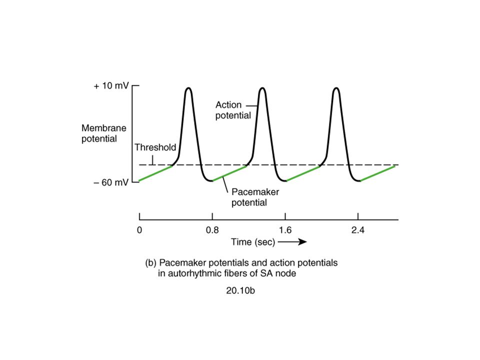
Acts as pacemaker because membrane leaks Na+ and membrane potential is -55 to -60mV When membrane potential reaches -40 mV, slow Ca++ channels open causing action potential. After msec Ca++ channels close and K+channels open more thus returning membrane potential to -55mV.

16
Fast Response Action Potential of Contractile Cardiac Muscle Cell

17
Pacemaker and Action Potentials of the Heart

18
Slow Response Action Potential (Pacemaker Potential)
")
19
Intrinsic rate and speed of conduction of the components of the system
SA node action potential /min (Pacemaker) AV node action potential /min Purkinje action potential /min Conduction Speed SA node: slow speed of conduction Ventricular and Atrial muscle: Moderate speed AV node: slowest speed of conduction Purkinje fibers: Fastest speed of conduction Ectopic Pacemaker- Abnormal site of pacemaker
 AV node action potential /min. Purkinje action potential /min. Conduction Speed. SA node: slow speed of conduction. Ventricular and Atrial muscle: Moderate speed. AV node: slowest speed of conduction. Purkinje fibers: Fastest speed of conduction. Ectopic Pacemaker- Abnormal site of pacemaker.")
20
Extrinsic Innervation of the Heart
20

21
Pacemaker Function

22
Autonomic neurotransmitters affect ion flow to change rate
Sympathetic – increases heart rate by Ca+2 & If channel (net Na+) flow Parasympathetic – decreases rate by K+ efflux & Ca+2 influx What part of the graph is not changed by autonomic influences?
 flow. Parasympathetic – decreases rate by K+ efflux & Ca+2 influx. What part of the graph is not changed by autonomic influences")
23
3 4 3 4 Effect of Sympathetic & Parasympathetic Stimulation
3 4 Sympathetic Parasympathetic Effect of Sympathetic & Parasympathetic Stimulation 3 4 Sympathetic Parasympathetic

24
Regulation of the heart beat
Sympathetic from the cardiac plexus supplies all parts of the heart (atria, ventricle and all parts of the conduction system) Parasympathetic from Vagus nerves supply mainly the atria, SA and AV nodes, very little supply to ventricles Sympathetic: increase the permeability of the cardiac cells to Na+ and Ca++ i.e Positive Chronotropic and positive Inotropic action Parasympathetic: Increase the permeability of the cardiac cells to K+ and decrease its permeability to Na+ and Ca++
 Parasympathetic from Vagus nerves supply mainly the atria, SA and AV nodes, very little supply to ventricles. Sympathetic: increase the permeability of the cardiac cells to Na+ and Ca++ i.e Positive Chronotropic and positive Inotropic action. Parasympathetic: Increase the permeability of the cardiac cells to K+ and decrease its permeability to Na+ and Ca++")
25
Sinus Node is Cardiac Pacemaker
Normal rate of discharge in sinus node is 70-80/min.; A-V node /min.; Purkinje fibers /min. Sinus node is pacemaker because of its faster discharge rate Intrinsic rate of subsequent parts is suppressed by “Overdrive suppression”

26
Ectopic Pacemaker This is a portion of the heart with a more rapid discharge than the sinus node. Also occurs when transmission from sinus node to A-V node is blocked (A-V block).
.")
27
Parasympathetic Effects on Heart Rate
Parasympathetic (vagal) nerves, which release acetylcholine at their endings, innervate S-A node and A-V junctional fibers proximal to A-V node. Causes hyperpolarization because of increased K+ permeability in response to acetylcholine. This causes decreased transmission of impulses maybe temporarily stopping heart rate.
 nerves, which release acetylcholine at their endings, innervate S-A node and A-V junctional fibers proximal to A-V node. Causes hyperpolarization because of increased K+ permeability in response to acetylcholine. This causes decreased transmission of impulses maybe temporarily stopping heart rate.")
28
Sympathetic Effects on Heart Rate
Releases norepinephrine at sympathetic ending Causes increased sinus node discharge (Chronotropic effect) Increases rate of conduction of impulse (Dromotropic effect) Increases force of contraction in atria and ventricles (Inotropic effect)
 Increases rate of conduction of impulse (Dromotropic effect) Increases force of contraction in atria and ventricles (Inotropic effect)")
29
Thank You

Similar presentations


>")


>")


>")
 – is found in the right atrium and initiates.>")


