Download presentation
Presentation is loading. Please wait.

1
Seizures Diagnosis and Management
Nisha Kanani, David Cherney 2004

2
Resources Primary Care: Epilepsy. Browne T. R., Holmes G. L. NEJM; 344: , Apr 12, 2001. Current Concepts: Patients with refractory seizures. Devinsky O. NEJM; 340: , May 20, 1999 Consensus statements: Medical management of epilepsy. Neurology; 51(5 suppl4): S39-43, Nov Textbook of clinical neurology. Greenberg Canadian Driving Guidelines Online
: S39-43, Nov Textbook of clinical neurology. Greenberg. Canadian Driving Guidelines Online.")
3
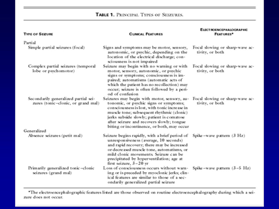
Objectives First seizure evaluation in adults Seizure classification
3. Management options

4
Case 32 y/o male taxi-driver is referred for evaluation of a “spell” while walking to the corner store, after which he was found on the ground. Brought in by EMS to the ER Subsequently sent home What are you going to do and tell the patient?

5
Definitions Seizure: transient disturbance in cerebral function caused by abnormal neuronal discharge Epilepsy: group of disorders represented by recurrent seizures (3% lifetime prevalence)
")
6
Evaluating seizures: Is this a seizure?
What type of seizure is this? (implications on treatment) Is there an underlying cause?
 Is there an underlying cause")
7
Is this a Seizure? Seizure Mimics:
Classic migraines include transient neurologic symptoms (as in partial seizures). epilepsy patients twice as likely to have migraines. Syncope Postural, flaccid paralysis, pre-syncope symptoms, no post-ictal state May have fasiculations (convulsive syncope) TIA Usually no LOC unless basilar stroke, usually negative findings not positive. Sometimes confusing if post-ictal Todd’s paralysis Pseudo-seizures 10-45% of patients with refractory epilepsy. Look for history of abuse. Patients can have both. 5) Movement disorders
. epilepsy patients twice as likely to have migraines. Syncope. Postural, flaccid paralysis, pre-syncope symptoms, no post-ictal state. May have fasiculations (convulsive syncope) TIA. Usually no LOC unless basilar stroke, usually negative findings not positive. Sometimes confusing if post-ictal Todd’s paralysis. Pseudo-seizures % of patients with refractory epilepsy. Look for history of abuse. Patients can have both. 5) Movement disorders.")
9
Is there an underlying cause?
(rule out secondary causes of seizures) 1o neurologic disorder Systemic disorder Head trauma Cancer Hemorrage Stroke Vascular malformations Meningitis/encephalitis Hypoglycemia Hyponatremia Hypocalcemia Uremia Hepatic encephalopathy Drug OD/withdrawal Hyperosmolar states Hyperthermia
 1o neurologic disorder. Systemic disorder. Head trauma. Cancer. Hemorrage. Stroke. Vascular malformations. Meningitis/encephalitis. Hypoglycemia. Hyponatremia. Hypocalcemia. Uremia. Hepatic encephalopathy. Drug OD/withdrawal. Hyperosmolar states. Hyperthermia.")
10
History Witness testimony is key!
Triggers, ictal behaviors, LOC, behaviour during seizure and the postictal state. Seizure precipitants or triggers: strong emotions, intense exercise, flashing lights, and loud music (often immediately before the seizure) fever, menstruation, lack of sleep, and stress
 fever, menstruation, lack of sleep, and stress.")
11
History Ask about . . . Drugs, alcohol, constitutional symptoms, HIV risk factors, fever, head trauma. Family History (absence and myoclonic seizures may be inherited)
")
12
Physical examination Generally unrevealing
Look for signs of disorders associated with seizures. Head trauma, meningismus, sinus infection. Focal or diffuse neurological abnormalities. Mental status abnormalities suggest lesions in the anterior frontal, parietal, or temporal lobes. Evaluate for lateralizing abnormalities: weakness, hyperreflexia, positive Babinski sign

13
Laboratory evaluation
Glucose, calcium, magnesium, hematology studies, renal function tests, lytes toxicology screens. Acute postictal changes: metabolic acidosis and leukocytosis, high CK LP if risk factors for infection (fever, HIV positive).
.")
14
Electroencephalography
Information provided: Presence of abnormal electrical activity Information of type of seizure disorder Location of seizure focus Perform study >48hrs after seizure Include recordings during sleep, photic stimulation, hyperventilation. 50% of patients with epilepsy have normal single EEG

15
Electroencephalography
If normal and high suspicion, repeat study after sleep deprivation 10% of persons with true seizure with have normal multiple EEG studies +EEG likelihood of second seizure over two years

16
Neuroimaging in adults with 1st seizure
Retrospective review of 148 patients studied within 30 days of the seizure Structural lesion was identified by CT in 55 (37 percent); 16 (11 percent) had metabolic seizures CT findings agreed with the results of neurological examination in 82 percent of cases. Ramirez-Lassepas, et al. Value of computed tomographic scan in the evaluation of adult patients after their first seizure. Ann Neurol 1984; 15:536.
; 16 (11 percent) had metabolic seizures. CT findings agreed with the results of neurological examination in 82 percent of cases. Ramirez-Lassepas, et al. Value of computed tomographic scan in the evaluation of adult patients after their first seizure. Ann Neurol 1984; 15:536.")
17
Neuroimaging All patients should receive neuroimaging.
MRI preferred over CT to identify small lesions such as cortical dysplasias, infarcts, or tumors. CT scan is suitable in emergency situations to exclude a mass lesion, hemorrhage, or large stroke.

18
When to initiate Antiepileptic drug therapy
Two or more seizures Single seizure secondary to identified CNS lesion with an epileptogenic focus Consider if significant occupational risk if patient suffers a second event. Consider if single seizure event with one or more risk factors for recurrent seizures Consider in the elderly patient with increased risk of seizure related morbidity (age, prolonged post-ictal state)
")
19
Risk of seizure recurrence in a patient with an apparently unprovoked or idiopathic seizure
31 to 71% risk in the first 12 months after the initial seizure. Risk factors associated with recurrent seizures include the following: (1) evidence of a structural lesion (2) EEG abnormalities (3) partial type seizure (4) family history of seizures (5) focal abnormalities on exam Most patients with one or more of these risk factors should be treated
 evidence of a structural lesion. (2) EEG abnormalities. (3) partial type seizure. (4) family history of seizures. (5) focal abnormalities on exam. Most patients with one or more of these risk factors should be treated.")
20
Antiepileptic Drugs of Choice
Primary Generalized Tonic-Clonic Partial Absence Atypical Absence, Myoclonic, Atonic First-Line Valproic acid Carbamezepine Phenytoin Carbamazepine Phenytoin Ethosuximide Valproate Valproic acid Alternatives Lamotrigine Primidone Phenobarbital Gabapentin Topiramate Tiagabine Primidone Phenobarbital Lamotrigine Clonazepam Lamotrigine Topiramate Clonazepam Felbamate

21
Principles of Treatment
Start with an average dose of a first line drug Poor control? Address compliance, maximize drug dose, confirm right diagnosis (partial complex v.s generalized) Majority of patients are controlled with single antiepileptic drug. This drug can be gradually withdrawn if seizure free for two years. Seizures recur in 25% of patients without risk factors and 50% of patients without risk factors. The drug can be reduced by 25% every two to four weeks.
 Majority of patients are controlled with single antiepileptic drug. This drug can be gradually withdrawn if seizure free for two years. Seizures recur in 25% of patients without risk factors and 50% of patients without risk factors. The drug can be reduced by 25% every two to four weeks.")
22
Principles of Treatment
20-35% of patients with epilepsy have persistent seizures despite medical therapy. If poor control with maximal dose, monotherapy with second drug. Continue to administer first drug until a full dose of second drug reached, then gradually withdraw first drug. If monotherapy with two drugs fail, patient may need re-evaluation (repeat MRI/EEG) before polytherapy commenced (1998 guidelines).
 before polytherapy commenced (1998 guidelines).")
23
Side effects Idiosyncratic toxicity:
rash, bone marrow suppression, or hepatotoxicity. Require laboratory tests (e.g., complete blood count and liver function tests) baseline during initial dosing and titration
 baseline. during initial dosing and titration.")
24
Other management issues:
Impact on independence, self-esteem, employment. Driving regulations: Private drivers cannot drive for 3 months after a single seizure. Private drivers can resume driving after being seizure free for 12 months on medication.

25
Side effects Canadian Guidelines

26
Neurologic Consultation (NEJM 2001)
Change in the type of seizure Uncertain diagnosis (e.g. normal EEG) Lack of seizure control in 3 months Failure of two monotherapies Patient is considering pregnancy Prolonged post-ictal state History of status epilepticus
 Lack of seizure control in 3 months. Failure of two monotherapies. Patient is considering pregnancy. Prolonged post-ictal state. History of status epilepticus.")
27
Summary Management after 1st seizure involves lots of discussion with patient about risks/benefits Remember impact on driving: tell the ministry! When in doubt about management (especially medications), get a neurologist involved
, get a neurologist involved.")
Similar presentations


 The only diagnostic test for absence seizures Ambulatory EEG monitoring over 24 hours may be useful to.>")


