Download presentation
Presentation is loading. Please wait.

1
Reflected Light Microscopy Francis, 2013
gold Reflected Light Microscopy Francis, 2013 gold gold arsenopyrite

2
Sulfide minerals and many oxides are opaque to transmitted light and can only be optically studied using reflected light. In addition, grains, seams, or inclusions whose dimensions are less than the thickness of a standard thin section ( 30 microns) can not be well resolved in transmitted light, but can be readily examined in reflected light. Furthermore, microprobe analysis requires an examination of the material of interest under reflected light to insure that surface defects will not degrade the analysis. gold
 can not be well resolved in transmitted light, but can be readily examined in reflected light. Furthermore, microprobe analysis requires an examination of the material of interest under reflected light to insure that surface defects will not degrade the analysis. gold.")
3
Concrete

4
Because of the limitations of reflected light, reflected light microscopy is a more qualitative art than transmitted light microscopy. The process is essentially one of using the features of easily identifiable minerals to constrain the identity of associated unknown minerals.

5
Reflectance and Colour
Reflectance is the measure of the ratio of the intensity of reflected light from a mineral’s surface to the intensity of incident plane-polarized light ( = 546 nm). Although reflectance can be quantitatively measured with suitable equipment, in general practice one qualitatively estimates reflectance by comparing the unknown mineral to a known mineral. Increasing reflectivity: sphalerite (17%) < magnetite (21%) < galena (43%) < pyrite (55%) < gold (75%) Colour is a more subtle feature in reflected light than in transmitted light, but can be very diagnostic. For example, Fe-oxides are commonly grey while many sulfides are distinctly yellowish in colour. Sphalerite and galena are exceptions, however, being grey and greyish-white respectively. Note: Sulfide minerals tarnish easily, so it is best to buff them gently on a cloth with 0.3 micron abrasive powder when first examining them.
. Although reflectance can be quantitatively measured with suitable equipment, in general practice one qualitatively estimates reflectance by comparing the unknown mineral to a known mineral. Increasing reflectivity: sphalerite (17%) < magnetite (21%) < galena (43%) < pyrite (55%) < gold (75%) Colour is a more subtle feature in reflected light than in transmitted light, but can be very diagnostic. For example, Fe-oxides are commonly grey while many sulfides are distinctly yellowish in colour. Sphalerite and galena are exceptions, however, being grey and greyish-white respectively. Note: Sulfide minerals tarnish easily, so it is best to buff them gently on a cloth with 0.3 micron abrasive powder when first examining them.")
6
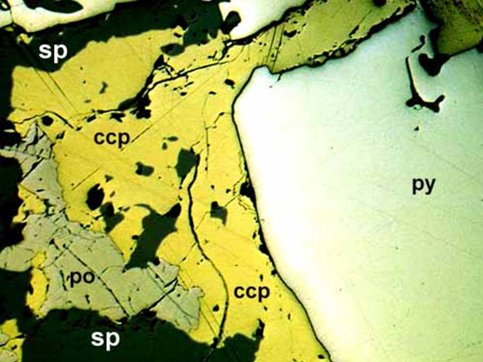
Reflectence / Colour pyrite > chalcopyrite
chalcopyrite > pyrrhotite pyrrhotite > sphalerite

7
pinkish brown to yellow
Some Common Opaque Minerals: listed in order of decreasing reflectance Mineral Formula Reflect. Colour Anisotropy Hardness Gold Au 74 bright yellow isotropic Pyrite FeS2 54 pale yellow Arsenopyrite FeAsS 52 white strong Pentlandite (Fe,Ni)9S8 l Light yellow Chalcopyrite CuFeS2 44 yellow weak Galena PbS 43 grey-white 2.5 Pyrrhotite Fe1-x S 34-40 pinkish brown to yellow 4.0 Chalcocite Cu2S 32 light grey Hematite Fe2O3 28 bluish grey Bornite Cu5FeS4 22 purplish brown 3.0 Magnetite Fe3O4 21 brownish grey 5.5 Ilmenite FeTiO3 17-20 pinkish grey Sphalerite ZnS 17 grey Chromite FeCr2O4 14 dark grey gold arsenopyrite pyrite chalco pyrite tetra hedrite dolomite chalcopyrite pyrite sphalerite
9S8. l. Light yellow Chalcopyrite. CuFeS yellow. weak. Galena. PbS. 43. grey-white Pyrrhotite. Fe1-x S pinkish brown to yellow Chalcocite. Cu2S. 32. light grey. Hematite. Fe2O bluish grey Bornite. Cu5FeS purplish brown Magnetite. Fe3O brownish grey Ilmenite. FeTiO pinkish grey. Sphalerite. ZnS. 17. grey. Chromite. FeCr2O dark grey. gold. arsenopyrite. pyrite. chalco. pyrite. tetra. hedrite. dolomite. chalcopyrite. pyrite. sphalerite.")
8
gold arsenopyrite pyrite pyrite gold galena gold gold arsenopyrite
bornite arsenopyrite chalcocite

9
Bireflectance and Reflection Pleochroism
As in transmitted light, isometric opaque minerals remain unchanged upon rotation of the microscope stage. Strongly anisotropic opaque minerals, however, may exhibit noticeable changes in reflectivity (bireflectance) or colour (pleochroism) upon rotation of the microscope’s stage. Anisotropy Isometric minerals appear either black under crossed polars, or remain dark grey upon rotation of the stage. Anisotropic minerals may exhibit a noticeable variation colour or brightness upon rotation of the stage, exhibiting 4 positions of extinction and 4 positions of maximum intensity or colour. These effects are often quite subtle and require careful observation. It sometimes helps to rotate the analyzer of the microscope slightly from the 90o crossed polar position to observe these features. Internal Reflections Minerals that are not totally opaque sometimes display coloured internal reflections under crossed polars when using bright illumination. Such internal reflections are characteristic of minerals such as sphalerite and the ruby-silver sulfosalts (eg. proustite – pyrargyrite Ag3AsS3 - Ag3SbS2). Internal reflections are also a good way of distinguishing silicate minerals in reflected light.
 or colour (pleochroism) upon rotation of the microscope’s stage. Anisotropy. Isometric minerals appear either black under crossed polars, or remain dark grey upon rotation of the stage. Anisotropic minerals may exhibit a noticeable variation colour or brightness upon rotation of the stage, exhibiting 4 positions of extinction and 4 positions of maximum intensity or colour. These effects are often quite subtle and require careful observation. It sometimes helps to rotate the analyzer of the microscope slightly from the 90o crossed polar position to observe these features. Internal Reflections. Minerals that are not totally opaque sometimes display coloured internal reflections under crossed polars when using bright illumination. Such internal reflections are characteristic of minerals such as sphalerite and the ruby-silver sulfosalts (eg. proustite – pyrargyrite Ag3AsS3 - Ag3SbS2). Internal reflections are also a good way of distinguishing silicate minerals in reflected light.")
10
Gold Gold soft hard arsenopyrite Cleavage
Cleavage is often easily seen in polished surfaces in reflected light as dark lines and straight sided pits, and can be characteristic of some minerals. For example, the polished surface of galena characteristically displays distinctive triangular pits because of its three directions of 90o cleavage. Hardness The opaque minerals vary greatly in polishing hardness. Polishing hardness can be judged by the quality of the polished surface (the hardest surfaces have the most mirror-like finishes) and can be tested with a needle or by measuring relative polishing reliefs of adjacent grains using the “Kalb line” test. The “Kalb line” is somewhat analogous to the “Becke line” in transmitted light. When using the high power objective, and a partly closed diaphragm, lowering the stage will cause the “Kalb line” to move from the grain boundary towards the softer of two adjacent mineral grains. Gold galena Gold soft hard arsenopyrite
 and can be tested with a needle or by measuring relative polishing reliefs of adjacent grains using the Kalb line test. The Kalb line is somewhat analogous to the Becke line in transmitted light. When using the high power objective, and a partly closed diaphragm, lowering the stage will cause the Kalb line to move from the grain boundary towards the softer of two adjacent mineral grains. Gold. galena. Gold. soft. hard. arsenopyrite.")
12
hematite chalcopyrite bornite

13
galena sphalerite

15
Lunar High-Ti Mare Basalts chromite magnetite magnetite magnetite
Feo chromite magnetite Lunar High-Ti Mare Basalts magnetite magnetite ilmenite ilmenite troilite

16
Meteorites troilite

Similar presentations


 Field outcrop observe relationship between rocks>")
 If one knows the chemical formula for a particular mineral, with a little help from.>")


