Download presentation
Presentation is loading. Please wait.

1
Goldmann Applanation Tonometry
Ted Barnett

2
Introduction Applanation tonometry: measures IOP by providing force which flattens the cornea. Variable force applanation tonometers (Goldmann, Perkins, Draeger, MacKay-Marg, and Tono-Pen and Pneumatonometer): area of the cornea being applanated held constant, variable for applied.
: area of the cornea being applanated held constant, variable for applied.")
3
Principles based on Imbert-Fick law:
pressure within a sphere (P) is roughly equal to the external force (f) needed to flatten a portion of the sphere divided by the area (A) of trhe sphere which is flattened: P = f / A applies to surfaces which are perfectly spherical, dry, flexible, elastic and infinitely thin
 is roughly equal to the external force (f) needed to flatten a portion of the sphere divided by the area (A) of trhe sphere which is flattened: P = f / A. applies to surfaces which are perfectly spherical, dry, flexible, elastic and infinitely thin.")
4
Principles (cont.) include force of cornea which pushes applanating surface away from eye (N), subtract surface tension of tear film toward the eye (M) since cornea has thickness, consider only flattening of inner corneal area (A1) P = f / A1 + M - N when A1 = 7.35, M and N cancel out so: P = f / 7.35 mm2
, subtract surface tension of tear film toward the eye (M) since cornea has thickness, consider only flattening of inner corneal area (A1) P = f / A1 + M - N. when A1 = 7.35, M and N cancel out so: P = f / 7.35 mm2.")
5
Principles (cont.) this internal area achieved when diameter of external area of corneal applanation is 3.06mm at this external diameter, grams of force applied multiplied by 10 is directly converted to mmHg measured pressure is 3% greater than IOP before applanation (not corrected) minimal displacement (0.5ul) of fluid or increase in IOP with applanation, thus unaffected by ocular rigidity
 minimal displacement (0.5ul) of fluid or increase in IOP with applanation, thus unaffected by ocular rigidity.")
7
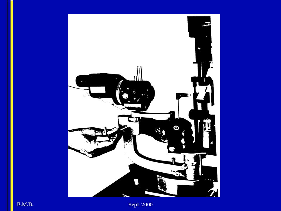
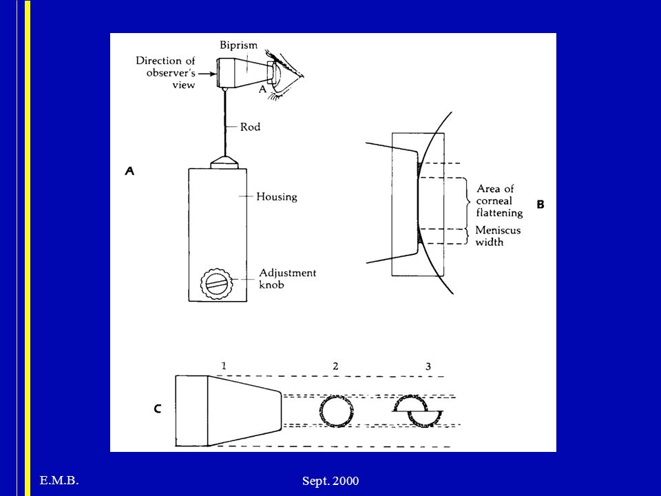
Technique of measurement
plastic biprism which contacts cornea creates two semicircles edge of corneal contact is visible after placing fluorescein into tear film & viewing with cobalt blue light manually rotate the dial calibrated in grams, force is adjusted by changing the length of a spring within the device. inner margins of semicircles touch when 3.06 mm of cornea is applanated.

9
Instructions to patient
press head firmly against chin and forehead rest. look straight ahead and fixate on a target (e.g. examiners opposite ear) breathe normally, do not hold your breath blink immediately prior to measurement to moisten cornea.
 breathe normally, do not hold your breath. blink immediately prior to measurement to moisten cornea.")
10
Measurement (cont.) position patient’s head with forehead rest well above eyebrows, allowing raising of eyebrows. anesthetic & fluorescein (0.25%) ,separately, or as mixture (preserved) placed in inferior cul-de-sac. with maximal illumination of biprism the lamp is moved toward the eye until the tip of biprism contacts the apex of the cornea stop moving forward when limbus shines with light, best observed with naked eye
 ,separately, or as mixture (preserved) placed in inferior cul-de-sac. with maximal illumination of biprism the lamp is moved toward the eye until the tip of biprism contacts the apex of the cornea. stop moving forward when limbus shines with light, best observed with naked eye.")
11
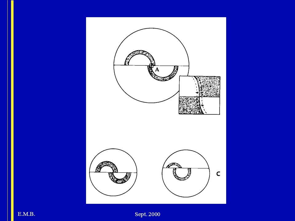
Measurement (cont.) After contact, semicircles visible through left (or right) ocular. Center in field of view. Adjust vertically until semicircles equal in size. Tension dial adjusted so that inner edge of upper and lower semicircles are aligned. Multiply dial reading (grams of force) by 10 to obtain IOP (mmHg) Read at median over which arcs glide to control for excursions due to ocular pulsations.
 by 10 to obtain IOP (mmHg) Read at median over which arcs glide to control for excursions due to ocular pulsations.")
13
Measurement (cont.) If slit-lamp moved too far toward patient the pressure arm will push against a spring which will press against the eye with a low inoffensive force. Mires (flattened area) too large, moving dial doen’t alter appearance. Solution: Draw back until regular pulsation noted and appearance of mires normalizes.
 too large, moving dial doen’t alter appearance. Solution: Draw back until regular pulsation noted and appearance of mires normalizes.")
14
Measurement (cont.) Blue central area represents applanated cornea, green semicircles are fluorescein-stained tears, inner border of ring is demarcation between flattened and non-flattened cornea. Without staining of tears, bright reflection from air-cornea interface is seen; leads to underestimation of IOP. Mires should be approximately 10% of circle width.

15
Errors in Measurement The fluorescein ring is too wide or too narrow:
Too wide: occurs if prism not dried after cleaning or lids touch prism. Overestimates IOP. Solution: dry prism Too narrow: inadequte fluorescein concentration may cause hypofluorescence. Underestimates IOP. Solution: patient blinks or additional fluorescein added.

16
Errors (cont.) thin corneas produces underestimate
thick cornea d/t increased collagen gives overestimate, if d/t edema gives underestimate. inadequate vertical alignment of semicircles leads to overstimate of IOP. distortion d/t irregular cornea influences accuracy, less useful with corneal scarring.

17
Errors (cont.) squeezing of eyelids, breath holding or other Valsalva maneuvers, pressure on globe, excessive EOM force applied to restricted globe, vertical gaze, tight collars, retreating patient, inaccurately calibrated tonometer. repeated tonometry may induce decline in estimated IOP.

18
Error d/t corneal curvature
increase of 1 mmHg for every 3D increase in corneal power. more fluid displaced under steep cornea, increases contribution of ocular rigidity in overestimating IOP. the steeper the cornea, the more cornea must be indented to produce standard area of contact. >3D astigmatism produces elliptical rather than circular area

19
Correction for astigmatism
With semicircles displaced horizontally, IOP underestimated by 1 mmHg for every 4D of WTR astigmatism, vice versa for ATR astigmatism. To minimize, prisms should be rotated so that axis of least corneal curvature is opposite red line on prism holder (i.e. align negative cylinder axis). Can average reading with vertical and horizontal alignment of prism.
. Can average reading with vertical and horizontal alignment of prism.")
20
Sterilization CDC recommendation (HIV, HSV, and adenovirus): wipe tip clean and disinfect tip only with bleach (1:10 dilution x 5”, changed once daily). Alternative is 3% H2O2, changed at least twice daily (affects tip less than bleach or ETOH). Alternative #2: wiping tip with 70% ETOH
: wipe tip clean and disinfect tip only with bleach (1:10 dilution x 5 , changed once daily). Alternative is 3% H2O2, changed at least twice daily (affects tip less than bleach or ETOH). Alternative #2: wiping tip with 70% ETOH.")
21
Reliability Goldmann applanation is standard against which others measured. Good accuracy in gas-filled eyes. Inter- and intraobserver variability (>30% varied by 2-3 mmHg), due to subjective nature of optical endpoint. Assume error of 2 mmHg.
, due to subjective nature of optical endpoint. Assume error of 2 mmHg.")
22
Calibration: Wessels & Oh (1990)
Tested tonometers in ophthalmologists offices. 19% outside range of manufacturers specifications (1mmHg of calibration), 4.5% > 2mmHg error. Annual recalibration in 86% of instruments. Practitioners who themselves performed calibration had the most accurate instruments. Less than 15% knew how to perform calibration check. Calibration here done 4 times/year
, 4.5% > 2mmHg error. Annual recalibration in 86% of instruments. Practitioners who themselves performed calibration had the most accurate instruments. Less than 15% knew how to perform calibration check. Calibration here done 4 times/year.")
Similar presentations


 Problem Measurement of eye pressure by Goldmann applanation tonometry is a technique first embodied in the.>")


