Download presentation
Presentation is loading. Please wait.

1
Musculoskeletal System & Movement
What are the types of movement? What are the different types of muscle tissue and skeletal systems? How do muscles exert force at the gross level? …at the cellular level? …at the molecular level? How do action potentials trigger muscle contraction in skeletal muscle? How do you quantify muscle activity? (force, work,power) Cost of transport vs. body size; vs. mode of transport
 Cost of transport vs. body size; vs. mode of transport.")
2
3 Basic Types of Movement
Ameboid Ciliary Locomotion Muscular Movement

3
Three Types of Muscle Smooth Cardiac STRIATED Skeletal
Slow twitch – red good for endurance Fast twitch – white good for sprints

4
Slow Twitch vs. Fast Twitch

5
Skeletal Systems Endoskeleton Exoskeleton Hydrostatic skeleton
Figure 46-18 Endoskeleton Skeletal Systems Exoskeleton Hydrostatic skeleton

6
Endo & Exoskeletons

7
Anatomy of Skeletal Muscle

8
Sliding Filaments are the basis of muscle shortening

9
Extended Contracted

10
Colors indicate protein subunits
Figure 46-21 Myosin head Colors indicate protein subunits ATP binding site Actin binding site

12
Conformational Change in Protein (myosin head) is “molecular motor” for movement
Figure 46-22 CHANGES IN THE CONFORMATION OF THE MYOSIN HEAD PRODUCE MOVEMENT. 1. ATP bound to myosin head. Head releases from thin filament. Myosin head of thick filament Actin in thin filament 4. ADP released. Cycle is ready to repeat. 2. ATP hydrolized. Head pivots, binds to new actin subunit. 3. Pi released. Head pivots, moves filament (power stroke).
.")
13
Myosin binding sites blocked
Figure 46-23 Tropomyosin and troponin work together to block the myosin binding sites on actin. Myosin head Myosin binding sites blocked Troponin Tropomyosin Actin Myosin binding sites Calcium ions When a calcium ion binds to troponin, the troponin-tropomyosin complex moves, exposing myosin binding sites. Myosin binding site exposed to myosin head Calcium ion Troponin-tropomyosin complex, moved

14
Figure 46-24 HOW DO ACTION POTENTIALS TRIGGER MUSCLE CONTRACTION?
Motor neuron Muscle cell HOW DO ACTION POTENTIALS TRIGGER MUSCLE CONTRACTION? Motor neuron Action potential 1. Action potential arrives; acetylcholine (Ach) is released. ACh ACh receptor Action potentials 2. ACh binds to ACh receptors on the muscle cell, triggering depolari-zation that leads to action potential. 3. Action potentials propagate across muscle cell’s plasma membrane and into interior of cell via T tubules. T tubule 4. Proteins in T tubules open Ca2+ channels in sarcoplasmic reticulum. Sarcoplasmic reticulum 5. Ca2+ is released from sarcoplasmic reticulum. Sarcomeres contract when troponin and tropomyosin move in response to Ca2+ and expose actin binding sites in the thin filaments (see Figure 46.23). Thick filaments (myosin) Thin filaments (actin) Ca2+ ions
 is released. ACh. ACh receptor. Action potentials. 2. ACh binds to ACh receptors on the muscle cell, triggering depolari-zation that leads to action potential. 3. Action potentials propagate across muscle cell’s plasma membrane and into interior of cell via T tubules. T tubule. 4. Proteins in T tubules open Ca2+ channels in sarcoplasmic reticulum. Sarcoplasmic reticulum. 5. Ca2+ is released from sarcoplasmic reticulum. Sarcomeres contract when troponin and tropomyosin move in response to Ca2+ and expose actin binding sites in the thin filaments (see Figure 46.23). Thick filaments (myosin) Thin filaments (actin) Ca2+ ions.")
15
HOW DO ACTION POTENTIALS TRIGGER MUSCLE CONTRACTION?
Figure 46-24a Motor neuron Muscle cell HOW DO ACTION POTENTIALS TRIGGER MUSCLE CONTRACTION? Motor neuron Action potential 1. Action potential arrives; acetylcholine (ACh) is released. ACh ACh receptor Action potentials 2. ACh binds to ACh receptors on the muscle cell, triggering depolari-zation that leads to action potential.
 is released. ACh. ACh receptor. Action potentials. 2. ACh binds to ACh receptors on the muscle cell, triggering depolari-zation that leads to action potential.")
16
HOW DO ACTION POTENTIALS TRIGGER MUSCLE CONTRACTION?
Figure 46-24b Motor neuron Muscle cell HOW DO ACTION POTENTIALS TRIGGER MUSCLE CONTRACTION? 3. Action potentials propagate across muscle cell’s plasma membrane and into interior of cell via T tubules. T tubule 4. Proteins in T tubules open Ca2+ channels in sarcoplasmic reticulum. Sarcoplasmic reticulum 5. Ca2+ is released from sarcoplasmic reticulum. Sarcomeres contract when troponin and tropomyosin move in response to Ca2+ and expose actin binding sites in the thin filaments (see Figure 46.23). Thick filaments (myosin) Thin filaments (actin) Ca2+ ions
. Thick filaments (myosin) Thin filaments (actin) Ca2+ ions.")
17
Quantify Performance in terms of Power (Watts; kcal/min;horsepower)
1 Watt = 6.12kg-m/min 1 Horsepower = 746 W 1 Kcal/min = W (70 Watts is close to the Basal Metabolic Rate for an average sized human) 1 Kcal/min x 60mins/hr x 24hrs/day = 1440 Kcal/day Total caloric expenditure = BMR + extra activity
 1 Kcal/min x 60mins/hr x 24hrs/day = 1440 Kcal/day. Total caloric expenditure = BMR + extra activity.")
18
Total Energy Expenditure (BMR + extra activity)
Skeletal Muscle tissue - largest consumer of extra calories Digestive, cardiovascular, immune can cycle on and off (N.B. = muscle is only 25% efficient so putting out 100 W on a machine requires burning calories at a rate of 400W)
")
19
The Cost of Movement How much force does it take to move 1kg of animal..?...

20
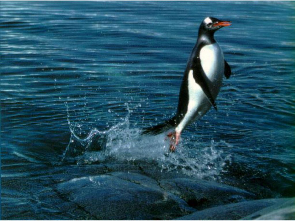
Body Shape of Swimming Animals

22
Some comparisons Most Cost effective mode of movement
Largest organisms Fastest speeds Water: 50 mph Land: 60 mph Air: mph

Similar presentations


 Bundle, fiber, myofibril, sarcomere Z-line, thick filament, thin filament Actin, myosin, sliding filament.>")


