Download presentation
Presentation is loading. Please wait.

1
Fetal Membranes

2
definition Fetal Membranes are structures which surround and support the fetus and aid in its growth and development

3
What constitute a Fetal Membrane Amnion Yolk sac Allantois Umbilical cord

4
Amnion Forms a membranous amniotic sac that surrounds the embryo and fetus Daily contribution of fluid from respiratory tract is 300-400 ml Fetus contributes to the amniotic fluid by excreting urine into the amniotic cavity (500mls) Half a liter of urine is added daily during the late pregnancy Amniotic fluid volume is 30 ml at 10 weeks, 350 ml at 20 weeks, 700-1000 ml at 37 weeks
 Half a liter of urine is added daily during the late pregnancy Amniotic fluid volume is 30 ml at 10 weeks, 350 ml at 20 weeks, ml at 37 weeks")
5
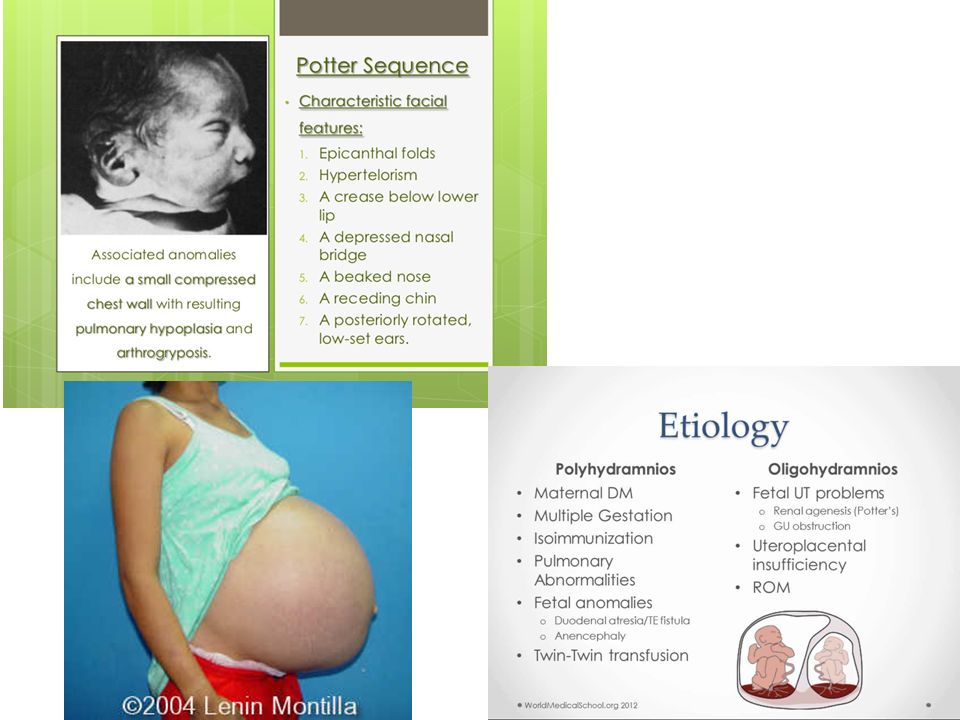
Disorders of Amniotic Fluid Volume Oligohydromnios – Renal agenesis – Obstructive uropathy Polyhydromnios – Esophageal atresia – Anencephaly

7
Composition of Amniotic Fluid High levels of alpha- phetoprotein (AFP) in amniotic fluid usually indicate the presence of a severe neural tube defect (anencephaly) Low levels of AFP may indicate chromosomal aberrations such as down’s syndrome
 in amniotic fluid usually indicate the presence of a severe neural tube defect (anencephaly) Low levels of AFP may indicate chromosomal aberrations such as down’s syndrome")
8
Significance of Amniotic Fluid Permits symmetrical external growth of the embryo and fetus Acts as a barrier to infection Permits normal fetal lung development Prevents adherence of amnion to fetus Cushions & protects the embryo and fetus Helps maintain the body temperature Enables the fetus to move freely

9
The Yolk Sac It attains largest size at 32 days Shrinks to 5mm pear shaped remnant by 10 th week & connected to the midgut by a narrow yolk stalk Becomes very small at 20 weeks Usually not visible thereafter

10
Significance of Yolk Sac Has a role in transfer of nutrients during the 2 nd and 3 rd weeks Blood development first occurs here Incorporated into the endoderm of embryo as a primordial gut Primordial germ cells appear in the endodermal lining of the wall of the yolk sac in the 3 rd week

11
The Allantois In the 3 rd week it appears as a sausage-like diverticulum from the caudal wall of yolk sac that extends into the connecting stalk During the 2 nd month, the extraembryonic part of the allantois degenerates Blood formation occurs in its wall during the 3 rd to 5 th week Its blood vessels persist as the umbilical vein and arteries

12
The Umbilical Cord Is attached to the placenta usually near the center of the fetal surface of this organ May attach to any other point Is usually 1-2 cm in diameter and 30-90 cm in length Long cord may cause prolapse or compression of the cord which may lead to fetal hypoxia Short cord may cause premature separation of the placenta from the wall of the uterus during delivery

13
Tight knot!!! (fatal)
")
14
The placenta & multiple gestation

15
15 PLACENTA This is a fetomaternal organ. It has two components: – Fetal part – develops from the chorionic sac ( chorion frondosum ) – Maternal part – derived from the endometrium ( functional layer – decidua basalis ) The placenta and the umbilical cord are a transport system for substances between the mother and the fetus.( vessels in umbilical cord ) Function Of The Placenta: 1.Protection 2.Nutrition 3.Respiration 4.Excretion 5.Hormone production
 – Maternal part – derived from the endometrium ( functional layer – decidua basalis ) The placenta and the umbilical cord are a transport system for substances between the mother and the fetus.( vessels in umbilical cord ) Function Of The Placenta: 1.Protection 2.Nutrition 3.Respiration 4.Excretion 5.Hormone production.")
16
16 FULL-TERM PLACENTA Cotyledons –about 15 to 20 slightly bulging villous areas. Their surface is covered by shreds of decidua basalis from the uterine wall. After birth, the placenta is always inspeced for missing cotyledons. Cotyledons remaining attached to the uterine wall after birth may cause severe bleeding. Maternal side

17
Major Causes of postpartum hemorrhage The 4T’s – Tone – Tissue (placenta) – Trauma – Thrombin 17
 – Trauma – Thrombin 17")
18
Placental membrane defects 18

19
Neonatal syphilis 19

20
Placental endocrine synthesis The syncytiotrophoblast synthesizes protein &steroid hormones The protein homones - human chorionic gonadotropin -h.c. thyrotropin (congenital hypothyroidism) -h.c. corticotropin ( congenital adrenal hyperplasia ) The steroid hormones: Progesterone & Estrogens
 -h.c. corticotropin ( congenital adrenal hyperplasia ) The steroid hormones: Progesterone & Estrogens.")
21
Congenital hypothyroidism & adrenal hyperplasia 21

22
22 placental anomalies: Third trimester bleeding is the common sign of these anomalies

23
Twins - 1 in 100 births Triplets are about 1 in 7,500 births Quadruplets are about 1 in 650,00 births

24
Predisposing Factors Maternal age and parity Maternal height and weight Genetic and racial factors Prior use of oral contraceptive agents Social class

25
Causes of Multiple Gestation Spontaneously In Vitro fertilization – Intrauterine insemination Fertility Drugs – Clomiphene citrate (clomid, serrophene) – Gonadotropins (GonalF, follistim, humagon)
 – Gonadotropins (GonalF, follistim, humagon)")
26
Twins Dizygotic twins (66%) – Dichorionic – separate chorion (placenta) – Diamniotic – separate amnion (amniotic sac) Monozygotic twin (33%) Ova division: < 72 hours: Dichorionic, diamniotic 4-8 days: Monchorionic, diamniotic 8-13 days: Monochorionic, monoamniotic > 13 days: conjoined twins
 – Dichorionic – separate chorion (placenta) – Diamniotic – separate amnion (amniotic sac) Monozygotic twin (33%) Ova division: < 72 hours: Dichorionic, diamniotic 4-8 days: Monchorionic, diamniotic 8-13 days: Monochorionic, monoamniotic > 13 days: conjoined twins")
27
Conjoined Twins (paraphagus)
")
28
28

29
Peripartum Complications Prematurity-major cause of neonatal death 50% of twins 90% of triplets and higher Spontaneous abortion Increased anomalies Cord Prolapse IUGR, discordant growth Intracranial Hemorrhage Locked Twins Description: Twins lock heads 1 st twin breech, 2 nd twin vertex

30
Second Twin Risks Asphyxia due to premature separation of placenta Fetus papyraceous - twin fetus that died in utero, become flattened and mummified Fetal transfusion Syndrome Placental AV shunt in monozygotic twins (~15%) Arterial twin pumps blood to other twin, starves self Other twin is bulky and plethoric
 Arterial twin pumps blood to other twin, starves self Other twin is bulky and plethoric")
31
Monozygotic twins (physical characteristics) Same sex Features alike, including teeth and ears Hair identical Eyes same color and shade Skin same texture and color Hands and feet same conformation and same size Anthropometric values closely agree
 Same sex Features alike, including teeth and ears Hair identical Eyes same color and shade Skin same texture and color Hands and feet same conformation and same size Anthropometric values closely agree")
32
Prevention Moniter treatment with fertility drugs Limit embryos transferred during IVF Counseling risks and long-term sequelae

33
THE END

Similar presentations


 MULTIPLE PARITY -Twins (two babies) -Monozygotic(Division of 1 ova fertilized.>")

 MULTIPLE PARITY -Twins (two babies) -Monozygotic(Division of 1 ova fertilized by the same sperm) -Dizygotic(Fertilization.>")


