Download presentation
Presentation is loading. Please wait.

1
PowerPoint ® Lecture Slides prepared by Betsy C. Brantley Valencia College C H A P T E R 1 An Introduction to Anatomy and Physiology © 2017 Pearson Education, Inc.

2
Common Functions of All Living Things (1-1) Responsiveness Growth Reproduction Movement Metabolism
 Responsiveness Growth Reproduction Movement Metabolism")
3
Responsiveness (1-1) Responsiveness Doing something in response to a change in the immediate environment Example: moving away from a painful stimulus Capacity to make longer–term adjustments (e.g., growing heavier coat of fur in winter) is adaptability
 Responsiveness Doing something in response to a change in the immediate environment Example: moving away from a painful stimulus Capacity to make longer–term adjustments (e.g., growing heavier coat of fur in winter) is adaptability")
4
Growth (1-1) Growth An increase in organism size accomplished by: Growth of cells or Addition of new cells Differentiation Process of individual cells becoming specialized for particular functions
 Growth An increase in organism size accomplished by: Growth of cells or Addition of new cells Differentiation Process of individual cells becoming specialized for particular functions")
5
Reproduction and Movement (1-1) Reproduction Creation of new generations of similar organisms Movement May be internal or external Internal: transporting blood, food, or other material within the body External: moving through the environment
 Reproduction Creation of new generations of similar organisms Movement May be internal or external Internal: transporting blood, food, or other material within the body External: moving through the environment")
6
Metabolism (1-1) Sum total of all chemical operations in the body Cells use materials absorbed from the environment for energy Nutrients from food Oxygen More complex organisms require specialized structures and systems for metabolic processes
 Sum total of all chemical operations in the body Cells use materials absorbed from the environment for energy Nutrients from food Oxygen More complex organisms require specialized structures and systems for metabolic processes")
7
Metabolic Processes (1-1) Respiration Absorption, transport, and use of oxygen by cells Digestion Breaking down complex foods into simpler compounds that can be absorbed Excretion Eliminating waste products generated by metabolic operations
 Respiration Absorption, transport, and use of oxygen by cells Digestion Breaking down complex foods into simpler compounds that can be absorbed Excretion Eliminating waste products generated by metabolic operations")
8
Anatomy (1-2) Greek origin Anatomy literally means “a cutting open” Study of: Internal and external structure Relationships between body parts Divided into gross anatomy or microscopic anatomy
 Greek origin Anatomy literally means a cutting open Study of: Internal and external structure Relationships between body parts Divided into gross anatomy or microscopic anatomy")
9
Gross Anatomy (1-2) Also called macroscopic anatomy Studies structures visible with unaided eye Includes: Surface anatomy Study of general form and superficial markings Regional anatomy Study of all the superficial and internal features of a specific region of the body Systemic anatomy Study of the structure of major organ systems
 Also called macroscopic anatomy Studies structures visible with unaided eye Includes: Surface anatomy Study of general form and superficial markings Regional anatomy Study of all the superficial and internal features of a specific region of the body Systemic anatomy Study of the structure of major organ systems")
10
Microscopic Anatomy (1-2) Studies structures that cannot be seen without magnification Includes: Cytology Study of internal structure of individual cells Histology Study of tissues, groups of specialized cells and cell products that work together to perform specific functions
 Studies structures that cannot be seen without magnification Includes: Cytology Study of internal structure of individual cells Histology Study of tissues, groups of specialized cells and cell products that work together to perform specific functions")
11
Physiology (1-2) Greek origin Study of function in living organisms Interrelated with anatomy Anatomy gives clues about function Physiology is explained in anatomical terms
 Greek origin Study of function in living organisms Interrelated with anatomy Anatomy gives clues about function Physiology is explained in anatomical terms")
12
Human Physiology (1-2) Human physiology studies functions of the human body Specialties include the study of: The functions of living cells – cell physiology Includes the chemical and molecular levels The physiology of specific organs – special physiology All aspects of the function of specific organ systems – systemic physiology The effects of diseases on organ or system functions – pathological physiology or pathology
 Human physiology studies functions of the human body Specialties include the study of: The functions of living cells – cell physiology Includes the chemical and molecular levels The physiology of specific organs – special physiology All aspects of the function of specific organ systems – systemic physiology The effects of diseases on organ or system functions – pathological physiology or pathology")
13
Levels of Organization (1-3) Chemical level Atoms are the smallest stable units of matter Atoms combine to form molecules Molecular shape defines function Cellular level Made up of cells, the smallest living units in the body Formed by interaction between different molecules
 Chemical level Atoms are the smallest stable units of matter Atoms combine to form molecules Molecular shape defines function Cellular level Made up of cells, the smallest living units in the body Formed by interaction between different molecules")
14
Levels of Organization (1-3) Tissue level Similar cells working together to perform a specific function form a tissue Organ level Two or more different tissues working together to perform specific functions form an organ
 Tissue level Similar cells working together to perform a specific function form a tissue Organ level Two or more different tissues working together to perform specific functions form an organ")
15
Levels of Organization (1-3) Organ system level Organs interacting to perform specific functions form organ systems Organism level All the organ systems of the body working together to maintain life and health form an organism
 Organ system level Organs interacting to perform specific functions form organ systems Organism level All the organ systems of the body working together to maintain life and health form an organism")
16
Figure 1-1 Levels of Organization

17
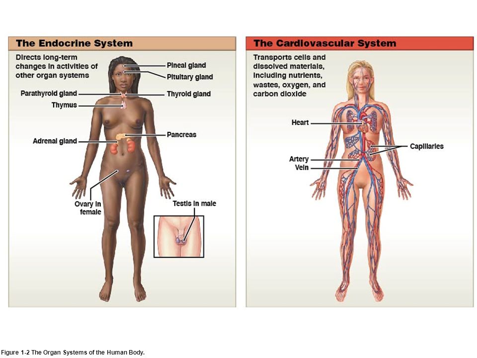
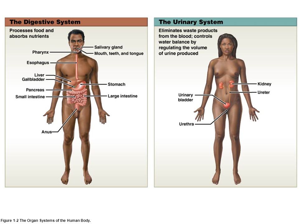
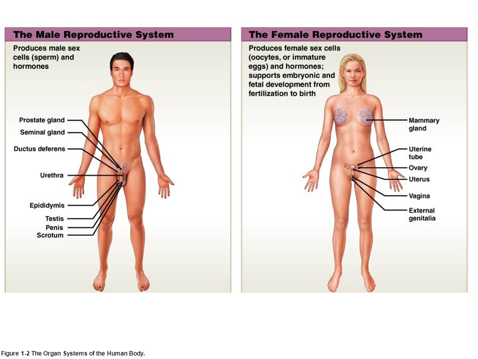
The 11 Organ Systems of the Human Body (1-4) 1.Integumentary 2.Skeletal 3.Muscular 4.Nervous 5.Endocrine 6.Cardiovascular 7.Lymphatic 8.Respiratory 9.Digestive 10.Urinary 11.Reproductive
 1.Integumentary 2.Skeletal 3.Muscular 4.Nervous 5.Endocrine 6.Cardiovascular 7.Lymphatic 8.Respiratory 9.Digestive 10.Urinary 11.Reproductive")
18
Figure 1-2 The Organ Systems of the Human Body.

24
Homeostasis (1-5) A state of internal balance or stable internal environment Must be maintained in order to survive Malfunction of organ systems when homeostatic responses are overwhelmed results in disease Accomplished by interdependent organ systems functioning together
 A state of internal balance or stable internal environment Must be maintained in order to survive Malfunction of organ systems when homeostatic responses are overwhelmed results in disease Accomplished by interdependent organ systems functioning together")
25
Homeostatic Regulation (1-5) Adjustments in physiological systems that preserve homeostasis Homeostatic regulation usually involves: A receptor that senses a particular change or stimulus A control center (integration center) that receives and processes information from the receptor An effector that responds to the control center commands This response may oppose or enhance the stimulus
 Adjustments in physiological systems that preserve homeostasis Homeostatic regulation usually involves: A receptor that senses a particular change or stimulus A control center (integration center) that receives and processes information from the receptor An effector that responds to the control center commands This response may oppose or enhance the stimulus")
26
Homeostatic Example (1-5) Thermostat is set at desired temperature Variation outside desired range triggers response Response negates the original stimulus Example of negative feedback Figure 1-3 The Control of Room Temperature.
 Thermostat is set at desired temperature Variation outside desired range triggers response Response negates the original stimulus Example of negative feedback Figure 1-3 The Control of Room Temperature.")
27
Negative Feedback (1-6) Most common form of homeostatic regulation Variations from normal trigger automatic response Response corrects situation back to normal range Example: thermoregulation Body temperature too high → responses to lower temperature Body temperature too low → responses to raise temperature
 Most common form of homeostatic regulation Variations from normal trigger automatic response Response corrects situation back to normal range Example: thermoregulation Body temperature too high → responses to lower temperature Body temperature too low → responses to raise temperature")
28
Negative Feedback: Thermoregulation (1-6) If body temperature is high, control center targets: Sweat glands Increases secretion (sweat) Body cools with evaporation of sweat Smooth muscle in blood vessels supplying skin Blood vessels dilate, increasing blood flow to body surface Heat is radiated from skin to environment Result: temperature reduced
 If body temperature is high, control center targets: Sweat glands Increases secretion (sweat) Body cools with evaporation of sweat Smooth muscle in blood vessels supplying skin Blood vessels dilate, increasing blood flow to body surface Heat is radiated from skin to environment Result: temperature reduced")
29
Figure 1-4a Negative Feedback in Thermoregulation.

30
Negative Feedback: Thermoregulation (1-6) If body temperature is low, control center targets: Sweat glands (decreasing activity) Smooth muscle in blood vessels supplying skin Blood vessels constrict, decreasing blood flow to body surface Decreasing heat loss to the environment Skeletal muscles Causes shivering, which produces heat Result: temperature increased
 If body temperature is low, control center targets: Sweat glands (decreasing activity) Smooth muscle in blood vessels supplying skin Blood vessels constrict, decreasing blood flow to body surface Decreasing heat loss to the environment Skeletal muscles Causes shivering, which produces heat Result: temperature increased")
31
Figure 1-4b Negative Feedback in Thermoregulation.

32
Positive Feedback (1-6) Response reinforces or exaggerates original stimulus Results in escalating cycle or positive feedback loop Involved in regulation of potentially dangerous or stressful processes requiring rapid completion Blood clotting Labor and delivery
 Response reinforces or exaggerates original stimulus Results in escalating cycle or positive feedback loop Involved in regulation of potentially dangerous or stressful processes requiring rapid completion Blood clotting Labor and delivery")
33
Figure 1-5 Positive Feedback.

34
Anatomical Terminology (1-7) Common language required for clear communication Called medical terminology Many terms based on Latin or Greek language Describes body regions, anatomical landmarks and directions, and body sections
 Common language required for clear communication Called medical terminology Many terms based on Latin or Greek language Describes body regions, anatomical landmarks and directions, and body sections")
35
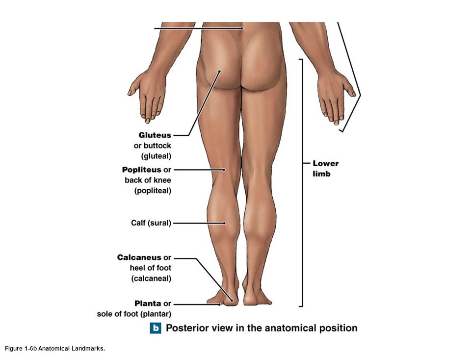
Anatomical Landmarks (1-7) Anatomical position Hands at the sides with the palms facing forward and feet together Lying down in anatomical position Supine (face up) Prone (face down) Right and Left according to subject’s orientation Anatomical regions referred to by their anatomical adjectives
 Anatomical position Hands at the sides with the palms facing forward and feet together Lying down in anatomical position Supine (face up) Prone (face down) Right and Left according to subject’s orientation Anatomical regions referred to by their anatomical adjectives")
36
Figure 1-6a Anatomical Landmarks.

38
Figure 1-6b Anatomical Landmarks.

40
Anatomical Regions (1-7) Surface of the abdominopelvic area can be mapped using two methods: 1.Abdominopelvic quadrants Used by clinicians to locate aches, pains, injuries 2.Abdominopelvic regions Used by anatomists for more precise location of internal organs
 Surface of the abdominopelvic area can be mapped using two methods: 1.Abdominopelvic quadrants Used by clinicians to locate aches, pains, injuries 2.Abdominopelvic regions Used by anatomists for more precise location of internal organs")
41
Figure 1-7a Abdominopelvic Quadrants and Regions. Abdominopelvic Quadrants

42
Figure 1-7b Abdominopelvic Quadrants and Regions. Abdominopelvic Regions

43
Figure 1-7c Abdominopelvic Quadrants and Regions. Abdominopelvic Regions

44
Anatomical Directions (1-7) Provide an orientation of structures relative to anatomical position Some terms can be used interchangeably Anterior – ventral Posterior – dorsal Left and right always refer to the left and right sides of the subject, not the observer
 Provide an orientation of structures relative to anatomical position Some terms can be used interchangeably Anterior – ventral Posterior – dorsal Left and right always refer to the left and right sides of the subject, not the observer")
45
Figure 1-8 Directional References. Superior Inferior RightLeft Anterior View

46
Figure 1-8 Directional References. Superior RightLeft Lateral View Inferior

47
Sectional Anatomy (1-7) Allows better understanding of three–dimensional aspect of the human body Required to interpret many imaging techniques looking at internal structures Described in reference to three primary sectional planes
 Allows better understanding of three–dimensional aspect of the human body Required to interpret many imaging techniques looking at internal structures Described in reference to three primary sectional planes")
48
Sectional Planes (1-7) 1.Frontal plane (or coronal plane) Divides body into anterior and posterior portions 2.Sagittal plane Divides body into left and right portions Midsagittal section divides into equal right and left halves 3.Transverse plane Divides body into superior and inferior portions Cut in this plane called transverse section or cross section
 1.Frontal plane (or coronal plane) Divides body into anterior and posterior portions 2.Sagittal plane Divides body into left and right portions Midsagittal section divides into equal right and left halves 3.Transverse plane Divides body into superior and inferior portions Cut in this plane called transverse section or cross section")
49
Figure 1-9 Sectional Planes.

51
Body Cavities (1-8) True body cavities Closed, fluid–filled spaces Lined by thin tissue layer called serous membrane Contain internal organs (viscera) suspended within them Protect internal organs Allow organs to change shape
 True body cavities Closed, fluid–filled spaces Lined by thin tissue layer called serous membrane Contain internal organs (viscera) suspended within them Protect internal organs Allow organs to change shape")
52
Body Cavities of the Trunk (1-8) Two major regions 1.Thoracic cavity 2.Abdominopelvic cavity Separated by the diaphragm Flat muscular sheet Figure 1-10a Relationships among the Subdivisions of the Body Cavities of the Trunk.
 Two major regions 1.Thoracic cavity 2.Abdominopelvic cavity Separated by the diaphragm Flat muscular sheet Figure 1-10a Relationships among the Subdivisions of the Body Cavities of the Trunk.")
53
Serous Membranes (1-8) Produce watery fluid Moistens opposing surfaces Reduces friction Parietal layer Lines walls of internal cavities Visceral layer Covers surfaces of visceral organs
 Produce watery fluid Moistens opposing surfaces Reduces friction Parietal layer Lines walls of internal cavities Visceral layer Covers surfaces of visceral organs")
54
Thoracic Cavity (1-8) Contains three internal chambers One pericardial cavity (contains the heart) Within central thoracic compartment, the mediastinum Two pleural cavities (one for each lung) Each cavity lined by a serous membrane Figure 1-10c Relationships among the Subdivisions of the Body Cavities of the Trunk.
 Contains three internal chambers One pericardial cavity (contains the heart) Within central thoracic compartment, the mediastinum Two pleural cavities (one for each lung) Each cavity lined by a serous membrane Figure 1-10c Relationships among the Subdivisions of the Body Cavities of the Trunk.")
55
Pericardial Cavity (1-8) Lined by serous membrane called pericardium Visceral pericardium is the layer covering the heart Parietal pericardium is the outer layer Pericardial fluid between two layers reduces friction Figure 1-10b Relationships among the Subdivisions of the Body Cavities of the Trunk.
 Lined by serous membrane called pericardium Visceral pericardium is the layer covering the heart Parietal pericardium is the outer layer Pericardial fluid between two layers reduces friction Figure 1-10b Relationships among the Subdivisions of the Body Cavities of the Trunk.")
56
Pleural Cavities (1-8) Each lung is found within its own pleural cavity Lined by serous membrane called pleura Visceral pleura is the layer covering the outer surfaces of a lung Parietal pleura lines the edge of the mediastinum and the inner body wall Figure 1-10c Relationships among the Subdivisions of the Body Cavities of the Trunk. Visceral pleura

57
Abdominopelvic Cavity (1-8) Extends from the diaphragm to the pelvis Subdivided into: Abdominal cavity (superior portion) Pelvic cavity (inferior portion) Contains the peritoneal cavity
 Extends from the diaphragm to the pelvis Subdivided into: Abdominal cavity (superior portion) Pelvic cavity (inferior portion) Contains the peritoneal cavity")
58
Peritoneal Cavity (1-8) Lined by serous membrane called peritoneum Visceral peritoneum covers internal organs Parietal peritoneum lines inner surface of body wall A few organs lie between peritoneal lining and wall of abdominal cavity Position called retroperitoneal http://teachmeanatomy.info/wp-content/uploads/Structure-of-the-Peritoneum-and-Peritoneal-Cavity.jpg retroperitoneal
 Lined by serous membrane called peritoneum Visceral peritoneum covers internal organs Parietal peritoneum lines inner surface of body wall A few organs lie between peritoneal lining and wall of abdominal cavity Position called retroperitoneal retroperitoneal")
Similar presentations