Download presentation
Presentation is loading. Please wait.

1
Choosing Wisely: Cardiology Jeffrey Ziffra D.O. Mercy Medical Center – North Iowa 10/14/2016

2
Financial Disclosures I have no active relevant financial disclosures

3
Objectives By the end of the session, participants will be able to: Risk stratify patients with or for development of coronary artery disease Be familiar with indications for cardiac testing Managing stable asymptomatic patients and discussing need or lack of need for cardiac testing

4
Objectives Characterize Types of Chest Pain Risk Stratify, Develop pre-test probability Indications for Cardiac Testing Stress Test Modalities Annual Monitoring Coronary CT Scans / Calcium Scoring

10
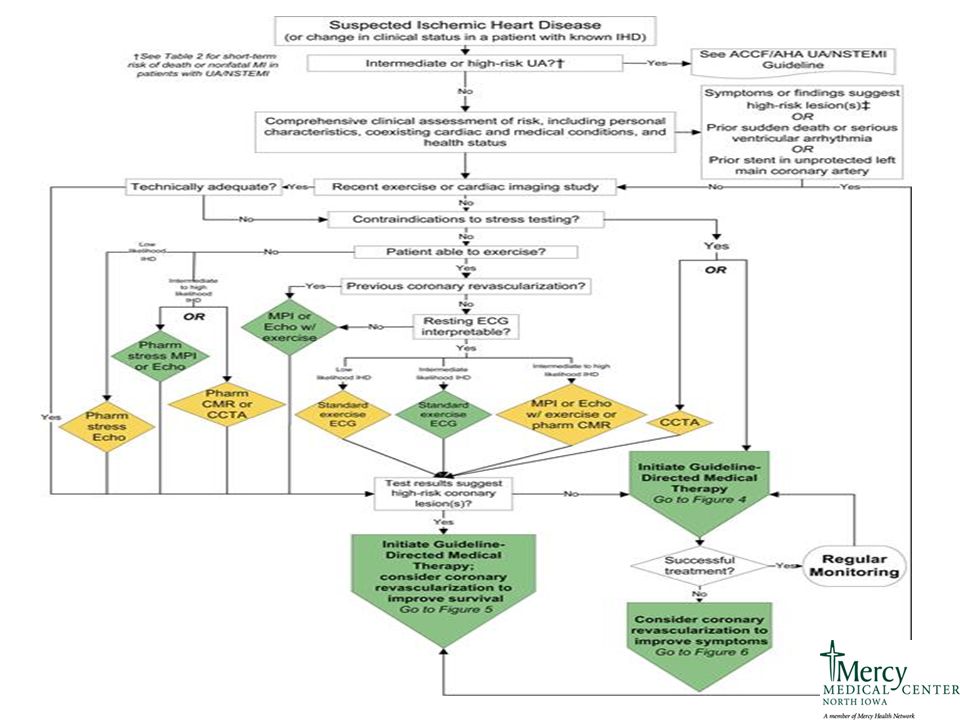
Date of download: 7/25/2016 Copyright © The American College of Cardiology. All rights reserved. From: 2012 ACCF/AHA/ACP/AATS/PCNA/SCAI/STS Guideline for the Diagnosis and Management of Patients With Stable Ischemic Heart Disease: A Report of the American College of Cardiology Foundation/American Heart Association Task Force on Practice Guidelines, and the American College of Physicians, American Association for Thoracic Surgery, Preventive Cardiovascular Nurses Association, Society for Cardiovascular Angiography and Interventions, and Society of Thoracic Surgeons J Am Coll Cardiol. 2012;60(24):e44-e164. doi:10.1016/j.jacc.2012.07.013 Spectrum of IHD Guidelines relevant to the spectrum of IHD are in parentheses. CABG indicates coronary artery bypass graft; CV, cardiovascular; ECG, electrocardiogram; IHD, ischemic heart disease; PCI, percutaneous coronary intervention; SCD, sudden cardiac death; SIHD, stable ischemic heart disease; STEMI, ST-elevation myocardial infarction; UA, unstable angina; UA/NSTEMI, unstable angina/non– ST-elevation myocardial infarction; and VA, ventricular arrhythmia. Figure Legend:
:e44-e164. doi: /j.jacc Spectrum of IHD Guidelines relevant to the spectrum of IHD are in parentheses. CABG indicates coronary artery bypass graft; CV, cardiovascular; ECG, electrocardiogram; IHD, ischemic heart disease; PCI, percutaneous coronary intervention; SCD, sudden cardiac death; SIHD, stable ischemic heart disease; STEMI, ST-elevation myocardial infarction; UA, unstable angina; UA/NSTEMI, unstable angina/non– ST-elevation myocardial infarction; and VA, ventricular arrhythmia. Figure Legend:.")
11
http://annals.org/article.aspx?articleid=1392193

12
http://bestpractice.bmj.com/best-practice/verify-user-north-american-access.html

13
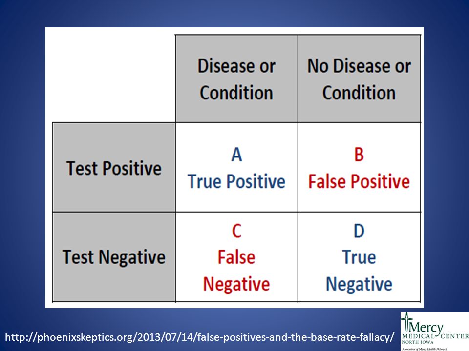
http://phoenixskeptics.org/2013/07/14/false-positives-and-the-base-rate-fallacy/
14
http://wiki.galenhealthcare.com/index.php/Galen_eCalcs_- _Calculator:_Framingham_Risk_for_General_CVD

15
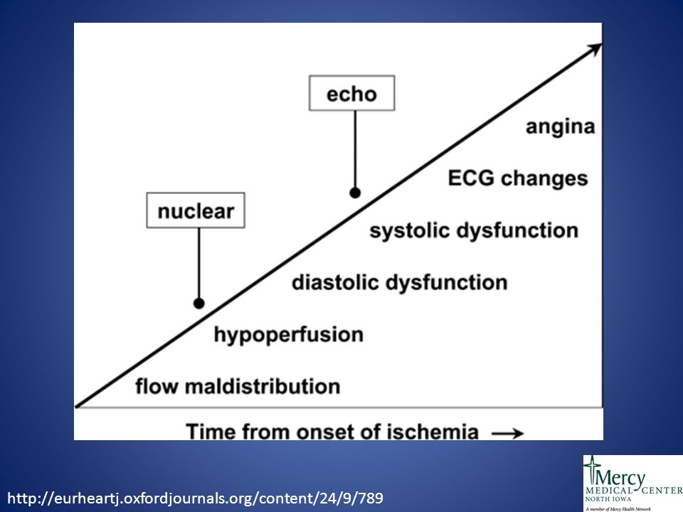
http://eurheartj.oxfordjournals.org/content/24/9/789
18
Stable vs Unstable

19
Diagnosis Resting EKG reasonable for the complaint of chest pain (1B)
")
20
Stable CAD – Follow-up Yearly follow-up at minimum Assessment of symptoms, monitoring risk factors, assess adequacy and adherance to lifestyle changes and medical therapy (1C). Periodic screening for important comorbidities (DM, CKD, Depression) might be reasonable. (Iib, C)
 might be reasonable. (Iib, C).")
21
Stable CAD – Follow-up A resting 12-lead ECG at 1-year or longer intervals between studies in patients with stable symptoms might be reasonable. (Iib, C)
.")
22
Stable CAD – EKGs Conclusion Don’t order annual EKGs or any other cardiac screening for low-risk patients without symptoms. With stable disease, there is no strong recommendation in regards to EKG

23
Echocardiogram

24
ECHO recommended to evaluate LV function and valvular function in known or suspected CAD and a prior MI, signs of heart failure, ventricular arrhythmia or murmur (IB)
")
25
Echocardiogram ECHO, MRI, Cardiac CT not recommended for routine monitoring of patients without signs of heart failure, MI or arrhythmia (III).
.")
26
Echocardiogram Routine LV function assessment (<1 year)not recommended if low risk and unless there is a change in clinical status or if it will change treatment plan (III).
not recommended if low risk and unless there is a change in clinical status or if it will change treatment plan (III).")
27
Echo - Conclusions Don’t perform echocardiography as routine follow-up for mild, asymptomatic native valve disease in adult patients with no change in signs or symptoms. ECHO reasonable if change in clinical status, chest pain, suspicion of worsening valvular pathology with symptoms

28
Transesophageal Echocardiogram http://ispub.com/IJA/23/2/3141

29
Pre-Operative Clearance http://www.jcomjournal.com/preoperative-cardiac-evaluation-for-noncardiac- surgery-a-critical-review/v

30
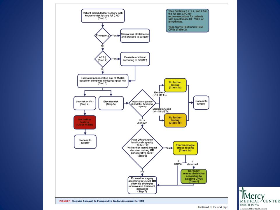
Perioperative Evaluation

32
http://www.aafp.org/afp/2012/0201/p239.html

33
Perioperative - Conclusion Don’t obtain baseline diagnostic cardiac testing (TTE, TEE, Cardiac stress test) in asymptomatic stable patients with known cardiac disease (e.g., CAD, valvular disease) undergoing low or moderate risk non-cardiac surgery.
 in asymptomatic stable patients with known cardiac disease (e.g., CAD, valvular disease) undergoing low or moderate risk non-cardiac surgery.")
34
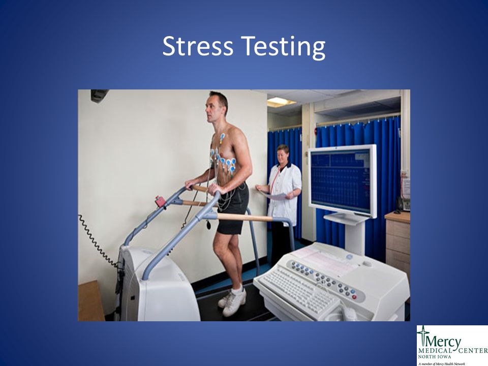
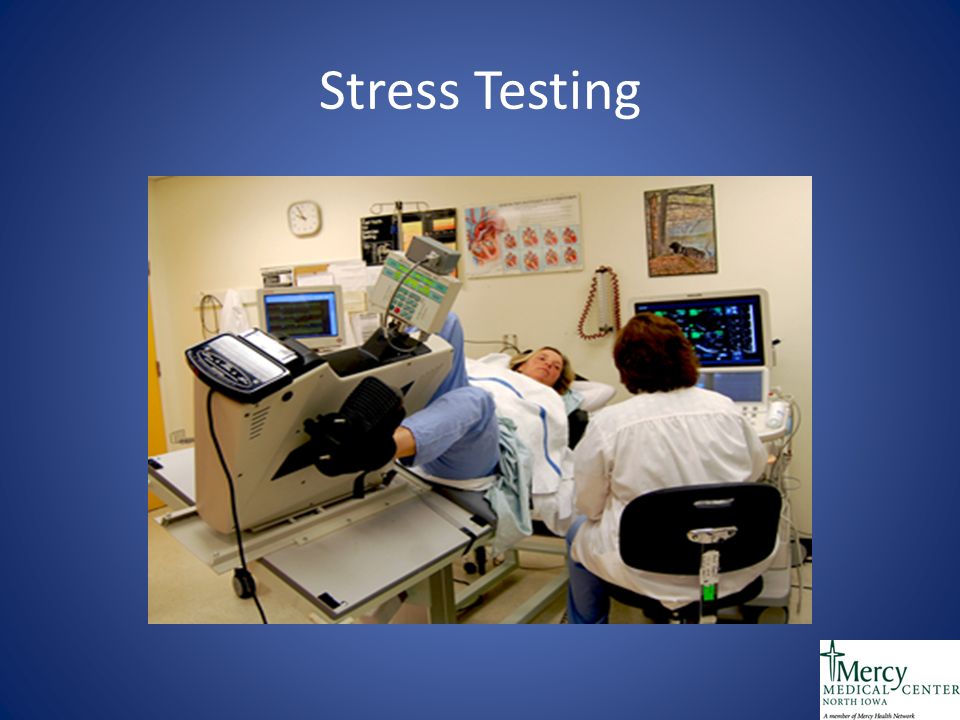
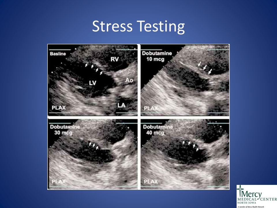
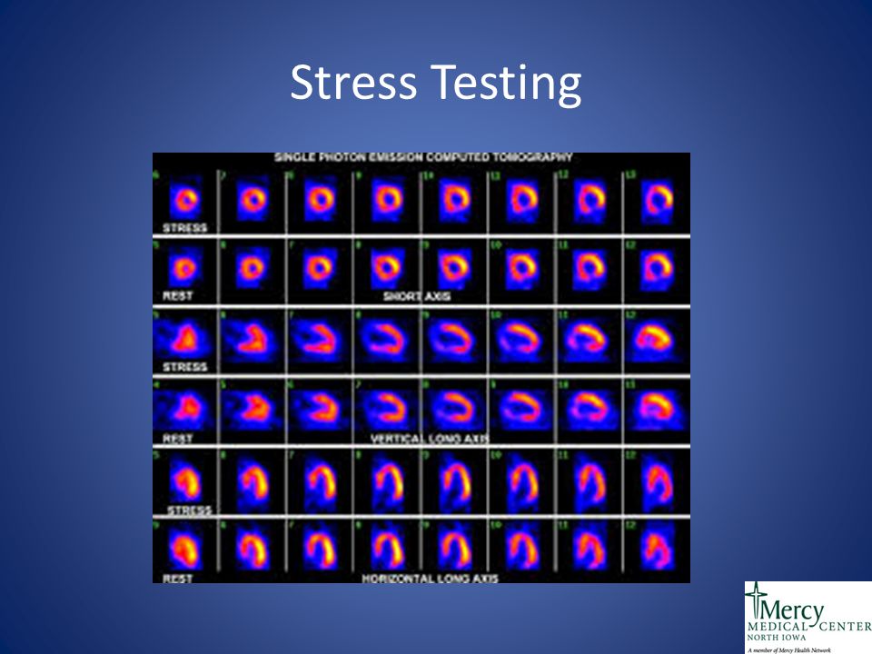
Stress Testing

39
http://www.revespcardiol.org/en/rational-use-of-noninvasive- cardiac/articulo/13052415/

40
Stress Testing Nuclear MRI, ECHO or MRI stress or CTA not recommended for stable patients specifically not within 5 years of CABG or 2 years of PCI without symptoms (III).
.")
41
Stress Testing In patients who have no new or worsening symptoms or no prior evidence of silent ischemia and not at high risk, the utility of annual surveillance exercise EKG testing is not well established (Iib,C)
")
42
Stress Testing Standard exercise ECG testing performed at 1- year or longer intervals might be considered in high risk patients who can walk and have interpretable ECG. (Iib, C) In patients who have no new symptoms, the usefulness of annual surveillance exercise ECG testing is not well established. (Iib, C)
 In patients who have no new symptoms, the usefulness of annual surveillance exercise ECG testing is not well established. (Iib, C).")
43
Stress Testing Exercise stress with nuclear MPI is not recommended as an initial test in low-risk patients who have an interpretable ECG (IIIC)
")
44
Stress Tests - Conclusion Don’t perform annual stress cardiac imaging or advanced non-invasive imaging as part of routine follow-up in asymptomatic patients. Don’t obtain screening exercise electrocardiogram testing in individuals who are asymptomatic and at low risk for coronary heart disease.

45
Stress Tests - Conclusion Don’t perform cardiac imaging for patients who are at low risk. Don’t perform stress cardiac imaging or advanced non-invasive imaging in the initial evaluation of patients without cardiac symptoms unless high-risk markers are present.

46
Coronary CT Calcium http://www.myvmc.com/investigations/ct-calcium-scoring/

47
Coronary CT Calcium Role is to change risk stratification from intermediate risk to either low/high risk Not as beneficial in those who already are low or high risk

48
CT Angiogram Inappropriate for : Post-Revascularization (PCI, CABG) to evaluate grafts or instent restenosis Uncertain benefit for: Intermediate risk patients undergoing intermediate or high risk surgery http://content.onlinejacc.org/article.aspx?articleid=1137956&_ga=1.185815593.1 895109799.1474037446
 to evaluate grafts or instent restenosis Uncertain benefit for: Intermediate risk patients undergoing intermediate or high risk surgery articleid= &_ga=")
49
CT Angiogram https://www.google.com/search?q=coronary+ct+calcium&biw=1696&bih=869&source=lnms&tb m=isch&sa=X&ved=0ahUKEwiltfKswJTPAhVNySYKHTHMA64Q_AUIBigB#tbm=isch&q=coronary+c t+stent&imgrc=jxSCcAQ4_ln2BM%3A

50
Cardiac CT - Conclusion Don’t use coronary artery calcium scoring for patients with known coronary artery disease (including stents and bypass grafts). Don’t order coronary artery calcium scoring for preoperative evaluation for any surgery, irrespective of patient risk.

51
Let’s Simplify Chest Pain – Stable or Unstable Risk stratification Initial EKG

52
Let’s Simplify Stable Chest pain, low probability – no testing High probability – likely angiography Intermediate probability – Stress Testing

53
Let’s Simplify Stress Test – Exercise preferred EKG preferred if no baseline changes Imaging (ECHO and Nuc) if intermediate to high risk
 if intermediate to high risk")
54
Let’s Simplify Stable Ischemic Heart Disease No benefit in routine stress tests or ECHO imaging unless change in clinical status Coronary Artery Calcium is for risk stratifying intermediate risk patients to low or high risk No annual EKG if low risk and asymptomatic

55
Let’s Simplify Perioperatively, no survival benefit for revascularization for procedure. No basic testing needed unless high risk patient

56
Questions?

57
Thank You

Similar presentations


 CAD is most common form of heart disease and causes premature death. In UK, 1 in 3 men and.>")


 38411524; +98(51)38472927.>")


