Download presentation
Presentation is loading. Please wait.

1
Lodish Berk Kaiser Krieger scott Bretscher Ploegh Matsudaira MOLECULAR CELL BIOLOGY SEVENTH EDITION The Eukaryotic Cell Cycle 2014-12-15 Copyright © 2013 by W. H. Freeman and Company

3
The master controllers of these events are a small number of heterodimeric protein kinases that contain a regulatory subunit and catalytic subunit. These kinases regulate the activities of multiple proteins involved in DNA replication and mitosis by phosphorylating them at specific regulatory sites, activating some and inhibiting others to coordinate their activities.

4
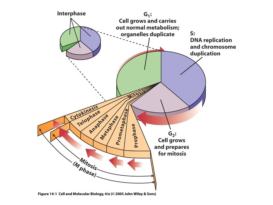
19.1. Overview of the Cell Cycle and Its Control The Cell Cycle Is an Ordered Series of Events Leading to Replication of Cells back

5
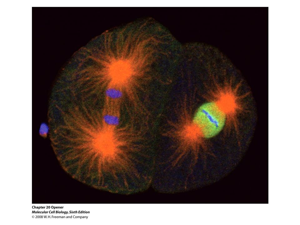
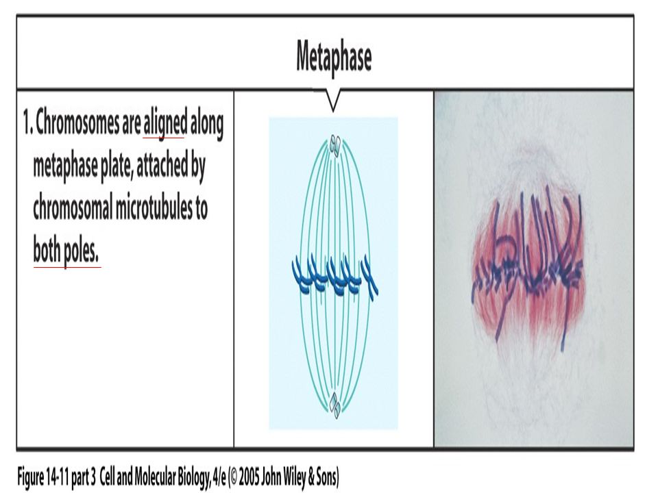
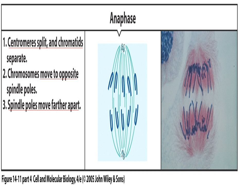
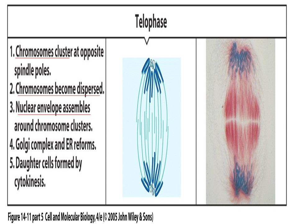
Figure 19.2 The stages of mitosis.

12
In vertebrates and diploid yeasts, cells in G1 have a diploid number of chromosomes (2n), one inherited from each parent. In haploid yeasts, cells in G1 have one of each chromosome (1n). Rapidly replicating human cells progress through the full cell cycle in about 24 hours: mitosis takes ≈30 minutes; G1, 9 hours; the S phase, 10 hours; and G2, 4.5 hours. only ≈90 minutes in rapidly growing yeast cells. Postmitotic cells in multicellular organisms can "exit" the cell cycle and remain for days, weeks, or in some cases (e.g., nerve cells and cells of the eye lens) even the lifetime of the organism without proliferating further. Most postmitotic cells in vertebrates exit the cell cycle in G1, entering a phase called G0. G0 cells returning to the cell cycle enter into the S phase; this reentry is regulated, thereby providing control of cell proliferation
. Rapidly replicating human cells progress through the full cell cycle in about 24 hours: mitosis takes ≈30 minutes; G1, 9 hours; the S phase, 10 hours; and G2, 4.5 hours. only ≈90 minutes in rapidly growing yeast cells. Postmitotic cells in multicellular organisms can exit the cell cycle and remain for days, weeks, or in some cases (e.g., nerve cells and cells of the eye lens) even the lifetime of the organism without proliferating further. Most postmitotic cells in vertebrates exit the cell cycle in G1, entering a phase called G0. G0 cells returning to the cell cycle enter into the S phase; this reentry is regulated, thereby providing control of cell proliferation.")
14
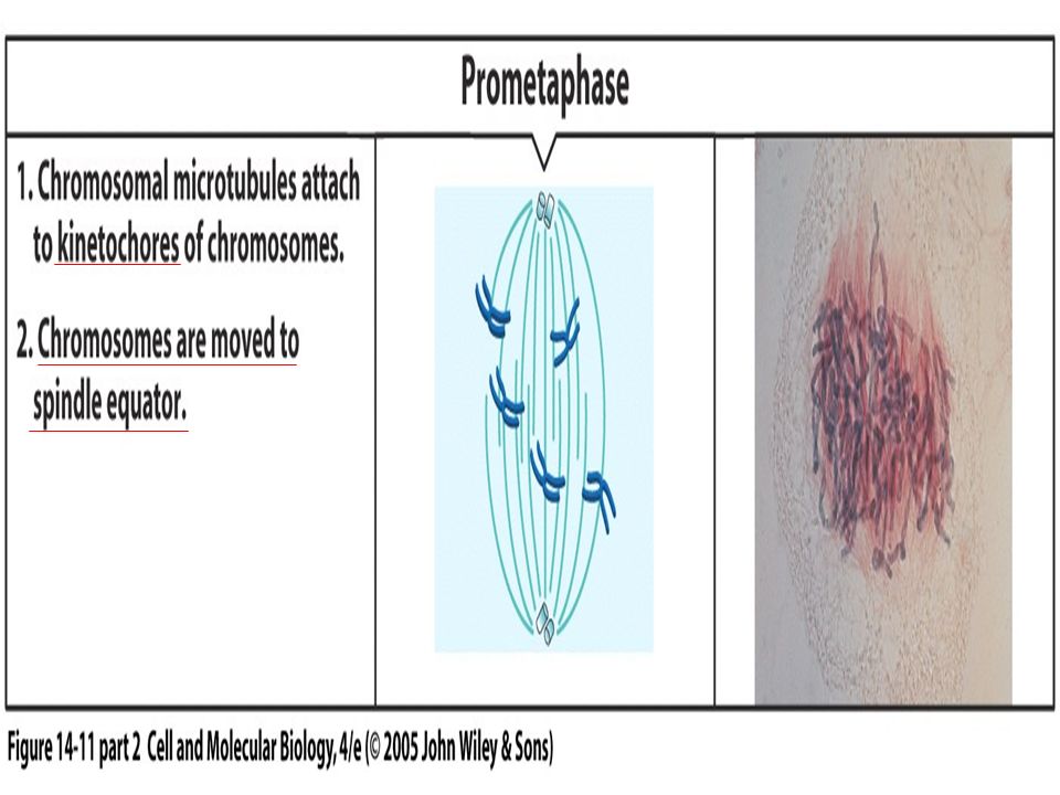
Diverse Experimental Systems Have Been Used to Identify and Isolate Cell-Cycle Control Proteins When interphase cells in the G1, S, or G2 stage of the cell cycle were fused to cells in mitosis, their nuclear envelopes vesiculated and their chromosomes condensed (Figure 20-3). diffusible componentinterphaseFigure 20-3 Similarly, when cells in G1 were fused to cells in the S phase and the fused cells exposed to radiolabeled thymidine, the label was incorporated into the DNA of the G1 nucleus, indicating that DNA synthesis began in the G1 nucleus shortly after fusion. However, when cells in G2 were fused to S-phase cells, no incorporation of labeled thymidine occurred in the G2 nuclei. Thus diffusible factors in an S-phase cell can enter the nucleus of a G1 cell and stimulate DNA synthesis, but these factors cannot induce DNA synthesis in a G2 nucleus. We now know that these factors are S-phase Cdk complexes, which can activate the pre-replication complexes assembled on DNA replication origins in early G1 nuclei.

15
Figure 19.3 The budding yeast S. cerevisiae.

16
Figure 19.4 The fission yeast S. pombe.

17
Genetic system The budding yeast Saccharomyces cerevisiae and the distantly related fission yeast Schizosaccharomyces pombe temperature-sensitive mutations cell-division cycle (CDC) nonpermissive temperature complementation Complementation: Human cDNA
 nonpermissive temperature complementation Complementation: Human cDNA")
20
Figure 19.6 Progesterone stimulates meiotic maturation of Xenopus oocytes.

21
Figure 19.7 Cell division patterns during the life cycle of Drosophila melanogaster.

22
Figure 19.8 Human cells undergoing mitosis.

23
MPF Promotes Maturation of Oocytes and Mitosis in Somatic Cells The process of oocyte maturation, from G2-arrested oocyte to the egg arrested in metaphase of meiosis II, can be studied in vitro by surgically removing G2-arrested oocytes from the ovary of an adult female frog and treating them with progesterone. When cytoplasm from eggs arrested in metaphase of meiosis II is microinjected into G2-arrested oocytes, the oocytes mature into eggs in the absence of progesterone. This system not only led to the initial identification of a factor in egg cytoplasm that stimulates maturation of oocytes in vitro in the absence of progesterone but also provided an assay for this factor, called maturation-promoting factor (MPF). As we will see shortly, MPF turned out to be the key factor that regulates the initiation of mitosis in all eukaryotic cells.
. As we will see shortly, MPF turned out to be the key factor that regulates the initiation of mitosis in all eukaryotic cells..")
24
Following fertilization, MPF activity falls again until the zygote (fertilized egg) enters the first mitosis of embryonic development. All the cells in early frog embryos undergo 12 synchronous cycles of mitosis. Throughout these cycles MPF activity is low in the interphase periods between mitoses and then rises as the cells enter mitosis. The cultured mammalian cells can be arrested in mitosis by treatment with compounds (e.g., colchicine) that inhibit assembly of microtubules. MPF ????
 that inhibit assembly of microtubules. MPF .")
25
Mitotic Cyclin Was First Identified in Early Sea Urchin Embryos As in early frog embryos, the initial cell cycles in the early sea urchin embryo occur synchronously, with all the embryonic cells entering mitosis simultaneously. In these studies, synchronously fertilized sea urchin eggs were incubated with a radiolabeled amino acid and samples were removed every 10 minutes. In subsequent experiments, a cDNA clone encoding sea urchin cyclin B was used as a probe to isolate a homologous cyclin B cDNA from Xenopus laevis. Western blotting of MPF purified from Xenopus eggs, using antibody prepared against the protein encoded by cyclin B cDNA, showed that one subunit of MPF is indeed cyclin B. The other subunit is the catalytic Cdk subunit, first identified in genetic experiments with yeasts discussed later.

26
Experimental Figure 19.9 Analysis of DNA content by flow cytometry. Propidium iodide

27
Cyclin B Levels and MPF Activity Change Together in Cycling Xenopus Egg Extracts Some unusual aspects of the rapid cell cycles in early animal embryos provided a way to study the role of mitotic cyclin in controlling MPF activity. Of particular importance, in the 12 rapid, synchronous cell cycles that occur following fertilization of Xenopus eggs, the G1 and G2 periods are minimized, and the cell cycle consists of alternating M and S phases. Once mitosis is complete, the early embryonic cells proceed immediately into the S phase, and once DNA replication is complete, the cells progress almost immediately into the next mitosis.

28
Figure 19.10 Regulation of cell cycle transitions.

29
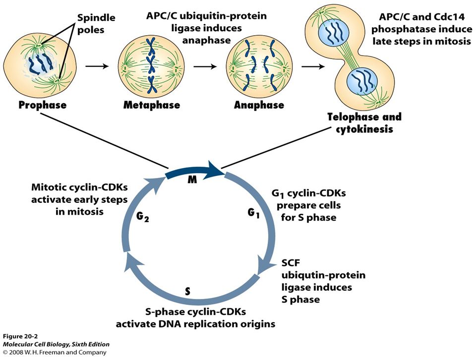
Figure 19.11 An overview of how CDKs regulate cell cycle progression.

30
Anaphase-promoting complex (APC) controls degradation of mitotic Cyclins and exit from mitosis APC specificity factor phosphatase
 controls degradation of mitotic Cyclins and exit from mitosis APC specificity factor phosphatase")
33
Figure 19.14 ATP analog–dependent CDK mutant.

34
Regulated Protein Phosphorylation and Degradation Control Passage through the Cell Cycle

35
20.6. Cell-Cycle Control in Mammalian Cells In multicellular organisms, precise control of the cell cycle during development and growth is critical for determining the size and shape of each tissue. Cell replication is controlled by a complex network of signaling pathways that integrate extracellular signals about the identity and numbers of neighboring cells and intracellular cues about cell size and developmental program. Most cells withdraw from the cell cycle during G1, entering the G0 state, to differentiate. Some differentiated cells (e.g., fibroblasts and lymphocytes) can be stimulated to reenter the cycle and replicate. Many differentiated cells, however, never reenter the cell cycle to replicate again; they are referred to as postmitotic cells. 負責教師 : 唐世杰 海洋大學生命科學系、生物科技所
 can be stimulated to reenter the cycle and replicate. Many differentiated cells, however, never reenter the cell cycle to replicate again; they are referred to as postmitotic cells. 負責教師 : 唐世杰 海洋大學生命科學系、生物科技所.")
36
Most studies of mammalian cell-cycle control have been done with cultured cells that require certain polypeptide growth factors (mitogens) to stimulate cell proliferation. Mammalian cells cultured in the absence of growth factors are arrested with a diploid complement of chromosomes in the G0 period of the cell cycle. If growth factors are added to the culture medium, these quiescent cells pass through the restriction point 14-16 hours later, enter the S phase 6-8 hours after that, and traverse the remainder of the cell cycle. Like START in yeast cells, the restriction point is the point in the cell cycle at which mammalian cells become committed to entering the S phase and completing the cell cycle. If mammalian cells are moved from a medium containing growth factors to one lacking growth factors before they have passed the restriction point, the cells do not enter the S phase. But once cells have passed the restriction point, they are committed to entering the S phase and progressing through the entire cell cycle, which takes about 24 hours for most cultured mammalian cells. Mammalian Restriction Point is Analogous to start in Yeast Cells

37
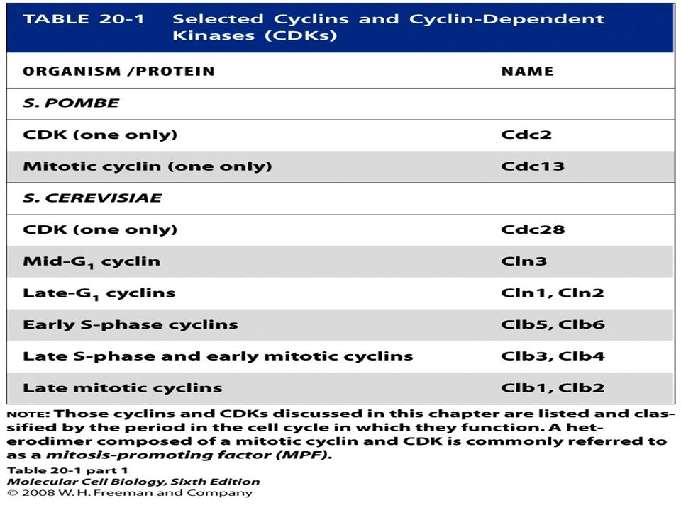
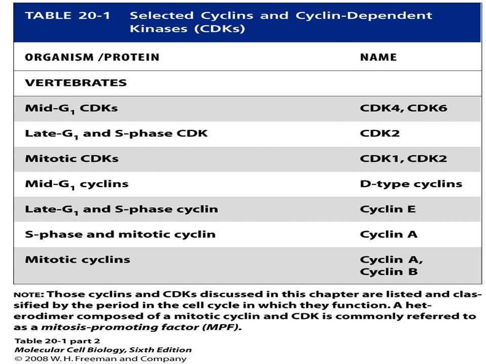
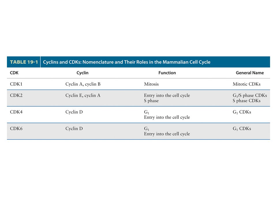
Multiple Cdks and Cyclins Regulate Passage of Mammalian Cells through the Cell Cycle The principle Cdks active in most mammalian cells have been named Cdk1, 2, 4, and 6 in order of their discovery. The first human Cdk to be discovered was identified by the ability of a cDNA clone encoding it to complement S. pombe cdc2 mutants; now designated Cdk1, this protein initially was called human Cdc2, a term that continues to be commonly used. A cDNA encoding mammalian Cdk2 was isolated by its ability to complement S. cerevisiae cdc28 mutants. Cdk4 and Cdk6 were isolated based on their homology to other Cdks. cDNAs for Cdk3 and Cdk5 also have been isolated, but the encoded proteins are not expressed at significant levels in most mammalian cells, and they function in processes other than cell cycle control.

38
Multiple Cdks and Cyclins Regulate Passage of Mammalian Cells through the Cell Cycle

39
Some delayed-response genes encode additional transcription factors such as E2Fs. Other delayed-response genes encode the D-type cyclins, cyclin E, Cdk2, Cdk4, and Cdk6. Cdk4, Cdk6, and the D-type cyclins are expressed first, followed by cyclin E and Cdk2. If growth factors are withdrawn before passage through the restriction point, transcription of these G1-phase Cdks and cyclins ceases. Since these proteins and the mRNAs encoding them are unstable, their concentrations fall precipitously. As a consequence, the cells do not pass the restriction point and do not replicate.

40
Passage Through the Restriction Point Depends on Phosphorylation of the Tumor-Suppressor Rb Protein E2F s activate genes encoding many of the proteins involved in DNA and deoxyribonucleotide synthesis. They also stimulate transcription of genes encoding the late-G1 cyclin (cyclin E), the S-phase cyclin (cyclin A), and the S-phase CDK (CDK2). In addition, E2Fs autostimulate transcription of their own genes. E2Fs function as transcriptional repressors when bound to Rb protein, which in turn binds histone deacetylase complexes. Rb protein was initially identified as the product of the prototype tumor-suppressor gene, RB (retinoblastoma).
, the S-phase cyclin (cyclin A), and the S-phase CDK (CDK2). In addition, E2Fs autostimulate transcription of their own genes. E2Fs function as transcriptional repressors when bound to Rb protein, which in turn binds histone deacetylase complexes. Rb protein was initially identified as the product of the prototype tumor-suppressor gene, RB (retinoblastoma)..")
41
Phosphorylation of Rb protein inhibits its repressing function, permitting activation of the genes required for entry into the S phase by E2Fs. Events in addition to Rb phosphorylation also contribute to control of passage through the restriction point as evidenced by the finding that RB -/- cells retain some dependence on mitogens, although greatly reduced mitogen concentrations suffice. Rb protein is maintained in the phosphorylated state throughout the S, G2, and M phases by Ckd2 -and Cdk1-cyclin complexes. After cells complete mitosis and enter early G1 or G0, the fall in Cdk-cyclin levels leads to dephosphorylation of Rb protein by the action of unopposed phosphatases. As a consequence, hypophosphorylated Rb protein is available to inhibit E2F activity during early G1 of the next cycle. ***

42
Figure 19.15 Control of the G 1 –S phase transition

43
Figure 19.16 Regulation of S phase and mitotic cyclin levels in budding yeast.

44
Figure 19.17 Control of S phase onset in S. cerevisiae by regulated proteolysis of the S phase inhibitor, Sic1. Cdk inhibitors

45
Figure 19.18 Six suboptimal G 1 /S phase CDK phosphorylation sites in Sic1 create a switch-like cell cycle entry.

46
Figure 19.19 The molecular mechanisms governing the initiation of DNA replication.

47
Unlinking of Sister Chromatids Initiates Anaphase in late anaphase, polyubiquitination of mitotic cyclin by the anaphase-promoting complex (APC ) leads to the proteasomal destruction of this cyclin (see Figure 21-10). Additional experiments with Xenopus egg extracts provided evidence that degradation of cyclin B, the Xenopus mitotic cyclin, and the resulting decrease in MPF activity are required for chromosome decondensation but not for chromosome segregation (Figure 21-18a, b)
.")
48
Immunoprecipitation studies with antibodies specific for Xenopus SMC proteins revealed that in cycling egg extracts some SMC proteins are part of a multiprotein complex called condensin, which becomes phosphorylated as cells enter mitosis. When the anti-SMC antibodies were used to deplete condensin from an egg extract, the extract lost its ability to condense added sperm chromatin. Other in vitro experiments showed that phosphorylated purified condensin binds to DNA and winds it into supercoils whereas unphosphorylated condensin does not.

50
Figure 19.20 Model for establishing cohesin linkage of sister chromatids.

51
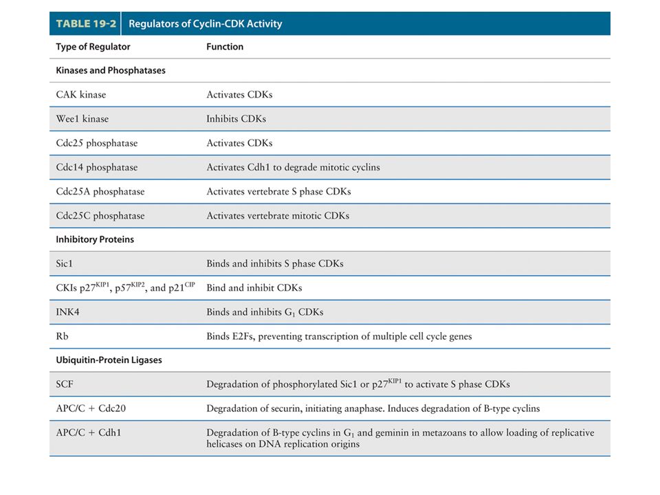
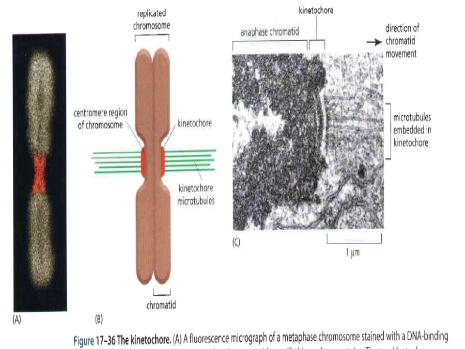
Once all chromosome kinetochores have attached to spindle microtubules, the APC is directed by a specificity factor called Cdc20 to polyubiquitinate securin, leading to the onset of anaphase. (This specificity factor is distinct from Cdh1, which directs the APC to polyubiquitinate B-type cyclins.) polyubiquitinated securin is rapidly degraded by proteasomes, thereby releasing separase.
 polyubiquitinated securin is rapidly degraded by proteasomes, thereby releasing separase..")
52
Figure 19.28 The protein phosphatase Cdc14 triggers exit from mitosis in budding yeast.

53
Figure 19.30 Fundamental processes in the eukaryotic cell cycle.

54
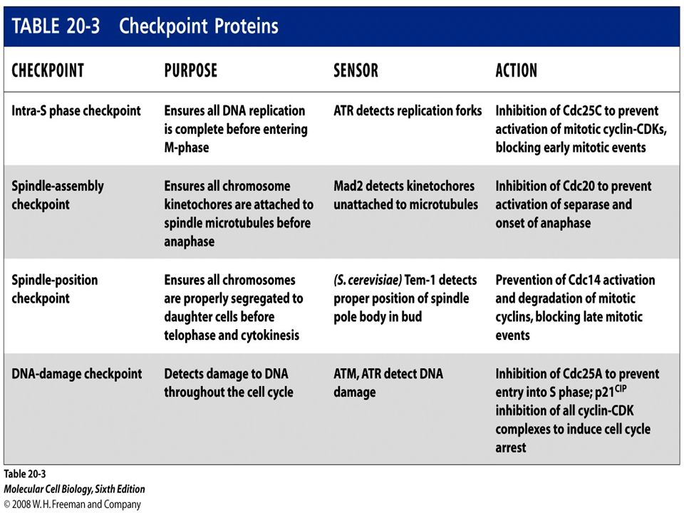
Figure 19.34 Overview of DNA damage checkpoint controls in the cell cycle.

Similar presentations


 Prepared by Cai Chunhui.>")


>")


