Download presentation
Presentation is loading. Please wait.

1
H EART Chapter 14 Seidel’s Guide to Physical Examination

2
O BJECTIVES To identify layers of heart To understand ventricles and atria To understand heart murmur To explain concept of cardiac output To explain cardiac cycle To explain ECG rhythms To identify 5 points for cardiac auscultation

3
K EY WORDS Pericardium Epicardium Myocardium Endocardium Aorta Atrioventricular Valves Semilunar Valves Purkinjie Fibers SA node AV node QRS complex Pericarditis Systole Diastole Electrocardiogram (EKG) Cardiac Output Atria Ventricles Heart Murmur Cardiac Cycle Heart Failure Myocardial Infarction Bruit
 Cardiac Output Atria Ventricles Heart Murmur Cardiac Cycle Heart Failure Myocardial Infarction Bruit")
4
A NATOMY & P HYSIOLOGY : H EART L OCATION In mediastinum Left of midline Above diaphragm Between medial/lower borders of lungs Behind sternum & 3 rd – 6 th intercostal cartilages Estimate size by percussion, begin tapping anterior axillary line Precordium: Area of chest overlying heart Base: broader upper portion of heart Apex: Narrower lower tip of heart

5
A NATOMY & P HYSIOLOGY : P OSITION V ARIANCE Body build Chest configuration Diaphragm level Dextrocardia

6
A NATOMY & P HYSIOLOGY : L AYERS OF HEART

7
A NATOMY & P HYSIOLOGY : H EART Pericardium Tough, double-walled, fibrous sac Encases and protects the heart Fluid between inner and outer layers of pericardium Epicardium Thin outermost muscle layer Covers surface of heart and extends over great vessels Myocardium Thick muscular, middle layer Responsible for pumping of heart Endocardium Inner most layer, covers valves

8
A NATOMY & P HYSIOLOGY : C HAMBERS Left atrium Left ventricle Right atrium Right ventricle Right/left atria – reservoirs Right/left ventricles - pumps Interventricular Septum – blood tight partition dividing the left and right heart Left Heart Right Heart

9
A NATOMY & P HYSIOLOGY : V ENTRICLES Right & left ventricles primary mass of heart Left ventricle mass greater than right ventricle Larger because higher pressure in systemic circulation requires greater contraction force Most anterior of heart – right ventricle Left ventricle’s contraction & thrust=apical impulse in 5 th intercostal space Aorta carries oxygenated blood to the body from the left ventricle

10
A NATOMY & P HYSIOLOGY : V ALVES Tricuspid Mitral Pulmonic Aortic Atrioventricular valves Semilunar valves

11
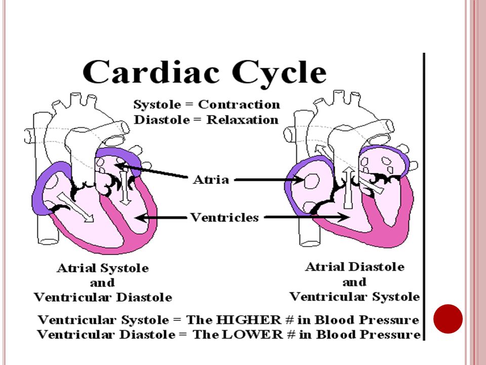
A NATOMY & P HYSIOLOGY : C ARDIAC CYCLE Systole – contraction of ventricles opens semilunar valves, causing blood to rush into pulmonary artery and aorta Diastole- relaxation of ventricles, valves close, draw blood into ventricles as atria contract https://www.youtube.com/watch?v=jLTdgrhpDCg https://www.youtube.com/watch?v=r32VObKw0gY

12
A NATOMY & P HYSIOLOGY : CARDIAC CYCLE Note: Pressures right ventricle, right atrium, and pulmonary artery lower than left side of heart

14
A NATOMY & P HYSIOLOGY : E LECTRICAL C ONDUCTION OF THE H EART

15
A NATOMY & P HYSIOLOGY : E LECTRICAL CONDUCTION Sinoatrial node AV node AV bundle (Bundle of His) Purkinje fibers
 Purkinje fibers")
16
A NATOMY & P HYSIOLOGY : ECG W AVES Electrocardiogram P Wave PR interval QRS complex ST segment T wave U wave

17
C ARDIAC R HYTHMS Normal Sinus Rhythm V-Tach Atrial Fibrillation (afib) & atrial flutter
 & atrial flutter")
18
H EART M URMURS Prolonged extra sounds heard during systole or diastole Cause- disruption blood flow into, through, or out of heart Swooshing or blowing sound heard auscultation Similar sound called a “bruit” heard over arteries Conditions contribute to heart murmurs Increased blood velocity Structural valve defects Valve malfunction Abnormal chamber openings https://www.youtube.com/watch?v=tGcAidBJCdM

19
T YPES OF H EART M URMURS Midsystolic Pansystolic Diastolic Exam & Findings Timing/duration Pitch Intensity Pattern Quality Location/radiation

20
C ARDIAC H EALTH A SSESSMENT Collecting Subjective Data History present health concern, chest pain, Palpations Past health history Family history Lifestyle and health practices Collecting Objective Data Inspect pulsations Palpate the apical impulse Palpate for abnormal pulsations Auscultate heart rate and rhythm If you detect an irregular rhythm, auscultate for a pulse rate deficit

21
E XAMINATION Inspect pulsations Palpate the apical impulse- most visible when patient is in the upright position Palpate for abnormal pulsations Auscultate heart rate and rhythm If you detect an irregular rhythm, auscultate for a pulse rate deficit

22
5 C ARDIAC A USCULTATION POINTS https://www.youtube.com/watch?v=83CBjj9dMRc

23
C ARDIAC A BNORMALITIES Angina Heart Failure Myocardial Infarction (MI) Pericarditis Common symptom with abnormalities especially in the older adult is fatigue Risk Factors: Smoking Hypertension Obesity High Cholesterol Level
 Pericarditis Common symptom with abnormalities especially in the older adult is fatigue Risk Factors: Smoking Hypertension Obesity High Cholesterol Level")
24
S UMMARY Anatomy heart Chambers Electrocardiogram Cardiac cycle 5 cardiac auscultation points Cardiac output Cardiac abnormalities

Similar presentations


>")


