Download presentation
Presentation is loading. Please wait.

1
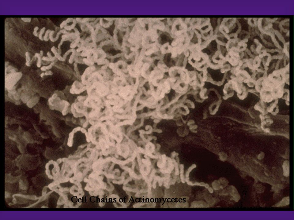
Actinomycetes Classification –Order – Actinomycetales Show fungus-like characteristics such as branching in tissues or in culture (look like mycelia). The filaments frequently segment during growth to produce pleomorphic, diphtheroidal, or club shaped cells. The cell wall and the internal structures are typical of bacteria rather than fungi. Some are aerobic and others are anaerobic. All are slow growing

2
Actinomycetes –The anaerobic genera: Actinomyces, Arachnia, and Bifidobacterium Morphology and cultural characteristics –G+ branching, or diphtheroid-like bacilli –Anaerobic and require CO 2 for growth –Non-sporing –Will grow on anaerobic BA or PEA. A israelii, the most commonly isolated species, produces rough, granular colonies that resemble molars. Biochemistry –ID by gas liquid chromatography (GLC) of metabolic by-products or fluorescent antibody studies
 of metabolic by-products or fluorescent antibody studies.")
3
Actinomycetes Clinical significance –Are part of the NF found in the cavities of humans and other animals. –All may cause actinomycosis or “lumpy jaw”. A cervicofacial infection that used to occur following tooth extractions or dental surgery which provided traumatized tissue for growth of the microorganism which may also invade the bone. –May cause thoracic or abdominal infections –May cause meningitis, endocarditis, or genital infections

4
Actinomycetes –Every kind of infection is characterized by draining sinuses, usually containing characteristic granules which are colonies of bacteria that look like dense rosettes of club- shaped filaments in radial arrangement Treatment –Penicillin

5
ACTINOMYCES Gram positive, branched filamentous bacteria (A) they are anaerobic to microaerophilic (B). Some will grow aerobically (facultative anaerobe) Superficially, they look like nocardiae. They are not acid fast, they are part of the normal flora Type species: Actinomyces israelii
 Superficially, they look like nocardiae. They are not acid fast, they are part of the normal flora Type species: Actinomyces israelii.")
7
Actinomyces israelii Causes 4 forms of Actinomycosis in humans (Almost always a mixed anaerobic infection; Granules in the pus is characteristic (A) 1. Cervicofacial form (B - Called lumpy jaw) 2. Thoracic form (anaerobic pulmonary abscess) 3. Abdominal form (often secondary to trauma) 4. Intra-uterine form (secondary to use of IUD) Drug of choice is penicillin may require surgical intervention
 2. Thoracic form (anaerobic pulmonary abscess) 3. Abdominal form (often secondary to trauma) 4. Intra-uterine form (secondary to use of IUD) Drug of choice is penicillin may require surgical intervention.")
8
Granules

9
Actinomycetes –The aerobic genera: Nocardia, Actinomadura, and Streptomyces. There are three clinically important species of Nocardia – N. asteroides, N. brasiliensis, and N. caviae Morphology and cultural characteristics –G+ branching bacillus that may fragment to bacillary or coccoid forms –Aerobic –Specimens should be inoculated onto 7H10 agar or Lowenstein-Jensen agar and brain heart infusion agar. Colonies produced are typically orange, dry, crumbly, and adherent. –The organisms are weakly acid fast or non acid fast

10
Actinomycetes Biochemistry –The organisms are identified based on sugar fermentations and hydrolysis reactions (caseine, tyrosine, etc.) Clinical significance –Mycetoma – organism enters the body through breaks in the skin and causes a localized infection involving skin, cutaneous, and subcutaneous tissue. The three most characteristic features seen are swelling, draining sinuses and granules. This disease can also be caused by fungi as well as Nocardia, Actinomadura, and Streptomyces.

11
Actinomycetes –Nocardiosis – is a localized or disseminated disease occurring after inhalation of organisms. Pulmonary infections resemble tuberculosis and can remain confined to the lungs or may disseminate, with a predilection for the brain and meninges. The disease is characterized by multiple confluent abscesses and intense suppuration. It is usually a disease of compromised hosts. Antimicrobic susceptibility/treatment –Mycetoma – aminoglycosides –Nocardiosis – sulfonamides or sxt

12
Nocardia Species

13
Nocardia is a genus of aerobic actinomycetes responsible for localized or disseminated infections in animals and humans. The genus is named after Edmond Nocard. who in 1888 described the isolation of an aerobic actinomycete from cattle with bovine farcy. The first human case of nocardiosis was reported by Eppinger in 1890. Cases of human disease have increased substantially in the past two decades, in association with an increasing population of immunocompromised hosts and improved methods for detection and identification of Nocardia in the clinical laboratory.

14
CLASSIFICATION The aerobic actinomycetes are a large and diverse group of gram-positive bacteria that appear on microscopy as branching filamentous cells. Members of the group are often only distantly related phylogenetica1ly. A subgroup, the “aerobic nocardiform actinomycetes.” is the most important cause of human infection and includes Mycobacterium, Corynebacterium, Nocardia, Rhodococcus, Gordona, and Tsukamurella. The putative cause of Whipple’s disease (Tropheryma whippeli) also belongs to this group. All members have cell walls containing mesodiaminopimelic acid, arabinose, galactose (type IV cell wall), and mycolic acids of various chain lengths.
 also belongs to this group. All members have cell walls containing mesodiaminopimelic acid, arabinose, galactose (type IV cell wall), and mycolic acids of various chain lengths..")
15
The last are responsible for varying acid fastness on appropriate staining. In addition, Nocardia spp. are characterized by an ability to form aerial hyphae, an ability to grow in media containing Lysozyme, and an inability to grow at 50˚ C. In the past few years the application of new molecular methods, particularly 16S ribosomal ribonucleic acid (rRNA) gene sequencing, has greatly expanded the spectrum of Nocardia with at least 30 species described and at least 13 of these documented to cause human infection. Of these, Nocardia asteroides sensu stricto, Nocardia farcinica, Nocardia nova, Nocardia brasiliensis, Nocardia pseudobrasiliensis, Nocardia otitidiscaviarum, and Nocardia transvalensis are the most important causes of human infection.
 gene sequencing, has greatly expanded the spectrum of Nocardia with at least 30 species described and at least 13 of these documented to cause human infection. Of these, Nocardia asteroides sensu stricto, Nocardia farcinica, Nocardia nova, Nocardia brasiliensis, Nocardia pseudobrasiliensis, Nocardia otitidiscaviarum, and Nocardia transvalensis are the most important causes of human infection..")
16
A Partially acid-fast (A), Gram Positive (B), beaded filamentous bacterium that fragments to form rods and cocci during growth AB
, Gram Positive (B), beaded filamentous bacterium that fragments to form rods and cocci during growth AB")
17
Nocardia acid fast stain

18
NOCARDIAL PHYSIOLOGY 1. Growth on simple medium 2. Grow at either ambient temperatures or 37 O C 3. Strictly aerobic 4. Produce dry wrinkled colonies 5. Produce aerial filaments (mycelia) on colonies 6. Species differentiated by growth characteristics 7. Susceptible to antibiotics (Sulfonamides Drugs of choice)
 on colonies 6. Species differentiated by growth characteristics 7. Susceptible to antibiotics (Sulfonamides Drugs of choice).")
19
ECOLOGY AND EPIDEMIOLOGY Nocardia is a ubiquitous environmental saprophyte, occurring in soil, organic matter, and water. Human infection usually arises from direct inoculation of the skin or soft tissues or by inhalation. Mycetoma due to N. brasiliensis is the most common nocardial infection reported from tropical regions, including the southern United States, Central and South America, and Australia. Worldwide, respiratory and disseminated infections are most often due to members of the N. asteroides complex.

20
Nocardia is a well-recognized cause of infection in animals, with bovine mastitis being the most common. There are no reports of animal-to-human transmission. Clusters of invasive nocardiosis acquired by patients in oncology and transplant units, presumed to be associated with inhalation of contaminated dust, have been described. Hospital construction work may have been a risk factor in separate clusters of postsurgical wound infections due to Nocardia. Pulsed-field gel electrophoresis and random amplifica tion of polymorphic deoxyribonucleic acid (DNA) (RAPD) fingerprinting have been successfully used for confirming clusters and defining common sources.
 (RAPD) fingerprinting have been successfully used for confirming clusters and defining common sources..")
21
Colonization Occasional instances of transient colonization of sputum and skin by Nocardia have been reported and appear to indicate aerosol contamination or soil-derived contamination. Colonization of the sputum is typically found in patients with underlying pulmonary pathology, who are not receiving steroid therapy, and requires no specific therapy. Significant isolates of Nocardia should be visible on Gram stain, produce a pure or predominant growth in culture, and be isolated repeatedly from clinical specimens. However, the extent to which spontaneously resolving or subclinical pulmonary’ infection occurs in the population is not well defined and at least one leading authority warns against dismissing positive sputum cultures as harmless.

22
Specific Virulence Determinants The nocardial envelope is structurally similar to that of other actinomycetes: 15% to 25% of the cell wall mass in rapidly growing organisms, and nearly twice that in stationary phase, is composed of peptidoglycan. Differences in cell wall ultrastructure and chemical composition are evident during logarithmic and stationary phases of growth. Intrastrain differences in toxicity to host cells as well as virulence in animal models may also relate to different virulence factor expression and influence specific cell tropisms. Mycolic acid polymers such as trehalose-6.6’-dimycolate (“cord factor”) are members of a group of biologically active cell wall glycolipids found in many actinomycetes including Nocardia and are associated with virulence.
 are members of a group of biologically active cell wall glycolipids found in many actinomycetes including Nocardia and are associated with virulence..")
23
They are toxic in vitro and in animal models, insert themselves into phospholipid bilayers in vitro, and contribute to inhibition of phagosome-lysosome fusion and acidification in macrophages. Nocardia contains no cell wall lipopolysaccharide, exopolysaccharide capsule, or surface fimbriae. However, strain-dependent specific adhesins and invasion properties influence the outcome of infection in animal models. Virulent strains of N. asteroides are relatively resistant to neutrophil-mediated killing, and organisms in the logarithmic growth phase are more toxic to macrophages. They inhibit phagosome-lysosome fusion more successfully in vitro, giving rise to L-forms, which can be isolated from within macrophages many days later. Cell wall-deficient forms (L-forms) of Nocardia have been isolated from serious human and animal infections and may explain occasional late relapse of nocardial infections.
 of Nocardia have been isolated from serious human and animal infections and may explain occasional late relapse of nocardial infections..")
24
B. AT LEAST FOUR BASIC FORMS OF DISEASE ARE RECOGNIZED IN HUMANS (Nocardial diseases in humans are often thought to be opportunistic, but this is not an absolute requirement since healthy humans can also be infected) 1. Pulmonary Nocardiosis 2. Localized extrapulmonary and systemic Nocardiosis (including infections of the brain) 3. Cutaneous Nocardiosis (sporotrichoid) 4. Mycetomas
 1. Pulmonary Nocardiosis 2. Localized extrapulmonary and systemic Nocardiosis (including infections of the brain) 3. Cutaneous Nocardiosis (sporotrichoid) 4. Mycetomas.")
25
CLINICAL MANIFESTATIONS Immunocompromise is a well-established risk factor for nocardiosis. Nocardia may therefore be considered as an opportunistic pathogen that causes serious and disseminated disease in settings such as organ transplantation and lymphoreticular neoplasia, with the relative risk of progressive disease reflecting the level of immunosuppression. A compilation of more than a thousand randomly selected cases from the literature showed that more than 60% of all reported nocardiosis is associated with preexisting immune compromise, ranging from alcoholism and diabetes to organ transplantation and acquired immunodeficiency syndrome (AIDS).
..")
26
Disseminated infection is characterized by widespread abscess formation. The most commonly reported sites include the CNS and eyes particularly the retinal, skin and subcutaneous tissues.

27
FIGURE 252-2. Computed tomography scan (A) and chest radiograph (B) from a patient demonstrating multiple abscesses due to Nocardia farcinica.
 and chest radiograph (B) from a patient demonstrating multiple abscesses due to Nocardia farcinica..")
28
Figure 252-3. Cerebral magnetic resonance scan showing a brain abscess due to Nocardia farcinica

29
LOCALIZED EXTRAPULMONARY AND SYSTEMIC NOCARDIOSIS (including infections of the brain) BRAIN ABSCESS EYE (RETINAL LESIONS)
 BRAIN ABSCESS EYE (RETINAL LESIONS)")
30
CUTANEOUS NOCARDIOSIS (SPOROTRICHOID)
")
31
MYCETOMAS

32
The microbiology laboratory should always be informed when nocardiosis is suspected, because the diagnosis may be missed by routine laboratory methods. Respiratory secretions, skin biopsies, or aspirates from deep collections are the most common specimens from which Nocardia is isolated. Direct smears from such specimens typically show gram- positive, beaded, branching filaments, which are usually acid fast. Standard blood culture media support the growth of Nocardia organisms, but prolonged incubation (up to 2 weeks) and blind subcultures may be required for their detection. LABORATORY DIAGNOSIS
 and blind subcultures may be required for their detection. LABORATORY DIAGNOSIS.")
33
In specimens containing mixed flora (e.g.. respiratory secretions). nocardial colonies are easily obscured by those of more rapidly growing bacteria, and the yield is increased by use of selective media, such as Thayer-Martin agar with antibiotics or paraffin agar. Buffered charcoal-yeast extract (BCYE) medium, which is commonly used for selective growth of Legionella spp. may also be used for isolation of Nocardia from respiratory specimens. Decontamination methods used for mycobacterial culture are too harsh for Nocardia and may substantially reduce the numbers of viable organisms present in the specimen. Most isolates are acid fast by a method such as the modified Kinyoun technique, but this characteristic may vary with strain and culture media used.
 medium, which is commonly used for selective growth of Legionella spp. may also be used for isolation of Nocardia from respiratory specimens. Decontamination methods used for mycobacterial culture are too harsh for Nocardia and may substantially reduce the numbers of viable organisms present in the specimen. Most isolates are acid fast by a method such as the modified Kinyoun technique, but this characteristic may vary with strain and culture media used..")
34
Isolates identified presumptively as Nocardia can be assigned to traditional groupings based on the hydrolysis of casein, tyrosine, xanthine, hvpoxanthine, and testosterone. These methods are relatively expensive, slow, and limited by their inability to differentiate members of the N. asteroides complex or to identify newly described species. Expanded biochemical tests and patterns of resistance to antibiotics can be used to differentiate N. farcinica and N. nova from N. asteroides sensu stricto. Although resistance to third-generation cephalosporins is considered characteristic of N. farcinica, this is variable and should not be used as the sole criterion for differentiating this species from N. asteroides sensu stricto.

35
Commercially available identification systems, including Microscan RA1/HNID panels, the 1D32C Yeast Identification System, and the API 20C may provide more rapid methods for differentiating Nocardia spp. Molecular techniques including restriction endonuclease analysis following PCR and l6S rRNA sequencing have been used to differentiate among the Nocardia and to characterize new species and are likely to become the methods of choice for identification of Nocardia spp. as they become more available. Immunodominant antigens of Nocardia have been described and serologic tests developed: these have proven useful in the diagnosis of N. braisiliensis mycetoma” but at present remain experimental.

36
TREATMENT: Sulfadiazine drug of choice. Other drugs may work such as: Bactrim (TMS), Tetracycline, Amikacin, Imepenem and Cefotaxime Penicillin is not a good choice since most nocardiae produce beta lactamase. Surgical intervention may be required.
, Tetracycline, Amikacin, Imepenem and Cefotaxime Penicillin is not a good choice since most nocardiae produce beta lactamase. Surgical intervention may be required..")
37
Duration of Therapy and Prognosis Lack of response to initial therapy may be due to primary drug resistance, inadequate penetration of drug into sites of infection (dependent on dose, bioavailability of oral drugs, abscess location and pathology, and on patient compliance), or the presence of a sequestered abscess requiring surgical drainage. Recommendations on the duration of therapy are necessarily empirical and based primarily on reports of relapse after sulfonamide therapy of different durations. In immunocompromised hosts, primary treatment failure may also be due to overwhelming nocardial infection or a coexisting or secondary opportunistic infection.

Similar presentations


>")


 Please click audio icon to hear Carol’s narration.>")
 Please click audio icon to hear Carol’s narration.>")

>")
 is an infectious disease caused by bacteria whose scientific name is Mycobacterium tuberculosis. It was first isolated.>")

