Download presentation
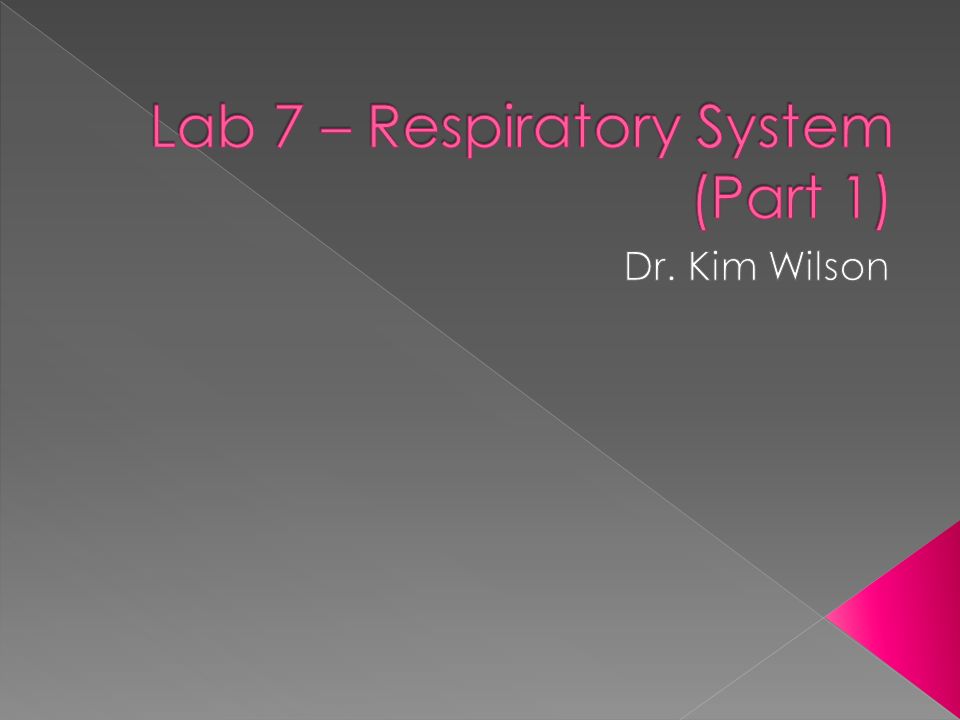
Presentation is loading. Please wait.

2
Exercise 36: Pp. 537-546 Refer to Figure 36.1 when studying structures of nose Website with flashcard animation › http://www.studystack.com/flashcard-921 http://www.studystack.com/flashcard-921

3
BIO 211 LAB #7 RESPIRATORY SYSTEM (PART 1): ANATOMY PART A: ANATOMY OF THE HUMAN RESPIRATORY SYSTEM INSTRUCTIONS: 1. Using the human torsos, larynx model, and the skulls, locate as many as possible of the following structures from the list, "Major Structures of the Human Respiratory System". Use your textbook for reference. 2. Answer related questions on the Questions Sheet.

4
UPPER RESPIRATORY TRACT 1. NOSE › EXTERNAL NOSE › INTERNAL NOSE (NASAL CAVITIES) › NASAL SEPTUM › SUPERIOR, MIDDLE, INFERIOR MEATI › SUPERIOR, MIDDLE, INFERIOR TURBINATES (CONCHAE) › ANTERIOR NARES › POSTERIOR NARES › OLFACTORY EPITHELIUM › PARANASAL SINUSES: FRONTAL, MAXILLARY, SPHENOID, › ETHMOID
 › NASAL SEPTUM › SUPERIOR, MIDDLE, INFERIOR MEATI › SUPERIOR, MIDDLE, INFERIOR TURBINATES (CONCHAE) › ANTERIOR NARES › POSTERIOR NARES › OLFACTORY EPITHELIUM › PARANASAL SINUSES: FRONTAL, MAXILLARY, SPHENOID, › ETHMOID .")
5
UPPER RESPIRATORY TRACT cont. 2. PHARYNX › NASOPHARYNX › OROPHARYNX › LARYNGOPHARYNX › EUSTACHIAN TUBE OPENINGS › FAUCES › PHARYNGEAL TONSILS (ADENOIDS) › PALATINE TONSILS › LINGUAL TONSILS
 › PALATINE TONSILS › LINGUAL TONSILS.")
6
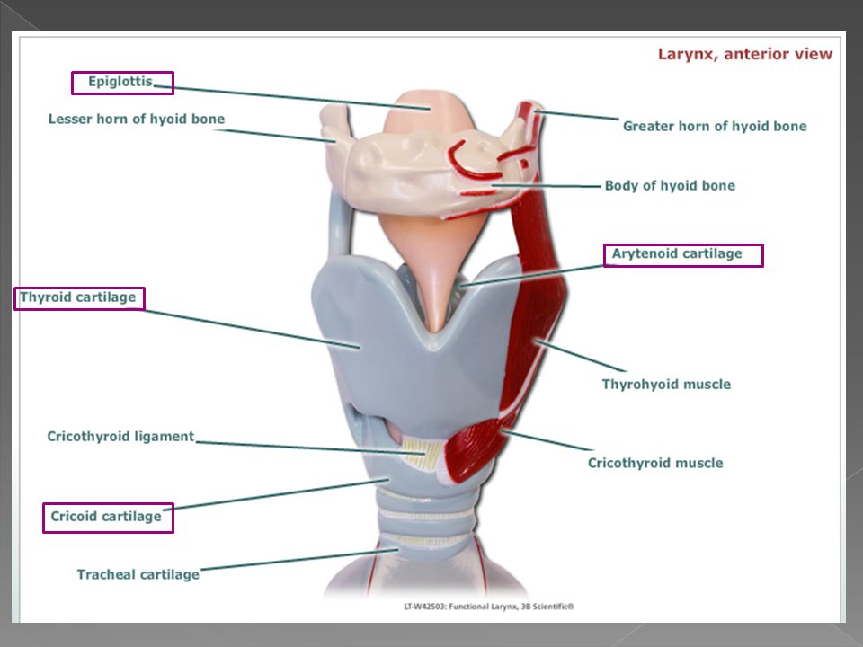
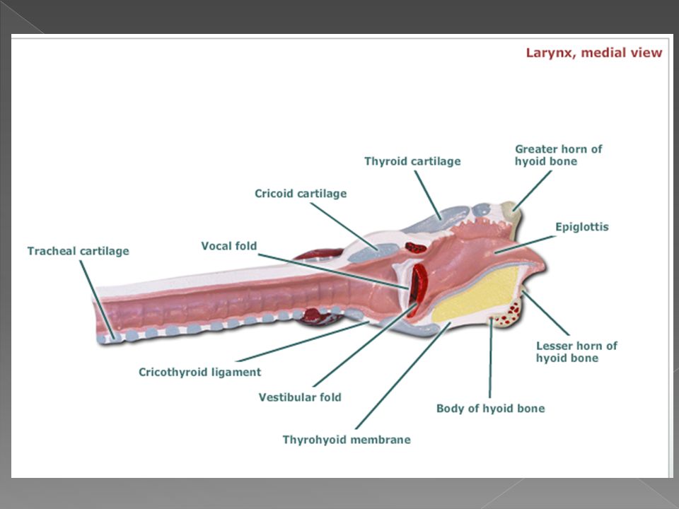
UPPER RESPIRATORY TRACT cont. 3. LARYNX › THYROID CARTILAGE (ADAM'S APPLE) › EPIGLOTTIS › CRICOID CARTILAGE › ARYTENOID CARTILAGES › TRUE AND FALSE VOCAL CORDS › GLOTTIS › DIVISIONS › VESTIBULE › VENTRICLE › INFRAGLOTTIC LARYNX
 › EPIGLOTTIS › CRICOID CARTILAGE › ARYTENOID CARTILAGES › TRUE AND FALSE VOCAL CORDS › GLOTTIS › DIVISIONS › VESTIBULE › VENTRICLE › INFRAGLOTTIC LARYNX.")
7
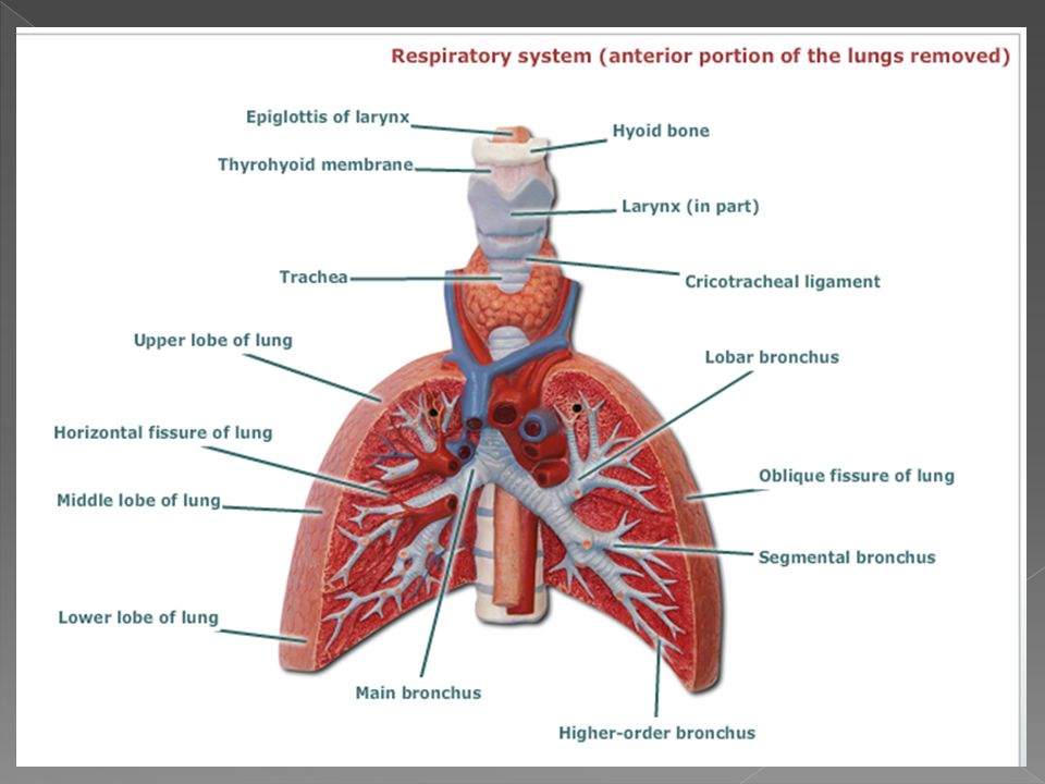
1. TRACHEA 2. PRIMARY BRONCHI (L&R) AND BRONCHIAL TREE 3. LUNGS › ROOT › HILUM › BASE › APEX › COSTAL SURFACE › LOBES › LEFT: SUPERIOR, INFERIOR › RIGHT: SUPERIOR, MIDDLE, INFERIOR OTHER › 1. PLEURA PARIETAL VISCERAL › 2. DIAPHRAGM

8
The left lung has 2 lobes and the right lung has 3 lobes. Source: http://home.comcast.net/~wnor/thoraxlesson2.htm

9
The pleural cavity is a closed space (like the inside of a balloon) within which the lung has grown. As the lung grows into the space, it picks up a layer of pleura (outside of balloon) and this is called the visceral pleura. The remainder of the pleura is called the parietal pleura. (parietal pleura in blue; visceral pleura in purple)
 and this is called the visceral pleura. The remainder of the pleura is called the parietal pleura. (parietal pleura in blue; visceral pleura in purple).")
18
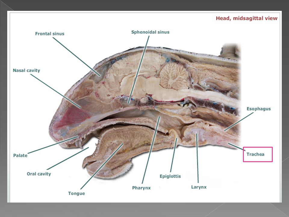
INSTRUCTIONS: 1. Obtain your assigned cat and locate each of the structures from the list, "Major Structures of the Cat Respiratory System". Use your lab manual (Color Photo Gallery) and the Rust lab manual for reference. (NOTE: Observe the cut on the demonstration cat that allows viewing of the epiglottis and related structures.) 2. Answer related questions on the Questions Sheet.
 and the Rust lab manual for reference. (NOTE: Observe the cut on the demonstration cat that allows viewing of the epiglottis and related structures.) 2. Answer related questions on the Questions Sheet..")
19
ANTERIOR NARES PHARYNX NASOPHARYNX OROPHARYNX LARYNGOPHARYNX LARYNX THYROID CARTILAGE EPIGLOTTIS CRICOID CARTILAGE ARYTENOID CARTILAGES TRUE AND FALSE VOCAL CORDS GLOTTIS

20
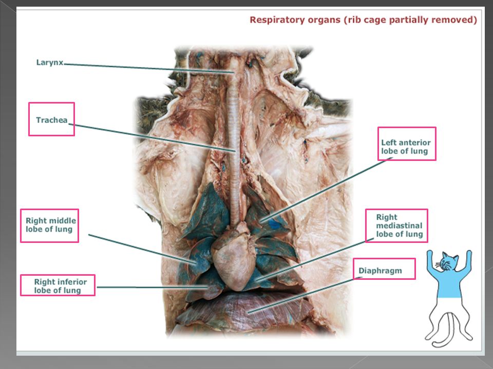
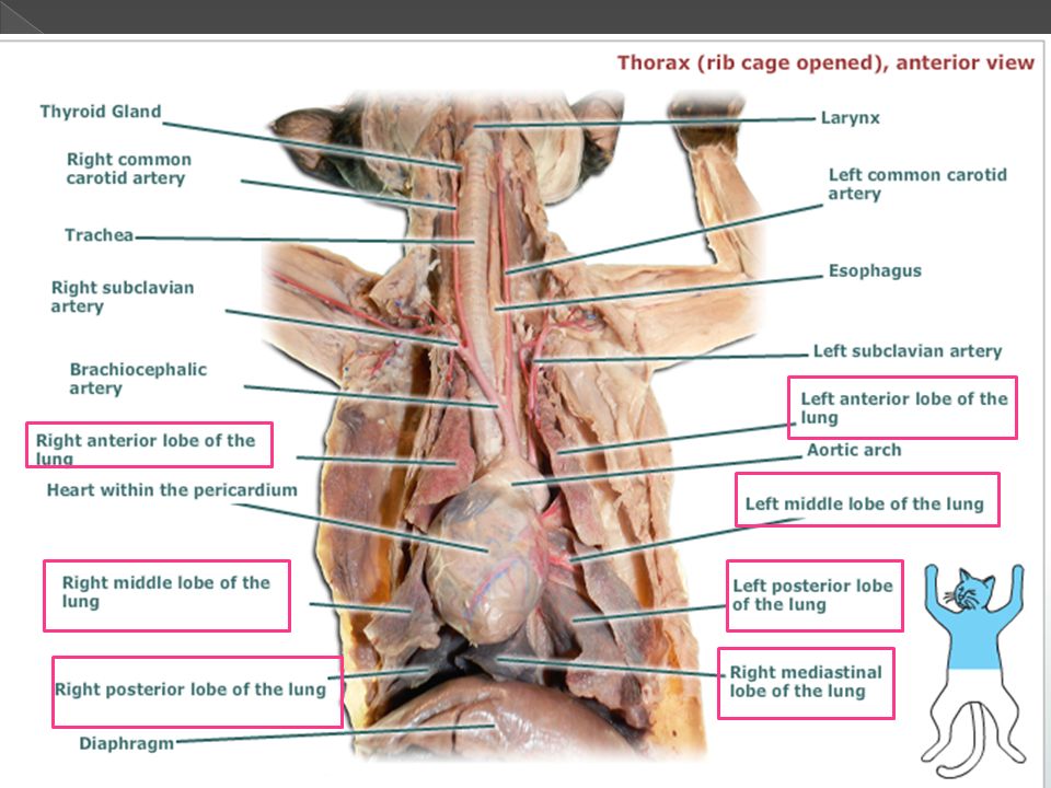
TRACHEA PRIMARY BRONCHI (L&R) AND BRONCHIAL TREE LUNGS BASE APEX COSTAL SURFACE LOBES (Compare # to Human) PLEURA › PARIETAL › VISCERAL DIAPHRAGM PHRENIC NERVES
 AND BRONCHIAL TREE LUNGS BASE APEX COSTAL SURFACE LOBES (Compare # to Human) PLEURA › PARIETAL › VISCERAL DIAPHRAGM PHRENIC NERVES")
22
trachea

27
There are R & L phrenic nerves, easily seen at diaphragm, attached to vena cava on R, in pleural folds on L. The right phrenic is attached to the inferior vena cava (image).
..")
28
The left phrenic nerve is enfolded in the pleural folds which are attached to the diaphragm.

Similar presentations


 nasal cavity.>")


 bronchus Right main (primary) bronchus Left lung.>")


 For maintenance, growth, defense, and division Krebs's cycle and the electron transport chain use oxygen.>")

