Download presentation
Presentation is loading. Please wait.

1
Klinefelter Syndrome and Turner Syndrome Robbins and Cotran Pathologic Basis of Disease, Chapter 5, 165-166

2
KLINEFELTER SYNDROME

3
Klinefelter Syndrome Klinefelter syndrome is best defined as male hypogonadism that occurs when there are two or more X chromosomes and one or more Y chromosomes. – It is one of the most frequent forms of genetic disease involving the sex chromosomes as well as one of the most common causes of hypogonadism in the male. – The incidence of this condition is approximately 1 in 660 live male births.

4
Klinefelter Syndrome Klinefelter syndrome can rarely be diagnosed before puberty, particularly because the testicular abnormality does not develop before early puberty. Most patients have a distinctive body habitus with an increase in length between the soles and the pubic bone, which creates the appearance of an elongated body. Also characteristic are eunuchoid body habitus with abnormally long legs; small atrophic testes often associated with a small penis; and lack of such secondary male characteristics as deep voice, beard, and male distribution of pubic hair. Gynecomastia may be present. The mean IQ is somewhat lower than normal, but mental retardation is uncommon. There is increased incidence of type 2 diabetes and the metabolic syndrome that gives rise to insulin resistance. Curiously, mitral valve prolapse is seen in about 50% of adults with Klinefelter syndrome. There is also an increased incidence of osteoporosis and fractures due to sex hormonal imbalance.

5
Klinefelter Syndrome

6
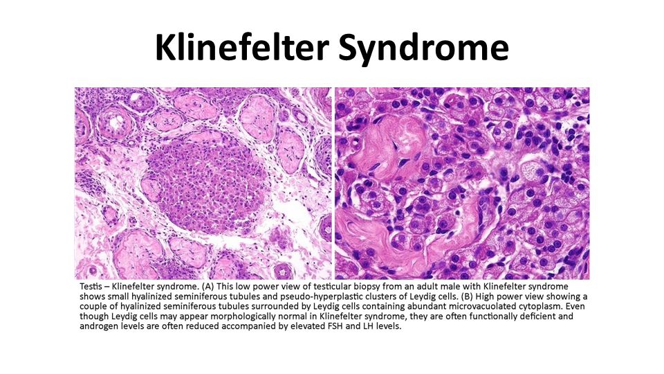
It should be evident that the clinical features of this condition are variable, the only consistent finding being hypogonadism. Plasma gonadotropin concentrations, particularly follicle- stimulating hormone, are consistently elevated, whereas testosterone levels are variably reduced. Mean plasma estradiol levels are elevated by an as yet unknown mechanism. The ratio of estrogens and testosterone determines the degree of feminization in individual cases.

7
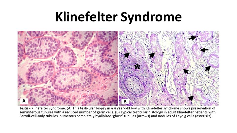
Klinefelter Syndrome Klinefelter syndrome is an important genetic cause of reduced spermatogenesis and male infertility. In some patients the testicular tubules are totally atrophied and replaced by pink, hyaline, collagenous ghosts. In others, apparently normal tubules are interspersed with atrophic tubules. In some patients all tubules are primitive and appear embryonic, consisting of cords of cells that never developed a lumen or progressed to mature spermatogenesis. Leydig cells appear prominent, as a result of the atrophy and crowding of the tubules and elevation of gonadotropin concentrations.

8
Klinefelter Syndrome

11
Patients with Klinefelter syndrome have a higher risk for: – Breast cancer (20 times more common than in normal males) – Extragonadal germ cell tumors – Autoimmune diseases such as systemic lupus erythematosus.
 – Extragonadal germ cell tumors – Autoimmune diseases such as systemic lupus erythematosus.")
12
Klinefelter Syndrome The classic pattern of Klinefelter syndrome is associated with a 47,XXY karyotype (90% of cases). This complement of chromosomes results from nondisjunction during the meiotic divisions in the germ cells of one of the parents. Maternal and paternal nondisjunction at the first meiotic division are roughly equally involved. There is no phenotypic difference between those who receive the extra X chromosome from their father and those who receive it from their mother. Maternal age is increased in the cases associated with errors in oogenesis. In addition to this classic karyotype, approximately 15% of patients with Klinefelter syndrome have been found to have a variety of mosaic patterns, most of them being 46,XY/47,XXY. Other patterns are 47,XXY/48,XXXY and variations on this theme.

13
Klinefelter Syndrome

14
As is the case with normal females, all but one X chromosome undergoes inactivation in patients with Klinefelter syndrome. Why then, do the patients with this disorder have hypogonadism and associated features? The explanation for this lies in genes on the X chromosome that escape lyonization and in the pattern of X inactivation.

15
Klinefelter Syndrome One pathogenic mechanism is related to uneven dosage compensation during X-inactivation. – In some cases about 15% of the X-linked genes escape inactivation. – Thus, there is an extra dose of these genes compared to normal males in whom only one copy of X is active, and it appears that “overexpression” of one or more of these genes leads to hypogonadism.

16
Klinefelter Syndrome A second mechanism involves the gene encoding the androgen receptor, through which testosterone mediates its effects. – The androgen receptor gene maps to the X chromosome and contains highly polymorphic CAG (trinucleotide) repeats. – The functional response of the receptor to any particular dose of androgen is dictated, in part, by the number of CAG repeats, as receptors with shorter CAG repeats are more sensitive to androgens than those with long CAG repeats. – In persons with Klinefelter syndrome, the X chromosome bearing the androgen receptor allele with the shortest CAG repeat is preferentially inactivated. – In XXY males with low testosterone levels, expression of androgen receptors with long CAG repeats exacerbates the hypogonadism and appears to account for certain aspects of the phenotype, such as small penis size.
 repeats. – The functional response of the receptor to any particular dose of androgen is dictated, in part, by the number of CAG repeats, as receptors with shorter CAG repeats are more sensitive to androgens than those with long CAG repeats. – In persons with Klinefelter syndrome, the X chromosome bearing the androgen receptor allele with the shortest CAG repeat is preferentially inactivated. – In XXY males with low testosterone levels, expression of androgen receptors with long CAG repeats exacerbates the hypogonadism and appears to account for certain aspects of the phenotype, such as small penis size..")
17
Klinefelter Syndrome

18
TURNER SYNDROME

19
Turner Syndrome Turner syndrome results from complete or partial monosomy of the X chromosome and is characterized primarily by hypogonadism in phenotypic females. It is the most common sex chromosome abnormality in females, affecting about 1 in 2500 live-born females.

20
Karyotypic Abnormalities of Turner Syndrome Approximately 57% are missing an entire X chromosome, resulting in a 45,X karyotype. Of the remaining 43%, approximately one third (approximately 14%) have structural abnormalities of the X chromosomes, and two thirds (approximately 29%) are mosaics.
 have structural abnormalities of the X chromosomes, and two thirds (approximately 29%) are mosaics..")
21
Karyotypic Abnormalities of Turner Syndrome The common feature of the structural abnormalities is to produce partial monosomy of the X chromosome. In order of frequency, the structural abnormalities of the X chromosome include – (1) an isochromosome of the long arm, 46,X,i(X)(q10) resulting in the loss of the short arm; – (2) deletion of portions of both long and short arms, resulting in the formation of a ring chromosome, 46,X,r(X); and – (3) deletion of portions of the short or long arm, 46X,del(Xq) or 46X,del(Xp).
 an isochromosome of the long arm, 46,X,i(X)(q10) resulting in the loss of the short arm; – (2) deletion of portions of both long and short arms, resulting in the formation of a ring chromosome, 46,X,r(X); and – (3) deletion of portions of the short or long arm, 46X,del(Xq) or 46X,del(Xp)..")
22
Karyotypic Abnormalities of Turner Syndrome The mosaic patients have a 45,X cell population along with one or more karyotypically normal or abnormal cell types. – Examples of karyotypes that mosaic Turner females may have are the following: (1) 45,X/46,XX; (2) 45,X/46,XY; (3) 45,X/47,XXX; or (4) 45,X/46,X,i(X)(q10).
 45,X/46,XX; (2) 45,X/46,XY; (3) 45,X/47,XXX; or (4) 45,X/46,X,i(X)(q10)..")
23
Karyotypic Abnormalities of Turner Syndrome Studies suggest that the prevalence of mosaicism in Turner syndrome may be much higher than the 30% detected by conventional cytogenetic studies. With the use of more sensitive techniques, the prevalence of mosaic Turner syndrome increases to 75%. Because 99% of conceptuses with an apparent 45,X karyotype are nonviable, many authorities believe that there are no truly nonmosaic Turner syndrome patients. While this issue remains controversial, it is important to appreciate the karyotypic heterogeneity associated with Turner syndrome, because it is responsible for significant variations in phenotype. In patients in whom the proportion of 45,X cells is high, the phenotypic changes are more severe than in those who have readily detectable mosaicism. The latter may have an almost normal appearance and may present only with primary amenorrhea.

24
Turner Syndrome

25
Five percent to 10% of patients with Turner syndrome have Y chromosome sequences either as a complete Y chromosome (e.g., 45,X/46,XY karyotype) or as fragments of Y chromosomes translocated on other chromosomes. These patients are at a higher risk for development of a gonadal tumor (gonadoblastoma).
..")
26
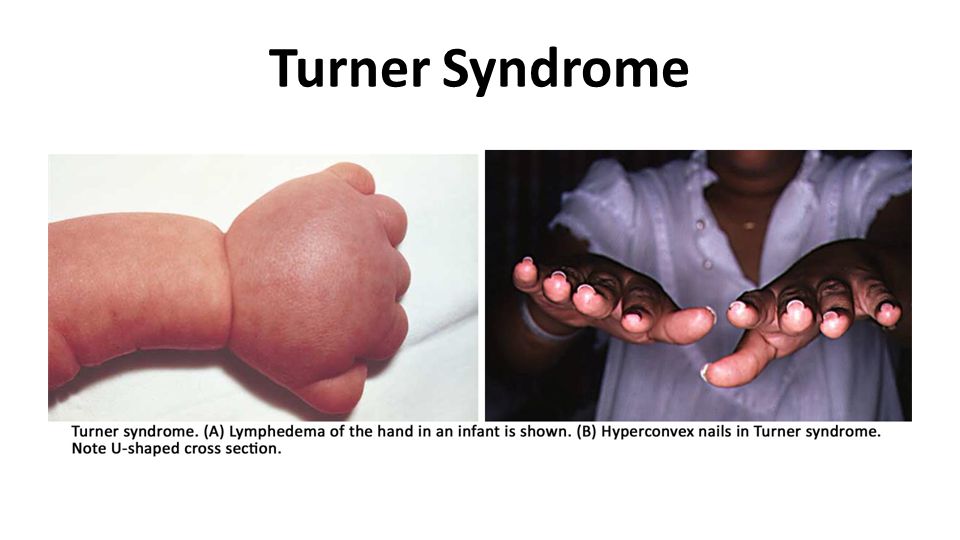
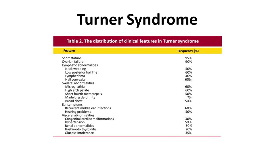
Turner Syndrome The most severely affected patients generally present during infancy with edema of the dorsum of the hand and foot due to lymph stasis, and sometimes swelling of the nape of the neck. The latter is related to markedly distended lymphatic channels, producing a so-called cystic hygroma ( Chapter 10 ).Chapter 10 As these infants develop, the swellings subside but often leave bilateral neck webbing and persistent looseness of skin on the back of the neck.
.Chapter 10 As these infants develop, the swellings subside but often leave bilateral neck webbing and persistent looseness of skin on the back of the neck..")
27
Turner Syndrome

28
Congenital heart disease is also common, affecting 25% to 50% of patients. Left-sided cardiovascular abnormalities, particularly preductal coarctation of the aorta and bicuspid aortic valve, are seen most frequently. Cardiovascular abnormalities are the most important cause of increased mortality in children with Turner syndrome.

29
The principal clinical features in the adolescent and adult are illustrated in Figure 5-22.Figure 5-22

30
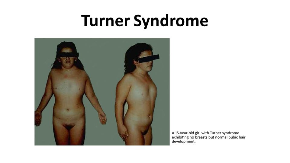
Turner Syndrome At puberty there is failure to develop normal secondary sex characteristics. The genitalia remain infantile, breast development is inadequate, and there is little pubic hair. The mental status of these patients is usually normal, but subtle defects in nonverbal, visual-spatial information processing have been noted. Of particular importance in establishing the diagnosis in the adult is the shortness of stature (rarely exceeding 150 cm in height) and amenorrhea.
 and amenorrhea..")
31
Turner Syndrome

34
Turner syndrome is the single most important cause of primary amenorrhea, accounting for approximately one third of the cases. For reasons not clear, approximately 50% of patients develop autoantibodies that react with the thyroid gland, and up to half of these develop clinically manifest hypothyroidism. Equally mysterious is the presence of glucose intolerance, obesity, and insulin resistance in a minority of patients. The last mentioned is significant, because therapy with growth hormone, commonly used in these patients, worsens insulin resistance.

35
Turner Syndrome The molecular pathogenesis of Turner syndrome is not completely understood, but studies have begun to shed some light. In approximately 75% of cases the X- chromosome is maternal in origin, thus suggesting that there is an abnormality in paternal gametogenesis. As mentioned earlier, both X chromosomes are active during oogenesis and are essential for normal development of the ovaries. During normal fetal development, ovaries contain as many as 7 million oocytes. The oocytes gradually disappear so that by menarche their numbers have dwindled to a mere 400,000, and when menopause occurs fewer than 10,000 remain.

36
Turner Syndrome In Turner syndrome, fetal ovaries develop normally early in embryogenesis, but the absence of the second X chromosome leads to an accelerated loss of oocytes, which is complete by age 2 years. In a sense, therefore, “menopause occurs before menarche,” and the ovaries are reduced to atrophic fibrous strands, devoid of ova and follicles (streak ovaries). Because patients with Turner syndrome also have other (nongonadal) abnormalities, it follows that some genes for normal growth and development of somatic tissues must also reside on the X chromosome.
. Because patients with Turner syndrome also have other (nongonadal) abnormalities, it follows that some genes for normal growth and development of somatic tissues must also reside on the X chromosome..")
37
Turner Syndrome

38
Among the genes involved in the Turner phenotype is the short stature homeobox ( SHOX ) gene at Xp22.33. This is one of several genes that remain active in both X chromosomes and has an active homologue on the short arm of the Y chromosome. Thus, both normal males and females have two copies of this gene. Haploinsufficiency of SHOX gives rise to short stature. Indeed, deletions of the SHOX gene are noted in 2% to 5% of otherwise normal children with short stature. In keeping with its role as a critical regulator of growth, the SHOX gene is expressed during fetal life in the growth plates of several long bones including the radius, ulna, tibia, and fibula. It is also expressed in the first and second pharyngeal arches.

39
Turner Syndrome Just as the loss of SHOX is always associated with short stature, excess copies of this gene are associated with tall stature. Whereas haploinsufficiency of SHOX can explain growth deficit in Turner syndrome, it cannot explain other clinical features such as cardiac malformations and endocrine abnormalities. Clearly several other genes located on the X chromosome are also involved.

40
Turner Syndrome

Similar presentations


. A feature of DNA.>")
. A feature of DNA.>")

