Download presentation
Presentation is loading. Please wait.

1
The Liver Objectives Describe, with the aid of diagrams and photographs, the histology and gross structure of the liver. Describe the formation of urea in the liver, including an outline of the ornithine cycle. Describe the roles of the liver in detoxification.

2
Position of the liver The liver is situated mostly in the top right portion of the abdominal cavity just under the diaphragm. It can be felt as a hardish mass just below the bottom right rib.

3
Structure of the liver

4
Liver basics Largest internal organ- holds 13% total blood at any one time- can store & release blood acting as a reservoir to compensate for smaller changes in blood volume Uses up to 20% total energy in body Composed of left & right lobes enclosed by fibrous capsule (Glissons Capsule) Each lobe formed from hexagonal lobules Dual blood supply: receives blood from 2 blood vessels The liver receives approximately 30% of resting cardiac output and is therefore a very vascular organ. The hepatic vascular system is dynamic, meaning that it has considerable ability to both store and release blood - it functions as a reservoir within the general circulation. In the normal situation, 10-15% of the total blood volume is in the liver, with roughly 60% of that in the sinusoids. When blood is lost, the liver dynamically adjusts its blood volume and can eject enough blood to compensate for a moderate amount of haemorrhage. Conversely, when vascular volume is increased, as when fluids are rapidly taken up, the hepatic blood volume expands, providing a buffer against increases in blood volume.
 Each lobe formed from hexagonal lobules. Dual blood supply: receives blood from 2 blood vessels. The liver receives approximately 30% of resting cardiac output and is therefore a very vascular organ. The hepatic vascular system is dynamic, meaning that it has considerable ability to both store and release blood - it functions as a reservoir within the general circulation. In the normal situation, 10-15% of the total blood volume is in the liver, with roughly 60% of that in the sinusoids. When blood is lost, the liver dynamically adjusts its blood volume and can eject enough blood to compensate for a moderate amount of haemorrhage. Conversely, when vascular volume is increased, as when fluids are rapidly taken up, the hepatic blood volume expands, providing a buffer against increases in blood volume.")
5
Gross Structure Key Questions; Why are there 2 blood supplies?
How does the structure allow the blood to flow past as many hepatocytes as possible?

6
Pulmonary Artery Aorta Pulmonary Vein Vena Cava Hepatic artery
Hepatic Vein Hepatic Portal vein HEART LUNG LIVER HPV: rich in sugars, amino acids insulin/glucagon (straight from pancresa) Delivers x3 blood volume than Hepatic artery. INTESTINE BODY
 Delivers x3 blood volume than Hepatic artery. INTESTINE. BODY.")
7
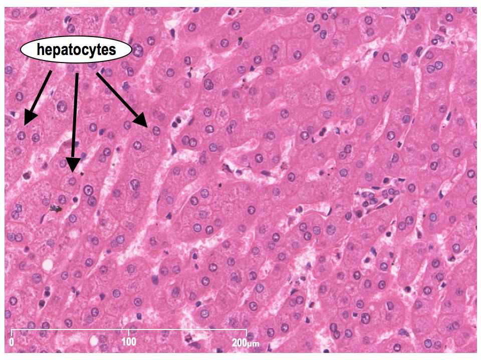
Hepatocytes Make up roughly 80% of the mass of the liver
Nuclei are distinctly round Hepatocytes are exceptionally active in synthesis of protein and lipids for export therefore have large quantities of both rough and smooth endoplasmic reticulum and Golgi apparatus. Lots of Glycogen granules and vesicles containing very low density lipoproteins

8
Hepatocytes

10
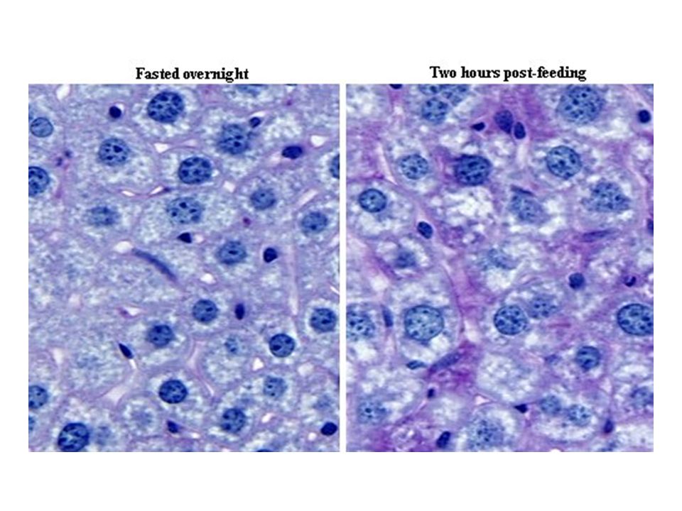
Glycogen granules within hepatocytes
Glycogen granules can make up to 8% mass of cells

12
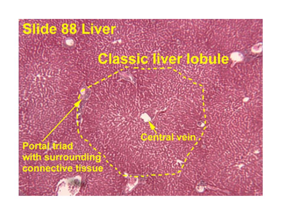
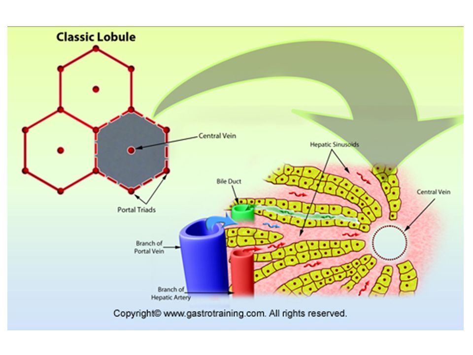
The hepatocytes are arranged into lobules

13
Branch of Hepatic Portal Vein
Branch of Hepatic Vein Branch of Hepatic Portal Vein Liver Lobule Branches of Hepatic Artery

14
Each lobule has a Hepatic vein in the centre and around the outside clusters containing a branch of the hepatic artery, hepatic-portal vein and bile duct

16
Blood moves from outside of lobule from HPV and HA HV
Blood from HPV and HA mix and enter channels called SINUSOIDS Hepatocytes lining the SINUSOIDS absorb products in blood and secrete products into the blood as it flows over them. Interlobular vessels: on outside of lobule: triad – three vessels: distinguishable as vein artery + other Hepatocytes lining the SINUSOIDS have microvilli to increase SA Interlobular vessels (triad) 16
 16.")
17
The arrangement of liver cells in a lobule
Stress Direction of blood flow outside inside Sinusoids are dilated capillaires: thin endothelial cells the body fluid filled space in between the hepatocytes and endothelium ois called the PERISINUSOIDAL SPACE or SPACE OF DISSE. Fluid percolate through the endothelial cells and make contact with the uunderlying hepatcytes. Hepatocytes have microvillis x 6 fold increase in surface area. 17

18
The sinusoids Direction of blood flow is outside inside lobule.
Kupffer cells in SINUSOID are phagocytic cells (derived from monocytes). They ingest bacteria from blood and breakdown bilirubin for absorption into hepatocytes Hepatocytes Release: plasma proteins (prothrombin, albumin, fibrinogen), lipoproteins and cholesterol Absorb: insulin, glucose, minerals, vitamins, blood borne toxins for detox.
. They ingest bacteria from blood and breakdown bilirubin for absorption into hepatocytes. Hepatocytes. Release: plasma proteins (prothrombin, albumin, fibrinogen), lipoproteins and cholesterol. Absorb: insulin, glucose, minerals, vitamins, blood borne toxins for detox.")
19
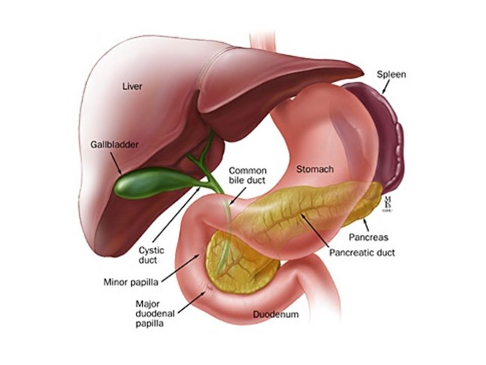
Bile Canaliculus Hepatocytes are separated by a second channel: bile canaliculus Hepatocytes close to a bile canaliculus are rich in Golgi apparatus for transport of bile constituents into the channels Transfers fluid inside outside of lobule Contents drain into hepatic bile duct: continuous with the common bile duct, which delivers bile into the duodenum, diverting through the gall bladder Drains up to 1 litre bile per day!

22
Composition of Bile Bile is a yellowish-green liquid that hepatic cells secrete It includes water, bile salts, bile pigments, cholesterol, and electrolytes. Bile salts made from cholesterol. Bile pigments are breakdown products from red blood cells Only the bile salts have a digestive function. Bile can make your repeated vomit/vomit on empty stomach that unpleasant colour green/yellow and very bitter. Liver and spleen break down some of the haemoglobin to haem and globin. Phagocytic cells of many parts of the body, including the liver take it up - Kuppfer cells ingest the Hb, they remove the iron from it. The iron is then combined with a plasma protein called transferrin. This complex may be taken up by the bone marrow cells for new Hb and red blood cell production or it may be taken up by the hepatocytes for storage. The non-iron part of the Hb is converted into a green-yellow pigment (bilirubin) and is released into the bile as an excretory product. •Globin hydrolysed to amino acids protein synthesis. •Neonatal jaundice if liver cannot excrete bilirubin accumulates in blood. UV light helps break down.
 and is released into the bile as an excretory product. •Globin hydrolysed to amino acids protein synthesis. •Neonatal jaundice if liver cannot excrete bilirubin accumulates in blood. UV light helps break down.")
23
the colour of bile comes from the breakdown product of haemaglobin
Hb is broken down to Haem and globin The non iron component of haem is bilirubin If the liver can not excrete the bilirubin quickly enough it builds up under the skin causing jaundice

24
Liver Histology

25
Summary Questions State the two sources of the blood supply to the liver and describe the primary physiological purpose of each supply. Briefly describe the role of the following structures in liver tissue: Bile canaliculi Phagocytic Kupffer cells Central vein Sinusoids

Similar presentations


 detoxification.>")


Amino acids to glucose. 1450 cm 3 of blood flows through.>")


