Download presentation
Presentation is loading. Please wait.

1
Fluorescence and Fluorochromes Peter O’Toole pot1@york.ac.uk Tel: 01904 328722

2
Main Principles Fluorescence Fluorophores, native or man made Excite with one colour (wavelength A) Emits with a different colour (wavelength B) Different fluorophores have different colour properties Use specialised filters to split colours to see specific fluorescent probes Use of new Fluorescent Proteins (XFPs e.g. GFP)
.")
3
Fluorescence - Photon Release Electron excited form ground state by absorption of light Fluorescence observed as electron decays - photon release Energy lost so light emitted at a longer wavelength

4
Fluorescence excitation emission non-radiative(quenching) Ground State Excited State
 Ground State Excited State")
5
Fluorescein – A Typical Fluorescent Probe

6
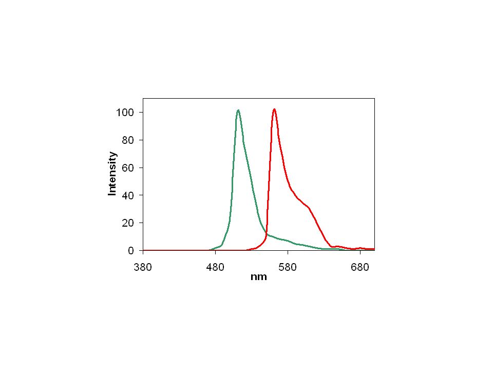
FITCPE

7
Fluorescent Properties Absorption Efficiency –Chose to suit lasers Emission Properties –Peak and broadness Quantum Efficiency – they do not always fluoresce! Environment Dependence –pH –Binding properties Bleaching

8
Why do we need fluorescence in flow cytometry? Many cells appear the same Fluorescence enables us to mark specific components/particles –Identify and characterise sub-populations Fluorescence enables quantification Enables specific discrimination –e.g. live/dead, cell cycle

9
Identify sub populations

10
Fluorescence Quantification e.g. DNA

11
Morphological Information …..but statistics?

12
Fluorochromes Used to label covalently other probes –e.g. fluorescein attached to an antibody Used to label cell components directly –e.g. propidium iodide which binds to DNA Used to explore their environment –e.g. pH sensitive dyes

13
Fluorochromes used to label nucleic acids Propidium Iodide (PI)blueredDNA DRAQ5orangeredDNA (viable cells) Chromomycin A3violetblueDNA (chromosome analysis) Hoechst 33258UVblueDNA (chromosome analysis) Hoechst 33342UVblueDNA (viable cells) DAPIUVblueDNA Acridine Orange (AO)bluegreenDNA redRNA FluorophoreExcited byEmitused for
blueredDNA DRAQ5orangeredDNA (viable cells) Chromomycin A3violetblueDNA (chromosome analysis) Hoechst 33258UVblueDNA (chromosome analysis) Hoechst 33342UVblueDNA (viable cells) DAPIUVblueDNA Acridine Orange (AO)bluegreenDNA redRNA FluorophoreExcited byEmitused for")
14
Typical Fluorochromes (antibodies labels) Fluorescein (FITC) 512 green Alexa 488515 green Phycoerythrin (PE) 565 yellow Cyanine 3(Cy3)570 yellow PE-Texas Red (ECD) 620 red PE-Cy5 (PC5)665 deep red Peridin-chlorophyll (PerCP)670 deep red PE-Cy5.5 (PC5.5)695 deep red PE-Cy7 (PC7)755 far red 488 nm excitation 633 nm Allophycyanin APC 660 Cy5 670 APC – Cy7 770 405 nm Alexa 405 440 Pacific Blue (PB) 440 Cascade Blue (CB) 440
 Fluorescein (FITC) 512 green Alexa green Phycoerythrin (PE) 565 yellow Cyanine 3(Cy3)570 yellow PE-Texas Red (ECD) 620 red PE-Cy5 (PC5)665 deep red Peridin-chlorophyll (PerCP)670 deep red PE-Cy5.5 (PC5.5)695 deep red PE-Cy7 (PC7)755 far red 488 nm excitation 633 nm Allophycyanin APC 660 Cy5 670 APC – Cy nm Alexa Pacific Blue (PB) 440 Cascade Blue (CB) 440")
18
FITCAPC

19
non-radiative(quenching) excited states ground state AB Energy transfer Molecule A absorbs light and is excited excitation transfer A passes the energy onto molecule B emission Molecule B emits light
 excited states ground state AB Energy transfer Molecule A absorbs light and is excited excitation transfer A passes the energy onto molecule B emission Molecule B emits light")
20
Energy transfer excitation emission transfer A B phycoerythrin-Texas RedECD phycoerythrin-cyanine5PC5

21
Fluorescence Many colours Many probes –Antibody stains, DNA stains, ion dyes… Many uses in flow cytometry –Immunophenotyping, cell cycle, calcium flux, apoptosis, transfection, receptor quantification, protein interaction, cell proliferation… Many uses in microscopy and spectroscopy

22
Lipid Droplets

Similar presentations


 Optical Light Scatter and Flow Cytometry.>")

Red laser (644nm,100mW) Alexa 700 APC Cy7 APC-Alexa750 Alexa 488 FITC CFSE SYTOX Green.>")


