Download presentation
Presentation is loading. Please wait.

1
Cellular Basis of Reproduction and Inheritance CH. 8 Ms. Haut Copyright © 2003 Pearson Education, Inc. publishing Benjamin Cummings

2
Connections between Cell Division and Reproduction Single-celled organisms reproduce asexually—individual parent divides into two genetically identical daughter cells Multicellular organisms reproduce sexually—each parent contributing genetic material to the offspring which is genetically unique

3
“Cells arise from preexisting cells” (Rudolf Virchow) Cell reproduction is called cell division Cell division has 3 major roles: 1.Asexual Reproduction 2.Growth and Development—multicellular organisms grow and develop from single cell (fertilize egg) 3.Repair and replacement—replace cells that die from normal wear & tear or accidents
 Cell reproduction is called cell division Cell division has 3 major roles: 1.Asexual Reproduction 2.Growth and Development—multicellular organisms grow and develop from single cell (fertilize egg) 3.Repair and replacement—replace cells that die from normal wear & tear or accidents")
4
Cell replacement (seen here in skin) Dead cells Figure 8.11B Dividing cells Epidermis, the outer layer of the skin Dermis Copyright © 2003 Pearson Education, Inc. publishing Benjamin Cummings

5
Prokaryotes Genes usually carried on a circular DNA molecule (simpler than eukaryotes) DNA has a few proteins and is attached to the plasma membrane at one point DNA not bounded by membrane (nucleoid region) Cells divide by binary fission
 DNA has a few proteins and is attached to the plasma membrane at one point DNA not bounded by membrane (nucleoid region) Cells divide by binary fission")
6
Binary Fission Before dividing, an exact copy of the chromosome is made The attachment point divides so the 2 new chromosomes are attached at separate parts of the membrane The cell elongates and a new plasma membrane is added and the attachment points move apart The plasma membrane and new cell wall pinch through the cell, separating the 2 chromosomes into two new, identical cells Copyright © 2001 Pearson Education, Inc. publishing Benjamin Cummings

7
Eukaryotic Cell Cycle and Mitosis Genome—a cell’s total hereditary endowment of DNA –Genome is specific to species Human DNA extends about 3 meters, so how is it possible to copy all of it and ensure cells get even distrubution? -DNA molecules are packaged into linear chromosomes which are more manageable

8
Before a cell starts dividing, the chromosomes are duplicated –This process produces sister chromatids Centromere Sister chromatids Figure 8.4B Copyright © 2003 Pearson Education, Inc. publishing Benjamin Cummings

9
When the cell divides, the sister chromatids separate –Two daughter cells are produced –Each has a complete and identical set of chromosomes Centromere Sister chromatids Figure 8.4C Chromosome duplication Chromosome distribution to daughter cells Copyright © 2003 Pearson Education, Inc. publishing Benjamin Cummings

10
Every eukaryotic organism has a characteristic number of chromosomes –Human somatic cells (all body cells except reproductive cells) contain 46 chromosomes (23 pairs) –Human reproductive cells, gametes—sperm and egg cells—have 23 chromosomes
 contain 46 chromosomes (23 pairs) –Human reproductive cells, gametes—sperm and egg cells—have 23 chromosomes")
11
Mitotic Cell Cycle In a dividing cell, the mitotic phase (M) phase alternates with interphase, a growth period. –Mitotic phase— usually the shortest part of cell cycle –Interphase— accounts for –90% of the cycle

12
Interphase Subphases G1 phase (first gap)—cell grows by producing proteins and cytoplasmic organelles S phase (synthesis of DNA)—cell continues to grow as in G1 phase, while duplicating chromosomes G2 phase (second gap)—grows more as it completes preparations for cell division
—cell grows by producing proteins and cytoplasmic organelles S phase (synthesis of DNA)—cell continues to grow as in G1 phase, while duplicating chromosomes G2 phase (second gap)—grows more as it completes preparations for cell division")
13
Mitosis Prophase Metaphase Anaphase Telophase

14
G2 of Interphase Nucleus well-defined and bounded by nuclear envelope Contains one or more nucleoli. 2 centrosomes (with centriole pairs) visible Chromosomes duplicated –Still seen as chromatin (DNA + protein) –No individual chromosomes seen
 visible Chromosomes duplicated –Still seen as chromatin (DNA + protein) –No individual chromosomes seen.")
15
Prophase Chromatin fibers become more tightly coiled, condensing into discrete chromosomes Nucleoli disappear Chromosomes appear as 2 identical sister chromatids joined together by centromere Mitotic spindle begins to form (made of microtubules), radiating from centrosomes Centrosomes move to opposite poles
, radiating from centrosomes Centrosomes move to opposite poles")
16
Late Prophase Nuclear envelope fragments-disintegrates Microtubules of spindle extend from poles and invade nucleus and interact with chromosomes Kinetochore forms on chromatids Some spindle fibers connect with kinetochores; some attach to opposite pole

17
Metaphase Centrosomes at opposite poles of cell Chromosomes convene on the metaphase plate Centromeres of all chromosomes are aligned with one another, and sister chromatids straddle metaphase plate Mitotic spindle completely formed

18
Anaphase Paired centromeres of each chromosome separate Each chromatid is now considered a full- fledged chromosome and move to opposite poles as kinetochore microtubules shorten

19
Telophase and Cytokinesis Nonkinetochore microtubules elongate the cell Daughter nuclei form at two poles of cell Nuclear envelopes arise from fragments of parent cell’s nuclear envelope and other portions of endomembrane system Chromatin fibers become less tightly coiled Cytokinesis—division of cytoplasm –Formation of cleavage furrow, which pinches cell in two

21
Cytokinesis in Plants No cleavage furrow During Telophase, vesicles derived from Golgi apparatus move along microtubules to middle of cell producing cell plate Cell plate enlarges until its surrounding membrane fuses with the plasma membrane

22
Mitosis collage, light micrographs Figure 8.6x1 Copyright © 2003 Pearson Education, Inc. publishing Benjamin Cummings

23
The Cell Cycle Review & Practice

24
Interphase

25
Prophase

26
Metaphase

27
Anaphase

28
Telophase

29
Metaphase

30
Interphase

31
Prophase

32
Metaphase

33
Anaphase

35
Telophase

36
Interphase

37
Telophase

38
Prophase

39
Anaphase Interphase

40
Metaphase Interphase Anaphase

41
Telophase

42
INTERPHASEPROPHASE Centrosomes (with centriole pairs) Chromatin NucleolusNuclear envelope Plasma membrane Early mitotic spindle Centrosome Chromosome, consisting of two sister chromatids Fragments of nuclear envelope Kinetochore Spindle microtubules Figure 8.6 Copyright © 2003 Pearson Education, Inc. publishing Benjamin Cummings

43
METAPHASETELOPHASE AND CYTOKINESIS Metaphase plate Spindle Daughter chromosomes Cleavage furrow Nucleolus forming Nuclear envelope forming ANAPHASE Figure 8.6 (continued) Copyright © 2003 Pearson Education, Inc. publishing Benjamin Cummings

44
Most animal cells divide only when stimulated, and others not at all In laboratory cultures, most normal cells divide only when attached to a surface –They are anchorage dependent Anchorage, cell density, and chemical growth factors affect cell division

45
Cells continue dividing until they touch one another –This is called density-dependent inhibition Cells anchor to dish surface and divide. Figure 8.8A When cells have formed a complete single layer, they stop dividing (density-dependent inhibition). If some cells are scraped away, the remaining cells divide to fill the dish with a single layer and then stop (density-dependent inhibition). Copyright © 2003 Pearson Education, Inc. publishing Benjamin Cummings
. If some cells are scraped away, the remaining cells divide to fill the dish with a single layer and then stop (density-dependent inhibition). Copyright © 2003 Pearson Education, Inc. publishing Benjamin Cummings.")
46
Growth factors are proteins secreted by cells that stimulate other cells to divide After forming a single layer, cells have stopped dividing. Figure 8.8B Providing an additional supply of growth factors stimulates further cell division.

47
Proteins within the cell control the cell cycle –Signals affecting critical checkpoints determine whether the cell will go through a complete cycle and divide Growth factors signal the cell cycle control system G 1 checkpoint M checkpointG 2 checkpoint Control system Figure 8.9A Copyright © 2003 Pearson Education, Inc. publishing Benjamin Cummings

48
Checkpoints integrate a variety of internal (intracellular) and external (extracellular) information For many cells, the G1 checkpoint is the “restriction point” –A go-ahead signal indicates that the cell will complete the cycle and divide –In the absence of a go-ahead signal, the cell may exit the cell cycle and remain in the non-dividing state called G0 phase Many human cells are in the G0 phase until they die—muscle and nerve cells
 and external (extracellular) information For many cells, the G1 checkpoint is the restriction point –A go-ahead signal indicates that the cell will complete the cycle and divide –In the absence of a go-ahead signal, the cell may exit the cell cycle and remain in the non-dividing state called G0 phase Many human cells are in the G0 phase until they die—muscle and nerve cells")
49
G1 Checkpoint: The binding of growth factors to specific receptors on the plasma membrane is usually necessary for cell division Growth factor Figure 8.8B Cell cycle control system Plasma membrane Receptor protein Signal transduction pathway G 1 checkpoint Relay proteins Copyright © 2003 Pearson Education, Inc. publishing Benjamin Cummings

50
G2 Checkpoint: Repair enzymes make sure DNA has been copied correctly There are plenty of proteins (growth factors) and organelles present
 and organelles present")
51
M Checkpoint: Anaphase does not begin until all chromosomes are attached to spindle at metaphase plate

52
Cancer cells have abnormal cell cycles –They divide excessively and can form abnormal masses called tumors Cancer cells do not respond normally to the body’s control mechanisms and divide excessively 1.Density-independent—make their own growth factors and continue to divide uncontrolled (“immortal”) 2.Anchorage-independent Radiation and chemotherapy are effective as cancer treatments because they interfere with cell division Growing out of control, cancer cells produce malignant tumors
 2.Anchorage-independent Radiation and chemotherapy are effective as cancer treatments because they interfere with cell division Growing out of control, cancer cells produce malignant tumors")
53
Abnormal cells that escape cell-cycle control are products of mutated or transformed normal cells 1.May proliferate to form a tumor—an unregulated growing mass of cells within normal tissue Benign tumor—if cells remain at the original site Malignant tumor—if mass impairs normal function of one or more organs of the body »Excessive proliferation »Cells with unusual number of chromosomes »Aberrant metabolism »Detaches and migrates through body (metastasis)
")
54
Malignant tumors can invade other tissues and may kill the organism Tumor Figure 8.10 Glandular tissue 123 A tumor grows from a single cancer cell. Cancer cells invade neighboring tissue. Lymph vessels Cancer cells spread through lymph and blood vessels to other parts of the body. Metastasis

55
When the cell cycle operates normally, mitotic cell division functions in: –Growth (seen here in an onion root) Review of the functions of mitosis: Growth, cell replacement, and asexual reproduction Figure 8.11A Copyright © 2003 Pearson Education, Inc. publishing Benjamin Cummings

56
Asexual reproduction (seen here in a hydra) Figure 8.11C Copyright © 2003 Pearson Education, Inc. publishing Benjamin Cummings

58
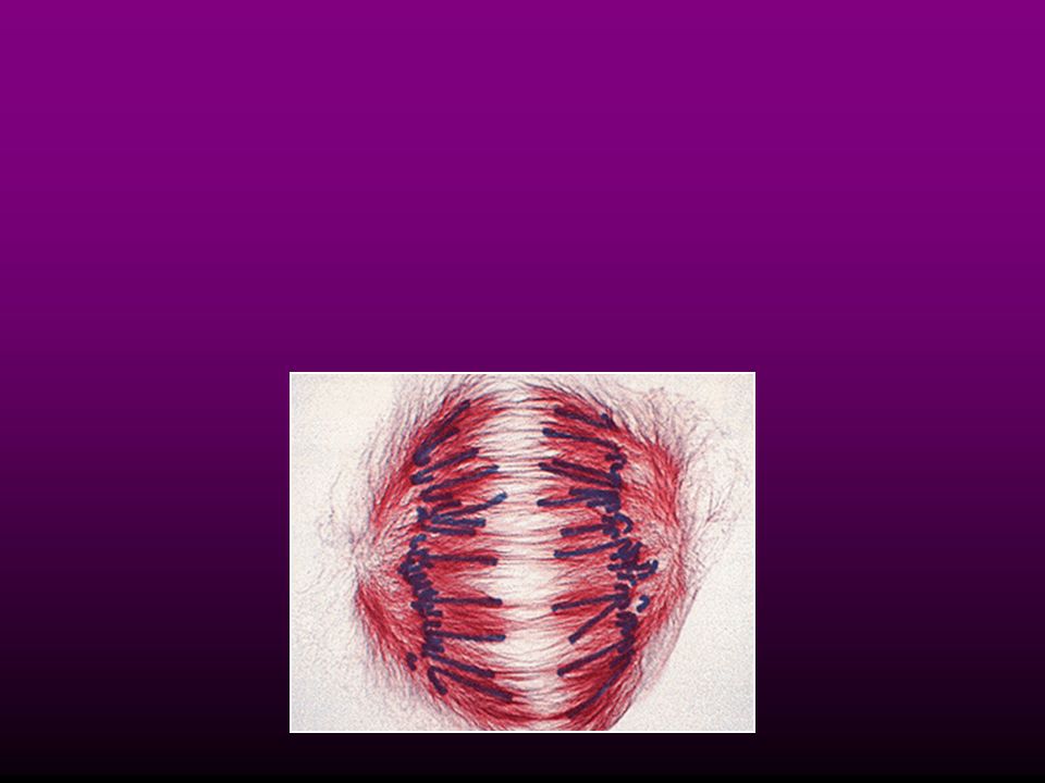
Mitotic spindle Figure 8.6x2

59
Acknowledgements Unless otherwise noted, illustrations are credited to Pearson Education which have been borrowed from BIOLOGY: CONCEPTS AND CONNECTIONS 4th Edition, by Campbell, Reece, Mitchell, and Taylor, ©2003. These images have been produced from the originals by permission of the publisher. These illustrations may not be reproduced in any format for any purpose without express written permission from the publisher. BIOLOGY: CONCEPTS AND CONNECTIONS 4th Edition, by Campbell, Reece, Mitchell, and Taylor, ©2001. These images have been produced from the originals by permission of the publisher. These illustrations may not be reproduced in any format for any purpose without express written permission from the publisher.

Similar presentations


>")


>")
