Download presentation
Presentation is loading. Please wait.

1
Diseases of the External Ear
Dr. Haitham Alnori

2
Perichondritis Definition: Is infection and inflammation of the perichondrium of auricular cartilage. Causes: Infection of haematoma auris. Complication of severe otitis externa. Laceration, mastoid surgery. High ear peircing. Clinical Picture: The pinna is uniformly enlarged and thickened and its surface is red and shiny. Severe pain and tenderness. Bacteriology: Pseudomonus aeruginosa

3
Treatment of Perichondritis
Vigorous parenteral antibiotic treatment. Application of soothing dressing such as ichthamol in glycerin. If subperichondrial abscess is formed it must be incised and drained with removal of all necrotic cartilage, followed by daily dressing.

4
Cauliflower deformity may result if the condition is not treated early.

5
Impacted Wax (Cerumen)
Wax is a mixture of secretions of ceruminous and sebaceous glands with desquamated skin cells. Cartilaginous portion of the EAM. Expelled outside the canal by movement of chewing and epithelial migration. Cerumen protects the ear from various bacterial and fungal infections, helps in cleaning and lubrication, and also protects the sensitive skin of the ear canal.

6
Symptoms: Deafness and discomfort Tinnitus and disturbance of balance Reflex cough: Arnold nerve Signs: Brown, yellowish or black plug obscuring the tympanic membrane. Treatment: Removal by: Ear wash: if the wax is hard it should be softened by 5% bicarbonate before ear wash tap water 27 C Jobson-Horn probe: hard piece of wax Suction: if there is TM perforation.

7
Keratosis Obturans The meatus on one or both sides become blocked in its deep portion by a mass consisting of wax, desquamated epithelium and cholesterol. This mass causes excessive erosion and expansion of the bony meatus and in this, it resembles a cholesteatoma of the middle ear. Aetiology unknown but it may be associated with bronchiectasis and sinusitis in young patients

8
Clinical Picture: Pain deafness and tinnitus. White glistening mass occluding the bony meatus. Treatment: Removal of the keratotic mass under G.A. Regular observation is advised as keratosis may reform.

9
Inflammatory conditions of the external auditory canal
Bacterial: 1- Diffuse OE 2- Localised OE: frunculosis 3- Malignant OE ( Necrotizing OE) Viral ( bullous- Herpes) Fungal: (Otomycosis) Non infective ( allergic OE, Seborrhoic OE)
 Viral ( bullous- Herpes) Fungal: (Otomycosis) Non infective. ( allergic OE, Seborrhoic OE)")
10
Diffuse otitis externa
Diffuse inflammation of the skin lining of the extenal auditory canal Pridisposing factors: - Skin laceration: Self inflicted Iatrogenic - Skin maceration: Hot humid atmosphere Swimmer ear

11
Increase on moving the jaw Why? Deafness Signs
Symptoms: Earache: Severe Why? Increase on moving the jaw Why? Deafness Signs Tenderness on moving the auricle or pressure on the tragus Tender pre and postauricular LN Otoscopic exam : Redness, edema, tenderness Scanty discharge TREATMENT Meticulous cleaning Packing of canal Antibiotic/ steroid drops Because skin is tightly adherent to underlying perichondrium Because the EAC Lies immediately behind Temporomandibular joint

13
Frunculosis Localized suppurative inflammation of a hair follicle in
the skin of the external auditory canal Organism Staph aureus PP factors: - scratching of ear canal - DM

14
Symptoms: Earache…. Why? Deafness Signs: External: Tenderness on moving the auricle or pressure on the tragus Tender pre and postauricular LN Otoscopic: Localised area of tenderness: difficult to examine the external canal by Otoscope No or scanty purulent otorrhea

15
Investigations: Blood glucose level especially in: Recurrent cases Bilateral cases DD acute mastoiditis Treatment Antibiotics ( flucloxacillin for 5 days ) Analgesics and heat application Aural toilet: removal of ear discharge when the furuncle has burst. Incision of a boil should be delayed until it is clearly pointing to the skin. Aural pack: by gauze strip soaked in ichthamol and glycerin and changed daily.
 Analgesics and heat application. Aural toilet: removal of ear discharge when the furuncle has burst. Incision of a boil should be delayed until it is clearly pointing to the skin. Aural pack: by gauze strip soaked in ichthamol and glycerin and changed daily.")
16
Malignant otitis externa (Necrotizing otitis externa)
Def: invasive potentially fatal infection of the external canal which extends to the base of the skull Incidence: elderly uncontrolled diabetic patient Organism: pseudomonas aeuruginosa Symptoms: Ear discharge and severe earache which does not respond to analgesics and increase at night Necrotizing otitis externa should be suspected when patients with DM complain of persistent external otitis that causes severe pain, especially at night

17
Signs External examination: Tenderness on pulling the AURICLE OR PRESSURE ON THE TRAGUS Otoscopic examination: Granulations at the floor of the external canal at the attachment of bony and cartilagenous part Scanty, sanginous and purulent otorrhea

18
CT scan of the temporal bone& skull base
Investigations: Blood glucose level CT scan of the temporal bone& skull base Radio-isotop scan ( Gallium99) to assess severity & prognosis Axial computed tomographic (CT) scan in a 65-year-old male patient with diabetes mellitus who had severe nocturnal otalgia for two months. This patient was referred because of facial nerve paralysis that developed despite oral treatment with ofloxacin (Floxin). The CT scan shows bony destruction of the right temporal bone. Note the missing posterior wall of the external auditory canal (short arrow). Mastoid air cells are secondarily involved and are opacified (long arrow) compared with the well-aerated left side.
 to assess severity & prognosis. Axial computed tomographic (CT) scan in a 65-year-old male patient with diabetes mellitus who had severe nocturnal otalgia for two months. This patient was referred because of facial nerve paralysis that developed despite oral treatment with ofloxacin (Floxin). The CT scan shows bony destruction of the right temporal bone. Note the missing posterior wall of the external auditory canal (short arrow). Mastoid air cells are secondarily involved and are opacified (long arrow) compared with the well-aerated left side.")
19
Complications Treatment Osteomyelitis of the temporal bone &skull base
Facial nerve paralysis at the stylomastoid foramen Last 4 cranial nerves paralysis at the jagular foramen Treatment Control of diabetes amikacin Ciprofloxacin 6 wk ! Analgesics Aural toilet+ topical AB Surgical excision or mastoidectomy is to be avoided or postponed.

20
(Otitis externa haemorrhagica)
Bullous myringitis (Otitis externa haemorrhagica) painful condition characterized by formation of haemorrhagic blebs on the TM. caused by viral infection. Clinical picture Severe extreme pain preventing sleep or work with almost normal hearing. Otoscopy: haemorrhagic bullae are found on the drum, dark bluish or red in colour. The bullae are prone to spontaneous rupture, with blood stained aural discharge. Treatment Analgesia and keeping the ear dry. Incision of the bullae is not indicated.
 painful condition characterized by formation of haemorrhagic blebs on the TM. caused by viral infection. Clinical picture. Severe extreme pain preventing sleep or work with almost normal hearing. Otoscopy: haemorrhagic bullae are found on the drum, dark bluish or red in colour. The bullae are prone to spontaneous rupture, with blood stained aural discharge. Treatment. Analgesia and keeping the ear dry. Incision of the bullae is not indicated.")
21
Herpes Zoster Oticus Etiology: Herpes zoster virus Clinically:
Pain in and around the ear Vesicles on the auricle and external canal Ramsay-Hunt syndrome: Vesicles+ facial nerve palsy + SNHL & Vertigo Treatment: - Antiviral - Corticosteroid if there is affection of VII nerve or VIII nerve

22
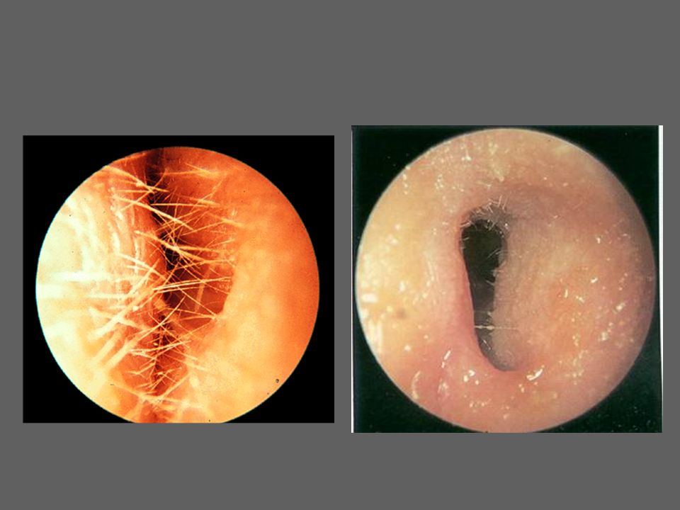
Otomycosis Fungal infection of the skin of the exernal canal Etiology:
Aspirigillus Niger Candida albicans Symptoms and signs: Itching is usually the only symptom Pain if there is secondary bacterial infection Deafness if the external canal is obstructed whitish mass with black spots like wet newspaper Treatment: Aural toilet: removal of the fungal mass by suction. Antifungal: nystatin, or salicylic acid (2%) as a keratolytic in alcohol as fungicidal
 as a keratolytic in alcohol as fungicidal.")
23
Osteoma and Exostosis Osteoma is a single tumor composed of cancelous bone located at outer part of bony canal. Exostosis is multiple growth composed of ivory bone located at deep meatus. Aetiology cold water swimmers. Symptoms: Hearing loss if the external canal is obstructed by large osteoma or wax. Treatment Asymptomatic: observation. If obstructing the canal: excision.

24
THANK YOU

Similar presentations


 Characteristic of drainage (acute,pulsatile) Systemic disea ses. Others(otalgia,neurologic deficit)>")


Congenital disorders B)Trauma to the auricle C)Inflammatory disorders.>")


