Download presentation
Presentation is loading. Please wait.

1
Dental Radiography RVT: Chapter 24

2
Objectives: Dental Radiography
Review dental terminology & tooth surfaces Review basic anatomy & formula for teeth, including number of roots Understand normal views & positioning for dental radiography Define parallel and bisecting angle techniques, and know when to use each Understand the differences between intraoral and extraoral views

3
Anatomical Directions: Oral Cavity

4
Positional Terminology: Oral Cavity
Directional Positional

5
Tooth Anatomy on Film What are the four basic types of teeth?

6
Assessing Age

7
Dental Formulas Carnassial tooth:
Dogs: Upper 4th premolar & lower 1st molar Cats: Upper 3rd premolar & lower 1st molar

8
Triadan Numbering System
Canine Feline 1st number = quadrant 2nd 2 numbers = tooth position

9
Number of Roots: Canine

10
Number of Roots: Feline

11
Normal Adult Canine Maxillary Incisors
7 – Incisive canal 9 – ___________ fissures (oval dark spaces)
")
12
Normal Adult Canine Mandibular Incisors
7 – Mandibular _______________ (dark black line)
")
13
Viewing Dental Radiographs
Film is exposed with the convex dot at the _________ end of the mouth Dot location will vary with right & left side radiographs When viewing- hold with convex side towards you Maxillary: point cusps toward the floor; apical portion of roots to ceiling Mandibular: point apical portion of roots towards the floor; cusps to the ceiling

14
Viewing Dental Radiographs

15
Film Mount Organize full mouth radiographs in a film mount: Hang as if looking at the animal, with animal’s right side on your left…

16
Dental Radiography Techniques
What makes dental rads tricky: getting the whole tooth! Must include complete __________ to be diagnostic Patient is always anesthetized Positions will include lateral, dorsal, or sternal Easier if skull is kept parallel to table top Paper towels and syringes/cases are your best friends Longer SID allows for wider primary beam Using size 0-2? Center the cone on the affected tooth Position yourself opposite from the cone to find the angle

17
Proper Oral Film Placement

18
Dental Radiography Techniques
Parallel Bisecting Angle

19
Parallel Technique Used for mandibular molars and pre-molars only
Film is placed inside of mouth & ____________ to the teeth Primary beam is placed perpendicular to film Only acceptable to use in one location!

20
Bisecting-angle Technique
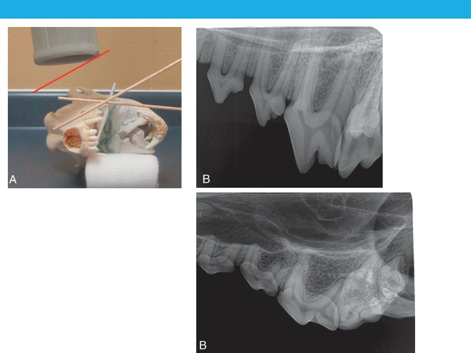
Technique for ALL maxillary teeth & ______________ canines and incisors Placing the beam perpendicular to tooth (or film) results in foreshortening or elongation Find the bisecting-angle: Place film parallel to tongue Draw an imaginary line along the tooth’s axis (use root) Draw another imaginary line along the axis of the film Find the angle that is exactly in the middle of these two Position the primary beam perpendicular to the “bisecting angle”
 results in foreshortening or elongation. Find the bisecting-angle: Place film parallel to tongue. Draw an imaginary line along the tooth’s axis (use root) Draw another imaginary line along the axis of the film. Find the angle that is exactly in the middle of these two. Position the primary beam perpendicular to the bisecting angle")
21
Bisecting Angle- Mandibular canine

22
Bisecting Angle- Mandibular incisors

23
Bisecting Angle- Maxillary incisors

24
Distortion: Foreshortening

25
Distortion: Elongation
FILM

Similar presentations
, you see the mouse holding an x-ray tubehead (see below),>")


>")


>")
. All rights reserved. No part of this product may be reproduced or transmitted.>")


