Download presentation
Presentation is loading. Please wait.

1
Aortic stenosis time and indication of surgery
Ankur kamra

2
Normal Aortic valve Three cusps, crescent shaped 3 commissures
3 sinuses supported by fibrous annulus 3.0 to 4.0 cm2

3
2D Echo-Long axis view Diastole Systole

4
Y or inverted Mercedes-Benz sign
2D Echo-Short axis view Diastole Systole Y or inverted Mercedes-Benz sign

5
Aortic stenosis- Causes
Most common :- Bicuspid aortic valve with calcification Senile or Degenerative calcific AS Rheumatic AS Less common:- Congenital Type 2 Hyperlipoproteinemia Onchronosis

7
HOW TO ASSESS AORTIC STENOSIS

8
Doppler assessment of AS
The primary haemodynamic parameters recommended (EAE/ASE Recommendations for Clinical Practice 2008) class 1 Peak transvalvular velocity Mean transvalvular gradient Valve area by continuity equation.
 class 1. Peak transvalvular velocity. Mean transvalvular gradient. Valve area by continuity equation.")
9
Peak transvalvular velocity
Continuous-wave Doppler ultrasound Multiple acoustic windows Apical and suprasternal or right parasternal most frequently yield the highest velocity rarely subcostal or supraclavicular windows may be required Three or more beats are averaged in sinus rhythm, with irregular rhythms at least 5 consecutive beats

10
Peak transvalvular velocity
AS jet velocity is defined as the highest velocity signal obtained from any window after a careful examination A smooth velocity curve with a dense outer edge and clear maximum velocity should be recorded The shape of the CW Doppler velocity curve is helpful in distinguishing the level and severity of obstruction. With severe obstruction, maximum velocity occurs later in systole and the curve is more rounded in shape With mild obstruction, the peak in early systole with a triangular shape of the velocity curve

12
Peak transvalvular velocity
The shape of the CWD velocity curve also can be helpful in determining whether the obstruction is fixed or dynamic Dynamic sub aortic obstruction shows a characteristic late peaking velocity curve, often with a concave upward curve in early systole

13
Mean transvalvular gradient
The difference in pressure between the left ventricle and aorta in systole Gradients are calculated from velocity information The relationship between peak and mean gradient depends on the shape of the velocity curve.

14
Pressure recovery The conversion of potential energy to kinetic energy across a narrowed valve results in a high velocity and a drop in pressure. Distal to the orifice, flow decelerates again. Kinetic energy will be reconverted into potential energy with a corresponding increase in pressure, the so-called PR

15
Pressure recovery Pressure recovery is greatest in stenosis with gradual distal widening Aortic stenosis with its abrupt widening from the small orifice to the larger aorta has an unfavorable geometry for pressure recovery PR= 4v²× (2EOA/AoA)x (1-EOA/AoA)
x (1-EOA/AoA)")
16
Comparing pressure gradients calculated from doppler velocities to pressures measured at cardiac catheterization.

17
Aortic valve area Continuity equation

19
Left ventricular systolic dysfunction
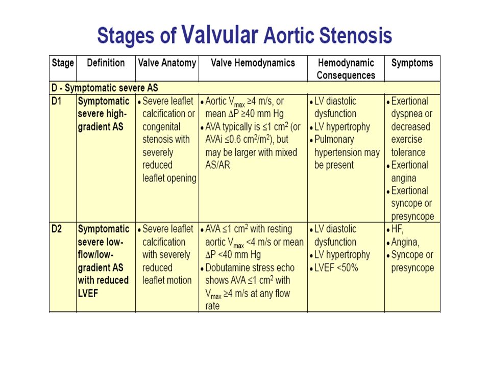
Low-flow low-gradient AS includes the following conditions: Effective orifice area < 1.0 Cm2 LV ejection fraction < 40% Mean pressure gradient < 30–40 mmHg Severe AS and severely reduced LVEF represent 5% of AS patients Vahanian A et al. Eur Heart J 2007;28:230–68.

20
Dobutamine stress Echo
Provides information on the changes in aortic velocity, mean gradient, and valve area as flow rate increases. Measure of the contractile response to dobutamine Helpful to differentiate two clinical situations Severe AS causing LV systolic dysfunction Moderate AS with another cause of LV dysfunction

21
Dobutamine stress Echo
A low dose starting at 2.5 or 5 ug/kg/min with an incremental increase in the infusion every 3–5 min to a maximum dose of 10–20 ug/kg/min The infusion should be stopped as soon as Positive result is obtained Heart rate begins to rise more than 10–20 bpm over baseline or exceeds 100bpm

22
Dobutamine stress Echo
Report should include AS velocity, mean gradient, valve area and ejection fraction preferably at each stage but at least at baseline and peak dose Findings recommend as reliable are: An increase in valve area to a final valve area 1.0 cm suggests that stenosis is not severe. Severe stenosis is suggested by AS jet 4.0 or a mean gradient 40 mmHg provided that valve area does not exceed 1.0 cm at any flow rate. Absence of contractile reserve (failure to increase SV or ejection fraction by 20%) is a predictor of a high surgical mortality and poor long-term outcome although valve replacement may improve LV function and outcome even in this subgroup.
 is a predictor of a high surgical mortality and poor long-term outcome although valve replacement may improve LV function and outcome even in this subgroup.")
23
PARADOXICAL LOW FLOW LOW GRADIENT
Some patients with severe aortic stenosis based on valve area have a lower than expected gradient (e.g. mean gradient < 30 mmHg) despite preserved LV ejection fraction (e.g. EF > 50%) It is due to deficient ventricular filling due to smaller cavity size rather than decrease lv function
 despite preserved LV ejection fraction (e.g. EF > 50%) It is due to deficient ventricular filling due to smaller cavity size rather than decrease lv function.")
24
Alternate measures of stenosis severity
(Level 2 EAE/ASE Recommendations )
")
25
Simplified continuity equation. AVA= CSA LVOT×VLVOT / VAV
Based on concept that in native aortic valve stenosis ratio of LVOT to aortic jet VTI is nearly identical to the ratio of the LVOT to aortic jet maximum velocity. AVA= CSA LVOT×VLVOT / VAV This method is less well accepted because results are more variable than using VTIs in the equation.

26
Velocity ratio Another approach to reduce error related to LVOT diameter measurements is removing CSA from the simplified continuity equation. This dimensionless velocity ratio expresses the size of the valvular effective area as a proportion of the CSA of the LVOT. Velocity ratio= VLVOT/VAV In the absence of valve stenosis, the velocity ratio approaches 1, with smaller numbers indicating more severe stenosis.

27
Aortic valve area -Planimetry
Planimetry may be an acceptable alternative when Doppler estimation of flow velocities is unreliable Planimetry may be inaccurate when valve calcification causes shadows or reverberations limiting identification of the orifice Doppler-derived mean-valve area correlated better with maximal anatomic area than with mean-anatomic area. Marie Arsenault, et al. J. Am. Coll. Cardiol. 1998;32;

28
Aortic valve area - Planimetry

29
Classification of AS severity (a ESC & bAHA/ACC Guidelines)
Aortic Sclerosis Mild Moderate Severe Aortic jet velocity (m/s) ≤ 2.5 m/s > 4 Mean gradient (mm Hg) < 20b(<30a) 20 – 40b (30 -50a) > 40 AVA (cm²) > 1.5 < 1.0 Indexed AVA (cm²/m²) > 0.85 0.60 – 0.85 < 0.6 Velocity ratio > 0.50 0.25 – 0.50 < 0.25
 ≤ 2.5 m/s > 4. Mean gradient (mm Hg) < 20b(<30a) 20 – 40b (30 -50a) > 40. AVA (cm²) > < 1.0. Indexed AVA (cm²/m²) > – < 0.6. Velocity ratio. > – <")
31
Classification Investigations Interventions

36
Diagnostic Testing–Initial Diagnosis: Recommendation
CLASS I TTE is recommended in the initial evaluation of patients with known or suspected VHD to confirm the diagnosis, establish etiology, determine severity, assess hemodynamic consequences, determine prognosis and evaluate for timing of intervention

37
CLASS IIa Low-dose dobutamine stress testing using echocardiographic or invasive hemodynamic measurements is reasonable in patients with stage D2 AS with all of the following Calcified aortic valve with reduced systolic opening; LVEF less than 50%; Calculated valve area 1.0 cm2 or less; and Aortic velocity less than 4.0 m per second or mean pressure gradient less than 40 mm Hg.

38
Diagnostic Testing Changing Signs or Symptoms
CLASS I TTE is recommended in patients with known VHD with any change in symptoms or physical examination findings. (Level of Evidence: C)
")
40
Diagnostic Testing Cardiac Catheterization
CLASS I Cardiac catheterization for hemodynamic assessment is recommended in symptomatic patients when non invasive tests are inconclusive or when there is a discrepancy between the findings on noninvasive testing and physical examination regarding severity of the valve lesion

41
Transaortic pressure gradients should be recorded for measurement of mean transaortic gradient along with Aortic valve area should be calculated with the Gorlin formula, using a Fick or thermodilution cardiac output measurement

42
EXERCISE TESTING: RECOMMENDATIONS
CLASS IIa Exercise testing is reasonable to assess physiological changes with exercise and to confirm the absence of symptoms in asymptomatic patients with a calcified aortic valve and an aortic velocity 4.0 m per second or greater or mean pressure gradient 40 mm Hg or higher.

43
Patients with symptoms provoked by exercise testing should be considered symptomatic even if the clinical history is equivocal. Exercise testing can brought out symptoms in 29% of patients who were considered asymptomatic before testing. patients with AS who manifested symptoms, an abnormal BP response (<20 mm Hg increase) or ST-segment abnormalities with exercise had a significantly reduced symptom-free survival at 2 years (19% compared with 85%) Lancellotti P et al. Prognostic importance of quantitative exercise, Doppler echocardiography in asymptomatic valvular aortic stenosis. Circulation 2005;112:I377–82.
 or ST-segment abnormalities with exercise had a significantly reduced symptom-free survival at 2 years (19% compared with 85%) Lancellotti P et al. Prognostic importance of quantitative exercise, Doppler echocardiography in asymptomatic valvular aortic stenosis. Circulation 2005;112:I377–82.")
44
CLASS III: Harm Exercise testing should not be performed in symptomatic patients with AS when the aortic velocity is 4.0 m per second or greater or mean pressure gradient is 40 mm Hg or higher

45
Medical Therapy: Recommendations
CLASS I Hypertension in patients at risk for developing AS (stage A) and in patients with asymptomatic AS (stages B and C) should be treated according to standard GDMT, started at a low dose and gradually titrated upward as needed with frequent clinical monitoring
 and in patients with asymptomatic AS (stages B and C) should be treated according to standard GDMT, started at a low dose and gradually titrated upward as needed with frequent clinical monitoring.")
46
Concern that antihypertensive medications might result in a fall in cardiac output has not found in studies on medical therapy, including small RCTs, likely because AS does not result in fixed valve obstruction until late in disease Briand M et al. Reduced systemic arterial compliance impacts significantly on left ventricular afterload and function in aortic stenosis: implications for diagnosis and treatment. J Am Coll Cardiol 2005;46:291–8.

47
There are no studies addressing specific antihypertensive medications in patients with AS, but diuretics should be avoided if the LV chamber is small, because even smaller LV volumes may result in a fall in cardiac output. ACE inhibitors may be advantageous due to the potential beneficial effects on LV fibrosis in addition to control of hypertension. Beta blockers are an appropriate choice in patients with concurrent CAD.

48
CLASS IIb Vasodilator therapy may be reasonable if used with invasive hemodynamic monitoring in the acute management of patients with severe decompensated AS (stage D) with NYHA class IV HF symptoms. (Level of Evidence: C) CLASS III: No Benefit Statin therapy is not indicated for prevention of hemodynamic progression of AS in patients with mild-to-moderate calcific valve disease (stages B to D)Level of Evidence: A (ASTRONOMER) trial. Circulation 2010 Cowell SJ, Newby DE, Prescott RJ, et al. A randomized trial of intensive lipid-lowering therapy in calcific aortic stenosis. N Engl J Med 2005
 with NYHA class IV HF symptoms. (Level of Evidence: C) CLASS III: No Benefit. Statin therapy is not indicated for prevention of hemodynamic progression of AS in patients with mild-to-moderate calcific valve disease (stages B to D)Level of Evidence: A. (ASTRONOMER) trial. Circulation Cowell SJ, Newby DE, Prescott RJ, et al. A randomized trial of intensive lipid-lowering therapy in calcific aortic stenosis. N Engl J Med")
49
TIMING AND CHOICE OF INTERVENTION

51
Timing of Intervention: Recommendations CLASS 1
1. AVR is recommended in symptomatic patients with severe AS(stage D1) (Level of Evidence: B): a. Decreased systolic opening of a calcified or congenitally stenotic aortic valve b. An aortic velocity 4.0 m per second or greater or mean pressure gradient 40 mm Hg or higher c. Symptoms of HF, syncope, exertional dyspnea, angina or presyncope by history or on exercise testing.
 (Level of Evidence: B): a. Decreased systolic opening of a calcified or congenitally stenotic aortic valve b. An aortic velocity 4.0 m per second or greater or mean pressure gradient 40 mm Hg or higher c. Symptoms of HF, syncope, exertional dyspnea, angina or presyncope by history or on exercise testing.")
52
Typical initial symptoms are dyspnea on exertion or decreased exercise tolerance.
Classical symptoms of syncope, angina and HF are late manifestations of disease. Calculation of valve area is not necessary when a high velocity/gradient is present and the valve is calcified and immobile. most patients will have a valve area 1.0 cm2 but some will have a larger valve area due to a large body size or coexisting aortic regurgitation (AR). Thus primary criterion for definition of severity of AS is based on aortic velocity or mean pressure gradient
. Thus primary criterion for definition of severity of AS is based on aortic velocity or mean pressure gradient.")
53
CLASS 1 2. AVR is recommended for asymptomatic patients with severe A (stage C2) and an LVEF less than 50% with decreased systolic opening of a calcified aortic valve with an aortic velocity 4.0 or greater or mean pressure gradient 40 mm Hg or higher (Level of Evidence: B) Depressed LVEF in many patients is caused by excessive afterload (afterload mismatch) and LV function improves after AVR in such patients
 and an LVEF less than 50% with decreased systolic opening of a calcified aortic valve with an aortic velocity 4.0 or greater or mean pressure gradient 40 mm Hg or higher (Level of Evidence: B) Depressed LVEF in many patients is caused by excessive afterload (afterload mismatch) and LV function improves after AVR in such patients.")
54
CLASS 1 3. AVR is indicated for patients with severe AS (stage C or D) when undergoing cardiac surgery for other indications when there is decreased systolic opening of a calcified aortic valve and an aortic velocity 4.0 m per second or greater or mean pressure gradient 40 mm Hg or higher (Level of Evidence: B) The additive risk of AVR at the time of other cardiac surgery is less than risk of reoperation within 5 years.
 when undergoing cardiac surgery for other indications when there is decreased systolic opening of a calcified aortic valve and an aortic velocity 4.0 m per second or greater or mean pressure gradient 40 mm Hg or higher (Level of Evidence: B) The additive risk of AVR at the time of other cardiac surgery is less than risk of reoperation within 5 years.")
55
CLASS IIa 1. AVR is reasonable for asymptomatic patients with very severe AS (stage C1) with (Level of Evidence: B): a. Decreased systolic opening of a calcified valve b. An aortic velocity 5.0 m per second or greater or mean pressure gradient 60 mm Hg or higher c. A low surgical risk.
 with (Level of Evidence: B): a. Decreased systolic opening of a calcified valve b. An aortic velocity 5.0 m per second or greater or mean pressure gradient 60 mm Hg or higher c. A low surgical risk.")
56
Several observational studies have shown higher rates of symptom onset and major adverse cardiac events in patients with very severe, compared with severe AS

57
CLASS IIa 2. AVR is reasonable in apparently asymptomatic patients with severe AS (stage C1) with (Level of Evidence: B): a. A calcified aortic valve b. An aortic velocity of 4.0 m per second to 4.9 m per second or mean pressure gradient of 40 mm Hg to 59 mm Hg c. An exercise test demonstrating decreased exercise tolerance or a fall in systolic BP.
 with (Level of Evidence: B): a. A calcified aortic valve b. An aortic velocity of 4.0 m per second to 4.9 m per second or mean pressure gradient of 40 mm Hg to 59 mm Hg c. An exercise test demonstrating decreased exercise tolerance or a fall in systolic BP.")
58
Patients without overt symptoms who Demonstrate
1) a decrease in systolic BP below baseline or a failure of BP to increase by at least 20 mm Hg or 2) a significant decrease in exercise tolerance compared with age and sex normal standards symptom onset within 1 to 2 years is high (about 60% to 80%)in these patients
 a decrease in systolic BP below baseline or a failure of BP to increase by at least 20 mm Hg or. 2) a significant decrease in exercise tolerance compared with age and sex normal standards. symptom onset within 1 to 2 years is high (about 60% to 80%)in these patients.")
59
CLASS IIa 3. AVR is reasonable in symptomatic patients with low-flow/low gradient severe AS with reduced LVEF (stage D2) (Level of Evidence: B): a. Calcified aortic valve with reduced systolic opening b. Resting valve area 1.0 cm2 or less c. Aortic velocity less than 4 m per second or mean pressure gradient less than 40 mm Hg d. LVEF less than 50% e. A low-dose dobutamine stress study that shows an aortic velocity 4 m per second or greater or mean pressure gradient 40 mm Hg or higher with a valve area 1.0 cm2 or less at any dobutamine dose.
 (Level of Evidence: B): a. Calcified aortic valve with reduced systolic opening b. Resting valve area 1.0 cm2 or less c. Aortic velocity less than 4 m per second or mean pressure gradient less than 40 mm Hg d. LVEF less than 50% e. A low-dose dobutamine stress study that shows an aortic velocity 4 m per second or greater or mean pressure gradient 40 mm Hg or higher with a valve area 1.0 cm2 or less at any dobutamine dose.")
60
LVEF typically increases by 10 LVEF units and may return to normal if afterload mismatch was the cause of LV systolic dysfunction. Some patients without contractile reserve may also benefit from AVR, but decisions in these high-risk patients must be individualized

61
CLASS IIa 4. AVR is reasonable in symptomatic patients with low-flow/low gradient severe AS (stage D3) with an LVEF 50% or greater, a calcified aortic valve with significantly reduced leaflet motion and a valve area 1.0 cm or less only if clinical, hemodynamic and anatomic data support valve obstruction as the most likely cause of symptoms and data recorded when the patient is normotensive (systolic BP <140 mm Hg) indicate (Level of Evidence: C): a. An aortic velocity less than 4 m per second or mean pressure gradient less than 40 mm Hg b. A stroke volume index less than 35 mL/m2 c. An indexed valve area 0.6 cm2/m2 or less.
 with an LVEF 50% or greater, a calcified aortic valve with significantly reduced leaflet motion and a valve area 1.0 cm or less only if clinical, hemodynamic and anatomic data support valve obstruction as the most likely cause of symptoms and data recorded when the patient is normotensive (systolic BP <140 mm Hg) indicate (Level of Evidence: C): a. An aortic velocity less than 4 m per second or mean pressure gradient less than 40 mm Hg b. A stroke volume index less than 35 mL/m2 c. An indexed valve area 0.6 cm2/m2 or less.")
62
Reason for this phenomena is low transaortic stroke volume with preserved LV systolic function due to small LV cavity occurs in 5% to 25% of patients with severe AS outcomes are worse when compared with patients with high-gradient severe AS.

63
CLASS IIa 5. AVR is reasonable for patients with moderate AS (stage B) with an aortic velocity between 3.0 m per second and 3.9 m per second or mean pressure gradient between 20 mm Hg and 39 mm Hg who are undergoing cardiac surgery for other indications. (Level of Evidence: C)
 with an aortic velocity between 3.0 m per second and 3.9 m per second or mean pressure gradient between 20 mm Hg and 39 mm Hg who are undergoing cardiac surgery for other indications. (Level of Evidence: C)")
64
CLASS IIb 1. AVR may be considered for asymptomatic patients with severe AS (stage C1) with an aortic velocity 4.0 m per second or greater or mean pressure gradient 40 mm Hg or higher if the patient is at low surgical risk and serial testing shows an increase in aortic velocity 0.3 m per second or greater per year. (Level of Evidence: C)
 with an aortic velocity 4.0 m per second or greater or mean pressure gradient 40 mm Hg or higher if the patient is at low surgical risk and serial testing shows an increase in aortic velocity 0.3 m per second or greater per year. (Level of Evidence: C)")
66
FLOW CHART

67
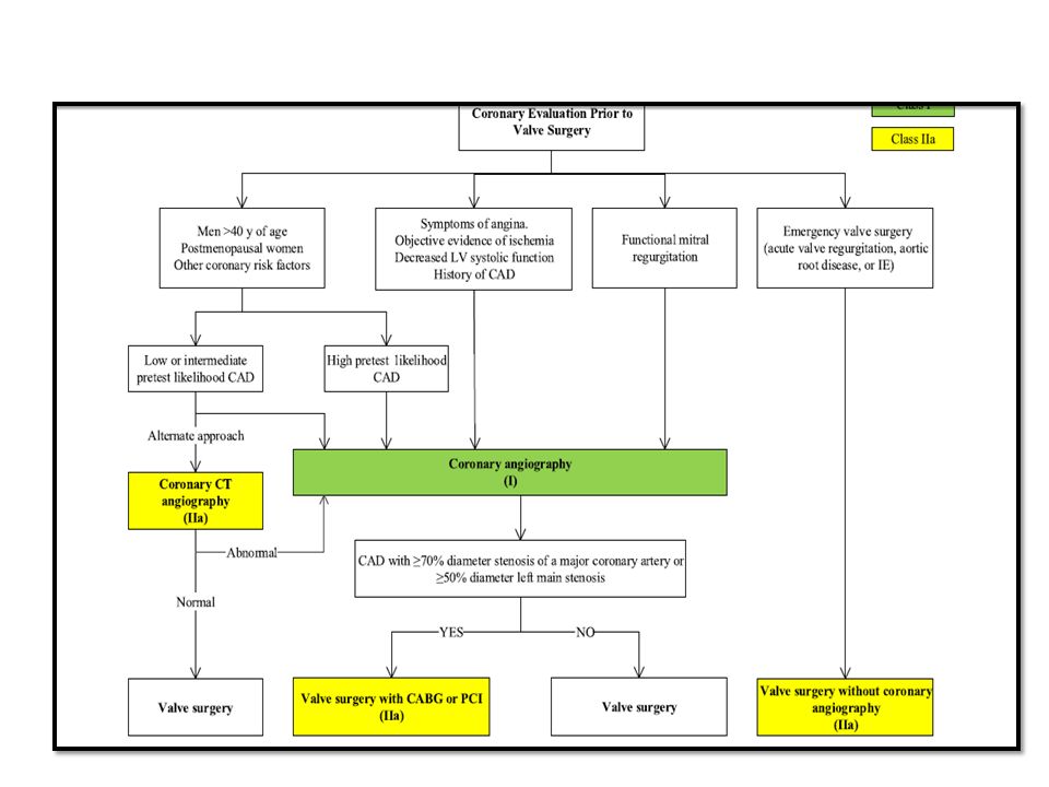
Choice of Intervention: Recommendations
The choice of surgery versus transcatheter AVR is based on multiple parameters including the risk of operation, patient frailty and comorbid conditions. Concomitant severe CAD may best served by AVR and CABG.

68
CLASS I Surgical AVR is recommended in patients who meet an indication for AVR with low or intermediate surgical risk (Level of Evidence: A)
")
69
CLASS1 2. For patients in whom TAVR or high-risk surgical AVR is being considered, a Heart Valve Team consisting of an integrated, multidisciplinary group of healthcare professionals with expertise in VHD, cardiac imaging, interventional cardiology, cardiac anesthesia, and cardiac surgery should collaborate to provide optimal patient care. (Level of Evidence: C)
")
70
Decision making is complex in the patient at high surgical risk with severe symptomatic AS.
The decision to perform surgical AVR, TAVR or to forgo intervention requires input from a Heart Valve Team

71
CLASS I 3. TAVR is recommended in patients who meet an indication for AVR who have a prohibitive risk for surgical AVR and a predicted post-TAVR survival greater than 12 months (Level of Evidence: B)
")
72
TAVR was compared with standard therapy in a prospective RCT of patients with severe symptomatic AS who were inoperable. Patient taken was Severe AS with NYHA class II to IV symptoms. Patients were considered to have a prohibitive surgical risk when predicted 30–day surgical morbidity and mortality were 50%

73
All-cause death at 2 years was lower with TAVR (43
All-cause death at 2 years was lower with TAVR (43.3%) compared with standard medical therapy (68%) with an HR for TAVR of 0.58 and p <0.02). There was a reduction in repeat hospitalization with TAVR (55% versus 72.5%; p<0.001). only 25.2% of survivors were in NYHA class III or IV 1year after TAVR, compared with 58% of patients receiving standard therapy (p<0.001). However, the rate of major stroke at 30 days was higher with TAVR (5.05% versus 1.0%; p<0.06) and remained higher at 2 years with TAVR compared with standard therapy (13.8% versus 5.5%;p<0.01). Leon MB; et al. Transcatheter aortic-valve implantation for aortic stenosis in patients who cannot undergo surgery. N Engl J Med 2010
 compared with standard medical therapy (68%) with an HR for TAVR of 0.58 and p <0.02). There was a reduction in repeat hospitalization with TAVR (55% versus 72.5%; p<0.001). only 25.2% of survivors were in NYHA class III or IV 1year after TAVR, compared with 58% of patients receiving standard therapy (p<0.001). However, the rate of major stroke at 30 days was higher with TAVR (5.05% versus 1.0%; p<0.06) and remained higher at 2 years with TAVR compared with standard therapy (13.8% versus 5.5%;p<0.01). Leon MB; et al. Transcatheter aortic-valve implantation for aortic stenosis in patients who cannot undergo surgery. N Engl J Med")
74
CLASS IIa 1. TAVR is a reasonable alternative to surgical AVR in patients who meet an indication for AVR and who have high surgical risk for surgical AVR. (Level of Evidence: B)
")
75
TAVR when compared with surgical AVR in a prospective RCT of patients with severe symptomatic AS who were high risk for surgery following results came. On an intention-to-treat analysis, all-cause death was similar in those randomized to TAVR (n 348) compared with surgical AVR (n 351) at 30 days, 1 year and 2 years (p 0.001) suggesting non inferiority of TAVR compared with surgical AVR. The composite endpoint of all-cause death or stroke at 2 years was 35% with surgical AVR compared with 33.9% with TAVR (p0.78). Smith CR et al. Transcatheter versus surgical aortic-valve replacement in high-risk patients. N Engl J Med 2011
 compared with surgical AVR (n 351) at 30 days, 1 year and 2 years (p 0.001) suggesting non inferiority of TAVR compared with surgical AVR. The composite endpoint of all-cause death or stroke at 2 years was 35% with surgical AVR compared with 33.9% with TAVR (p0.78). Smith CR et al. Transcatheter versus surgical aortic-valve replacement in high-risk patients. N Engl J Med")
76
CLASS IIb Percutaneous aortic balloon dilation may be considered as a bridge to surgical AVR or TAVR in patients with severe symptomatic AS. (Level of Evidence: C)
")
77
Mechanism is by fracture of calcify deposits within the valve leaflets and to a minor degree by stretching of the annulus and separation of the calcified or fused commissures. Immediate hemodynamic results include a moderate reduction in the transvalvular pressure gradient, but post dilation valve area rarely exceeds 1.0 cm symptomatic improvement occurs. However, serious acute complications, including acute severe AR and restenosis and clinical deterioration, occur within 6 to 12 months in most patients. Therefore in patients with AS, balloon dilation is not a substitute for AVR.

78
Aortic balloon dilation should be consider as a “bridge” to AVR, as improved hemodynamic state may reduce the risks of TAVR or surgery. Palliative balloon dilation in patients in whom AVR cannot be done because of serious co morbid conditions are less well established, with no data suggest improved longevity; however, some patients do report a decrease in symptoms. Asymptomatic severe AS who require urgent non cardiac surgery can undergo surgery at reasonably low risk with anaesthetic monitoring and attention to fluid. Balloon dilation is not recommended for these. If preoperative correction of AS is needed, they should be considered for AVR.

79
CLASS III: No Benefit 1. TAVR is not recommended in patients in whom existing comorbidities would preclude the expected benefit from correction of AS (Level of Evidence: B)
")
80
PARTNER (Placement of Aortic Transcatheter Valve) study, survival benefit of TAVR was seen in those with an STS score <5% and in those with an STS score between 5% and 14.9% but not in those with an STS score 15% TAVR is not recommended in patients with 1) a life expectancy of <1 year, even with a successful procedure 2) those with a chance of “survival with benefit” of <25% at 2 years
 a life expectancy of <1 year, even with a successful procedure. 2) those with a chance of survival with benefit of <25% at 2 years.")
81
Summary of Recommendations for AS: Choice of Surgical or Transcatheter Intervention.

83
MCQ 1 All are true about standard dobutamine stress echocardiography for evaluation of AS severity in setting of LV dysfunction except? A) Uses low dose of dobutamine starting at 2.5 or 5ủg/kg/min B) Maximum dose of dobutamine used is 10–20 ủg/kg/min C) The infusion should be stopped when the heart rate begins to rise more than 10–20 bpm over baseline D) Failure of LVEF to ↑ by 40% is a poor prognostic sign e) None of the above D
 Uses low dose of dobutamine starting at 2.5 or 5ủg/kg/min. B) Maximum dose of dobutamine used is 10–20 ủg/kg/min. C) The infusion should be stopped when the heart rate begins to rise more than 10–20 bpm over baseline. D) Failure of LVEF to ↑ by 40% is a poor prognostic sign. e) None of the above. D.")
84
MCQ-2 By definition Low-flow low-gradient AS includes the following conditions except Anatomic orifice area < 1.0 Cm2 LV ejection fraction < 40% Mean pressure gradient < 30–40 mmHg d) none d
 none. d.")
85
MCQ -3 Which is false about Severe AS? Aortic jet velocity > 4 m/s
Velocity ratio > 0.50 Indexed AVA < 0.6 cm²/m² Mean gradient > 40 mm Hg None of the above b

86
MCQ4 Partner trial compares TAVR VS AVR TAVR VS MEDICAL THEARPY
BOTH OF ABOVE NONE OF ABOVE c

87
MCQ5 PATIENT WITH SYMPTOMATIC SEVERE AORTIC STENOSIS POSTED FOR HERNIA SURGERY BEST STRATGEY CONSIDER FOR AVR BRIDGING THEARPY BY PABD TMT FOR RISK ASSESMENT DIRECT TO SURGERY a

88
MCQ6 Patient with prohibitive risk for surgical AVR and post TAVR survival more then 12 month should be consider for AVR TAVI MEDICAL THEARPY Risk assessment and then to heart valve team for decision b

89
MCQ7 Asymptomatic severe AS undergoes exercise test which demonstrate fall in systolic BP to do AVR comes under. Class 1 Class 2a Class 2b Class 3 B

90
MCQ8 50 year old male Known case of aortic stenosis with IE with hemodynamic instability AVR is planned to under go AVR without angiography Class 1 Class 2a Class 2b Class 3 B

Similar presentations