Download presentation
Presentation is loading. Please wait.

1
Clinical Urinalysis and Body Fluids Review, part 1 Austin Community College Medical Laboratory Technology Clinical II Spring 09

2
Body Fluids other than urine What general purposes do the body fluids serve? Nutrition Waste removal Lubrication Cushioning / protection

3
Body Fluids Types CSF Serous (transudate versus exudate) Peritoneal Pleural Pericardial Synovial Seminal Amniotic Sweat, gastric, feces, etc.
 Peritoneal Pleural Pericardial Synovial Seminal Amniotic Sweat, gastric, feces, etc.")
4
Body Fluids Testing Gross and hematological Cell counts Differential Chemistry TP Glucose Enzymes? Amylase, Lipase and LDH Microbiology – cultures Serology? Cytology ?

5
Body Fluids CSF Why evaluate?

6
Body Fluids CSF Why evaluate? Diagnose meningitis Evaluate for intracranial hemorrhage Diagnose malignancies, leukemia Investigate central nervous system disorders

7
Body Fluids CSF What normal term(s) are used to describe the color and clarity of CSF specimens? Identify terms used for ‘abnormal’ colors and clarity. What is that ‘special’ color term unique to CSF? How do you differentiate between traumatic tap and cerebral hemorrhage?

8
Body Fluids CSF Reference ranges / normal or expected values

9
Body Fluids What is the term used when there is increased numbers of cells in CSF? Pleocytosis Can be prefixed as Neutrophilic pleocytosis Lymphocytic pleocytosis, etc.

10
Body Fluids Why evaluate serous and synovial? To determine the reason for increased production What’s the term that is used to indicate increased production of these fluids?

11
Body Fluids What’s the term ? Effusion – an increase in the serous fluid due to some disruption in production and/ re-absorption processes.

12
Seminal fluid In what tissue are sperm produced? Four reasons for analyzing seminal fluids 12

13
Seminal fluid In what tissue is sperm produced? Spermatozoa - produced in the testes, mature in the epididymis. Four reasons for analyzing seminal fluids Infertility issues Post- vasectomy Forensic analysis Sperm donors 13

14
Body Fluids other than Urine What are the collection requirements? Normal appearance? Expected lab values? Cell count Motility Morphology 14

15
Body Fluids other than Urine What is the formula for calculating body fluid cells counted in a hemacytometer? 15

16
Body Fluids other than Urine What is the basic formula? What additional calculation is needed for sperm counts and why? 16

17
Body Fluids other than Urine What additional calculation is needed and why? the standard calculation provides results as per microliter (uL) And sperm normal value is per milliliter (mL) So you have to take the results and multiply X 1000. 17
 And sperm normal value is per milliliter (mL) So you have to take the results and multiply X")
18
Body Fluids other than Urine What seminal fluid substances can be used for forensic and rape evaluations? 18

19
Body Fluids other than Urine What seminal fluid substances can be used for forensic and rape evaluations? Acid phosphatase enzyme DNA UV light ABO /HLA 19

20
Body Fluids other than Urine Amniotic Fluid Of what is it composed? What is the name of the collection procedure? What purpose does it serve? Why is it sometimes analyzed?

21
Body Fluids other than Urine Amniotic Fluid Indications for analysis Chromosomal abnormalities, such as Down’s syndrome Metabolic disorders, such as Tay Sachs Neural tube defects – such as spinal bifida or an encephalic Determination of extent of HDN Others - gestational age, fetal maturity, etc.

22
Body Fluids other than Urine Why do we sometimes analyze sweat? What is the name of that procedure? What are normal values?

23
Body Fluids other than Urine Why do we analyze gastric fluids? How are the specimens collected? What lab procedures are performed?

24
Body Fluids other than Urine Indications for testing Drug analysis Peptic ulcer evaluation Specimen collection Nasal or oral intubation Fasting and avoid swallowing saliva Laboratory procedures Gastric acidy (acid <7.0) Peptic ulcers, diagnosis and treatment Zollinger-Ellison Syndrome – tumor of pancreas causing gastric over-secretion Decreased gastric secretion Anacidity – inability to produce acid Drug screening
 Peptic ulcers, diagnosis and treatment Zollinger-Ellison Syndrome – tumor of pancreas causing gastric over-secretion Decreased gastric secretion Anacidity – inability to produce acid Drug screening")
25
Body Fluids other than Urine Feces What is it and why do we care? What is the significance of different stool colors?

26
Body Fluids other than Urine What test is performed as a screen for colorectal cancer?

27
Body Fluids other than Urine What patient preparation is needed for this test to be most useful?

28
Body Fluids other than Urine What other tests are done on fecal samples?

29
Cells of the Body Fluids What cells can be found in body fluids? Cells seen in the peripheral blood, but they sure don’t look so good in the BFs.

30
Cells of the Body Fluids (other than Urine) More often they look like these: Lymphocytes and monocytes Cerebrospinal Fluid (CSF)
 More often they look like these: Lymphocytes and monocytes Cerebrospinal Fluid (CSF)")
31
Cells of the Body Fluids (other than Urine) Lymphocytes and eosinophils – CSF Patient had shunt
 Lymphocytes and eosinophils – CSF Patient had shunt")
32
Cells of the Body Fluids Lymphocytes, macrophages and basophil 32

33
Cells of the Body Other cells Ependymal cells - on left Mesothelial cells - on right 33

34
Cells of the Body Fluids Then there are the inclusions, and the ‘new’ names

35
Cells of the Body Fluids And the ‘other’ cells, ‘unclassified’ – those referred to the cytologist / pathologist. 35

36
Cells of the Body Fluids Intracellular inclusions: The yeast 36

37
Cells of the Body Fluids The yeast - Cryptococcus in CSF 37

38
Cells of the Body Fluids Bacteria 38

39
Cells of the Body Fluids For synovial fluid Crystals Calcium pyrophosphate Monosodium urate 39

40
Serous and Synovial Fluids LE cells – 40

41
Clinical Urinalysis and Body Fluids Review, part 2 Austin Community College Medical Laboratory Technology Clinical II Spring 09

42
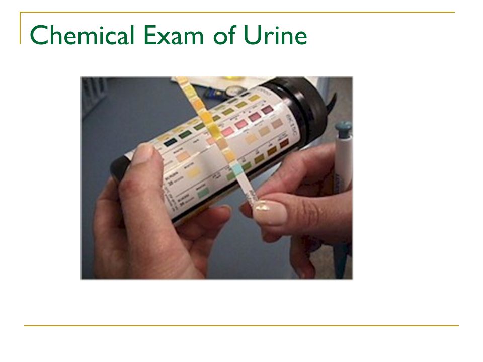
Chemical Exam of Urine

44
Reagent strip manufactures Bayer Corporation- Diagnostics Division (formerly Ames) produces Multistix Boehringer-Mannheim Corporation which produces Chemstrip Behring Diagnostics which produces Rapignost
 produces Multistix Boehringer-Mannheim Corporation which produces Chemstrip Behring Diagnostics which produces Rapignost")
45
Chemical Exam of Urine Recall normal dipstick procedure What are sources of error?

46
Chemical Exam of Urine Sources of error (and preventions) Testing cold specimens would result in a slowing down of reactions; test specimens when fresh or bring them to RT before testing Inadequate mixing of specimen could result in false reduced or negative reactions to blood and leukocyte tests mix specimens well before dipping Over-dipping of reagent strip will result in leaching of reagents out of pads; briefly, but completely dip the reagent strip into the urine
 Testing cold specimens would result in a slowing down of reactions; test specimens when fresh or bring them to RT before testing Inadequate mixing of specimen could result in false reduced or negative reactions to blood and leukocyte tests mix specimens well before dipping Over-dipping of reagent strip will result in leaching of reagents out of pads; briefly, but completely dip the reagent strip into the urine")
47
Chemical Exam of Urine Inadequate blotting and Failure to keep strip horizontal will result in over-run or mixing of reagents between the different reaction pads; blot excess urine off the strip and keep strip horizontal. If dipping from the tube, can run the side of the strip along the rim to remove excess urine. Improper timing of tests over development of reagent pad colors leading to falsely increased results; follow manufacturer’s recommendations

48
Chemical Exam of Urine Inadequate light misinterpretation of results; use good lighting Mis-using color chart misinterpretation of results; hold strip just over color chart and match colors as close as possible, consider use of back- up tests, if needed, especially if urine’s color masks reaction colors.

49
Chemical Exam of Urine Handling and Storage Keep strips in original container, stored at RT Protect from moisture and volatile fumes Use before expiration date Do not touch reagent pad areas

50
Chemical Exam of Urine Quality Control - use appropriate, commercially prepared positive and negative controls. Use commercially prepared pos and neg controls, at least once per 24 hours, and anytime a new bottle is opened, or question of validity of results. Readings should agree with published results ± one color block.

51
Urine Glucose Testing Normal : no glucose detected Clinical significance of abnormal results (Glucosuria) Sensitivity Approximately 50-100 mg (compared to Clinitest’s 250) SO- Can have a positive dipstick but a neg Clinitest Specificity - is specific for glucose only. not affected by other sugars or reducing substances.

52
Urine Glucose Testing Interfering substances High specific gravity and high pH may depress color. Ascorbic acid-false neg Bleach or peroxide may give false positive

53
Urine Bilirubin Testing Normal : no bilirubin detected Clinical significance of abnormal results (Bilirubinuria) Jaundice types
 Jaundice types")
54
Urine Bilirubin Testing Testing method Urine dipsticks for bilirubin – a diazo reaction Impregnated with stabilized diazotized 2,4 dichloraniline Color goes from buff to brown also shades of pink – violet If urine is strongly colored, look for change in pad color after dipping. Use Ictotest for backup.

55
Urine Bilirubin Testing Interfering substances Medication metabolites, pigments and indican may obscure readings False negatives due to aged specimens, especially those exposed to light and oxidation.

56
Urine Ketone Testing Ketone Bodies Origin - not normally present Products of fat catabolism - breakdown of fat into CO2 and H2O

57
Urine Ketone Testing Ketone Bodies What are the 3 ketone bodies? What effect do they have on the body? What is the testing methodology?

58
Urine Ketone Testing Acetone 2%. -Acetone is volatile and excreted primarily through the lungs Diacetic Acid (Acetoacetic) the first formed, 20 % of the total the form detected by most ketone test procedures Beta hydroxybutyric Acid majority formed, but not detected by routine tests. Only Hart’s test, an old ‘wet chemical’ test will detect this one.
 the first formed, 20 % of the total the form detected by most ketone test procedures Beta hydroxybutyric Acid majority formed, but not detected by routine tests. Only Hart’s test, an old ‘wet chemical’ test will detect this one..")
59
Urine Ketone Testing Clinical effects Metabolic acidosis Lowering of blood and urine pH Brain toxicity

60
Urine Ketone Testing Testing Most use nitroprusside Detects diacetic acid and a small amount of acetone, but does not detect β -hydroxybutyric acid. Produces purple color. Can be used on urine or blood.

61
Urine Specific Gravity Testing The specific gravity is a measure of the weight of urine compared to an equal amount of water. Water has an assigned value: 1.000 Specific gravity is related / proportional to urine osmolality; both are measures of concentration.

62
Urine Specific Gravity Testing Specific gravity between 1.002 and 1.035 on a random sample is normal if kidney function is normal.

63
Urine Specific Gravity Testing Dilute urine will have values less than 1.010. Fixed specific gravity = 1.010; isothenuria Diabetes insipidus End-stage renal disease And concentrated urine will have values usually over 1.020. Usually due to dehydration and can be seen in well population as well as sick.

64
Urine Specific Gravity Testing Increased urine specific gravity may be seen in: Dehydration Diarrhea Excessive sweating Glucosuria Heart failure (related to decreased blood flow to the kidneys) Renal arterial stenosis Syndrome of inappropriate antidiuretic hormone secretion (SIADH) Vomiting Water restriction
 Renal arterial stenosis Syndrome of inappropriate antidiuretic hormone secretion (SIADH) Vomiting Water restriction")
65
Urine Specific Gravity Testing Decreased urine specific gravity may indicate / be seen in: Excessive fluid intake Diabetes insipidus – central or nephrogenic Renal failure (that is, loss of ability to reabsorb water) Pyelonephritis
 Pyelonephritis")
66
Urine Specific Gravity Testing Specific gravity > 1.035 (refractometer) Could have very high glucose levels Could contain radiographic dye
 Could have very high glucose levels Could contain radiographic dye")
67
Urine Specific Gravity Testing Testing Polyelectrolytes, pH indicator (bromthymol blue measures the pH change) and alkaline buffer
 and alkaline buffer")
68
Urine Specific Gravity Testing Interfering substances False elevation of results may be seen in samples with increased protein concentration. Some reports of reduced specific gravity results on alkaline specimens. Lipids may also effect results

69
Urine Blood Testing Normally not found in urine Hemoglobinuria – free hemoglobin in urine Circulating free hemoglobin normally picked up by haptoglobin preventing loss in urine When serum levels of hemoglobin > 100 mg/dL threshold is exceeded Hematuria – RBCs in the urine Trauma / irritation of renal organs

70
Urine Blood Testing

71
Testing dipstick reaction

72
Urine Blood Testing ‘Blood’ test detects Free Hemoglobin RBCs – get lysed on the pad and their hemoglobin reacts Myoglobin – muscle hemoglobin Principle based on the peroxidase-like activity of the heme portion of the molecule

73
Urine Blood Testing Sensitivity – can detect at levels of 5-10 cells/uL Interfering substances Ascorbic acid Nitrates Oxidizing agents (ie bleach) Contaminate blood (menstrual)
 Contaminate blood (menstrual)")
74
Urine pH Testing Normal: kidneys capable of 4.5 – 8.0 Factors effecting pH Diet – general and specific foods Time of day Metabolic disorders Drugs / medications Dipstick capable: 4.5 – 9.0

75
Urine pH Testing Test method Dipstick indicators – methyl red and bromthymol blue Range 4.5-9.0 Caution – other chemicals on dipstick can effect pH reading

76
Urine Protein Testing Normally not found in measurable amounts on dipstick (<150 mg/dL /day) Permeability of glomerulus Damage to glomerular capillaries Changes in glom blood flow Albumin excretions may be increased temporarily due to exercise, UTI, and acute illness with fever. Dipstick results of > 1+ (30mg/dL) would equal to approximately 500 mg/dL (clinical proteinuria)
 would equal to approximately 500 mg/dL (clinical proteinuria).")
77
Urine Protein Testing Only albumin detectable by dipstick Sensitivity (approximately15-30 ml/dL)
")
78
Urine Protein Testing New testing for microalbumin and creatinine Results: Protein 20-200 mg/dL (30-300 mg/dL /24 hr) Creatinine 10-300 mg/dL Albumin/Creatinine ratio Normally albumin in the urine is less than 30 mg/ gram creatinine
 Creatinine mg/dL Albumin/Creatinine ratio Normally albumin in the urine is less than 30 mg/ gram creatinine")
79
Urine Protein Testing Principle - Protein error of indicators at fixed pH, certain indicators show one color in the presence of protein and another in absence of protein - the “error” of the indicator. Indicator – tetrabromphenol blue - can be hard to read at the trace end Citrate Buffer – maintains pH 3 -quite acid

80
Urine Protein Testing Sources of error Sensitive only to albumin Urine with strong / unusual color makes reading difficult Highly alkaline or buffered urine will neutralize acid buffer and lead to increased erroneous results. Urine container contamination would interfere

81
Urine Protein Testing Urine back up test 3% sulfosalicylic acid Added to the supernatant to detect any kind of protein. Urine will turn cloudy if protein is present.

82
Urine Urobilinogen Testing Normally found in small amounts, especially in early afternoon Increased amounts may indicate liver disease or be seen as result of hemolytic disorders Decreased amounts: If intestinal bacteria destroyed Liver doesn’t conjugate bilirubin Biliary obstruction – failure of bilirubin to reach small intestine

83
Urine Urobilinogen Testing Test principle based on Ehrilich’s reaction Para-dimethylaminobenzaldehyde = Ehrlich's reagent. Must protect specimen from light and test immediately

84
Urine Nitrate Testing Nitrate Detects presence of certain types of bacteria screening for presence of UTI. Certain species of bacteria convert nitrate (normal constituent of urine) to nitrite Escherichia - most common cause of UTI Klebsiella Proteus Pseudomonas Enterobacter Citrobacter
 to nitrite Escherichia - most common cause of UTI Klebsiella Proteus Pseudomonas Enterobacter Citrobacter.")
85
Urine Nitrate Testing Aromatic amine in reagent strip reacts with nitrite; producing a diazonium salt The diazonium salt reacts with sulfanilic acid and acetic acid to produce a pink azo dye

86
Urine Nitrate Testing Limitations reported as positive or negative Not all UTI causing bacteria convert nitrate to nitrite Haemophilus Staphylococcus Streptococcus

87
Urine Nitrate Testing Fresh first morning specimen is preferred - besides being the most concentrated specimen, the urine has been in the bladder longer, allowing bacteria time and opportunity to convert the nitrates to nitrites.

88
Urine Leukocyte Testing Leukocyte esterase testing is another test used as a means of screening for urinary tract infection. Does not measure concentration of leukocytes Will detect presence of lysed leukocytes as well as intact WBCs

89
Urine Leukocyte Testing test principle: Leukocyte esterase, an enzyme present in granulocytes, hydrolyzes indoxylcarbonic acid esterase to produce indoxyl, which reacts with a diazonium salt to create a purple color usually in 2 min.

90
Urine Leukocyte Testing Reaction interference False positives - oxidizing detergents False negatives - greatly increased glucose, protein, or specific gravity- increased sp gr could cause WBC to crenate preventing their releasing their esterase, So it is possible for the dipstick to be negative when there are WBCs present.

91
Microscopic Sediment A number of slides with microscopic elements RBC, WBC, yeast

92
Microscopic Sediment – Epithelial Cells Squamous epithelial cells ( stained with Sternheimer-Malbin)
")
93
Microscopic Sediment – Epithelial Cells Transitional epithelial cells Spherical, polyhedral and caudate are terms describing shapes. All have distinct centrally located nuclei. Sometimes called bladder cells, may be more often found in elderly. Can be found as fragments or as reactive.

94
Microscopic Sediment – Epithelial Cells RTEs; 250x magnification Also WBC and RBCs

95
Microscopic Sediment – Casts

96
RBC cast

97
Microscopic Sediment A number of slides with microscopic elements CRYSTALS, Uric acid

98
Microscopic Sediment – Crystals Calcium oxalate – envelope; may be dumbbell shaped. Usually appear as a square with a retractile cross

99
Microscopic Sediment A number of slides with microscopic elements Alkaline

100
Microscopic Sediment A number of slides with microscopic elements The rarely ever seen, abnormal crystals Cystine Tyrosine Leucine Bilirubin (occasionally seen in premies) Cholesterol
 Cholesterol")
101
Microscopic Sediment A number of slides with microscopic elements Mucous threads Bacteria, yeast, Trichomonas sperm Lots of artifacts

102
Microscopic What are oval fat bodies? How can you (quickly, cheaply) provide tentative proof they contain fat?
 provide tentative proof they contain fat .")
103
Microscopic Sediment – Miscellaneous Oval Fat Bodies This frame shows an oval fat body ("B") next to several transitional epithelial cells ("A"). Note the drops of lipids that appear to be contained within the cell.” – U of IA

Similar presentations

 Professor Austin Community College>")

 LECTURE TWO Dr. Essam H. Jiffri.>")


 LECTURE ONE>")


SC>")

