Download presentation
Presentation is loading. Please wait.

2
Alpha & Gamma Motor Neuron Functions Alpha & Gamma Motor Neuron Functions By Dr. Khaled Ibrahim By

4
The Efferent Neurons The efferent neurons of the spinal cord include: (i) Somatic efferents, which join the somatic nerves to innervate skeletal muscles. (ii) Visceral efferents, which join the autonomic nerves for innervation of visceral organs and blood vessels. In each segment of the spinal cord, several thousand somatic efferent motor neurons are located in the anterior horns of the cord gray matter. These neurons are of two types ; “alpha” motor neurons and “gamma’ motor neurons.
 Visceral efferents, which join the autonomic nerves for innervation of visceral organs and blood vessels. In each segment of the spinal cord, several thousand somatic efferent motor neurons are located in the anterior horns of the cord gray matter. These neurons are of two types ; alpha motor neurons and gamma’ motor neurons..")
9
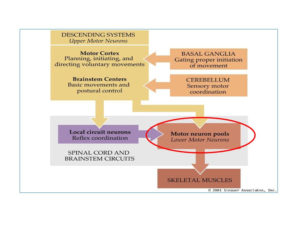
Alpha Motor Neurons The -motor neurons are large neurons that give rise to type A-alpha nerve fibers (about 14 m in diameter), which innervate the large skeletal muscle fibers (extrafusal fibers). The - motor neurons, are called the “final common motor pathway”, because all the motor commands originating anywhere in the nervous system have to pass through it before reaching skeletal muscles.

10
Regulation of - Motor Neuron discharge The firing rate of an - motor neuron is determined by “the magnitude of the membrane potential at the initial segment of its axon”. Such potential is ever- changing, and at any given time it is determined by the net summation of all the excitatory and inhibitory inputs to the - motor neuron at that time. Alpha motor neurons receive excitatory presynaptic input from many sources in the nervous system to be activated and discharge efferent motor signals.

12
The sources of this presynaptic excitation include : (i)spinal cord interneurons, (ii) sensory afferents from muscle spindles, and (iii) descending pathways from the higher motor centers in the brain and brain stem.
spinal cord interneurons, (ii) sensory afferents from muscle spindles, and (iii) descending pathways from the higher motor centers in the brain and brain stem.")
13
Whereas, inhibition of -MNs originates from inhibitory interneurons, that can be activated by: (i)afferent signals from peripheral receptors such as the Golgi tendon organs, and (ii) by signals from the inhibitory descending motor tracts. The - motor neurons, are called the “final common motor pathway”, because all the motor commands originating anywhere in the nervous system have to pass through it before reaching skeletal muscles.

14
Gamma Motor Neurons Gamma motor neurons ( -motor neurons) are located in the ventral horns of the spinal gray matter, intermingled with the -motor neurons. The -motor neurons are smaller in size than the -motor neurons and transmit their discharge along “A-gamma axons”, that innervate the “intrafusal fibers” of muscle spindles to regulate their function

15
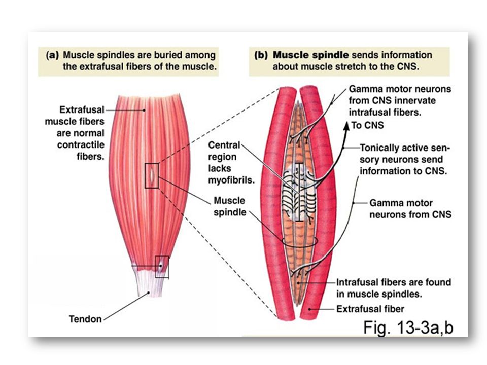
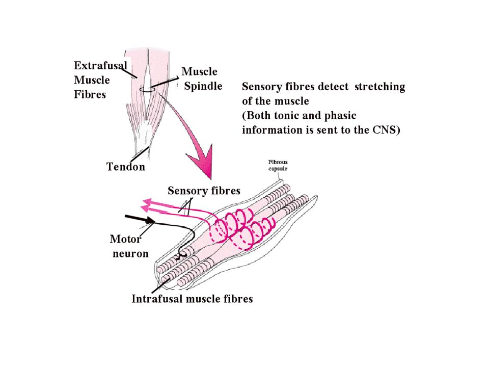
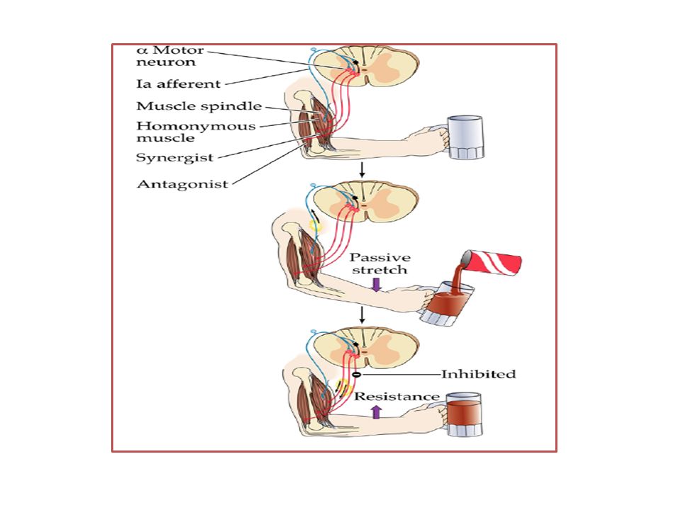
Muscle Spindles

17
Central part Peripheral part - Non-contractile - Contains the nuclei of the fibers & sensory endings arise form it. So, its stretch initiates stretch reflex. - Contractile - Striated (contains myofilaments). So, when contracted ------> stretch of the central part -----> stretch reflex.
. So, when contracted > stretch of the central part -----> stretch reflex..")
18
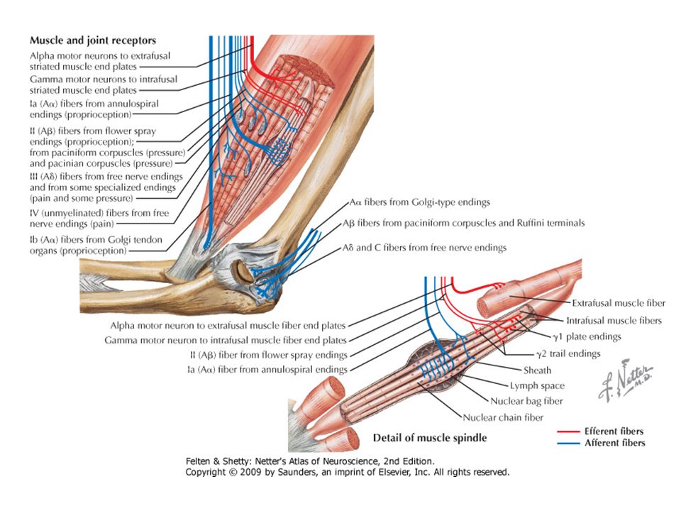
Efferent (Motor) Innervation of muscle spindle - Supplies the peripheral contractile parts on the intrafusal muscle fibers. - Their cell bodies are located in gamma motor neurons ( -MNs) which are the small AHCs (30% of AHCs). - Their axons are thin (diameter 3-6 µm). - According to their functions, Gamma motor neurons ( -MNs) are of 2 types: a) Static - MNs which innervate the nuclear chain fibers b) Dynamic - MNs which innervate the nuclear-bag fibers.
 which are the small AHCs (30% of AHCs). - Their axons are thin (diameter 3-6 µm). - According to their functions, Gamma motor neurons ( -MNs) are of 2 types: a) Static - MNs which innervate the nuclear chain fibers b) Dynamic - MNs which innervate the nuclear-bag fibers..")
19
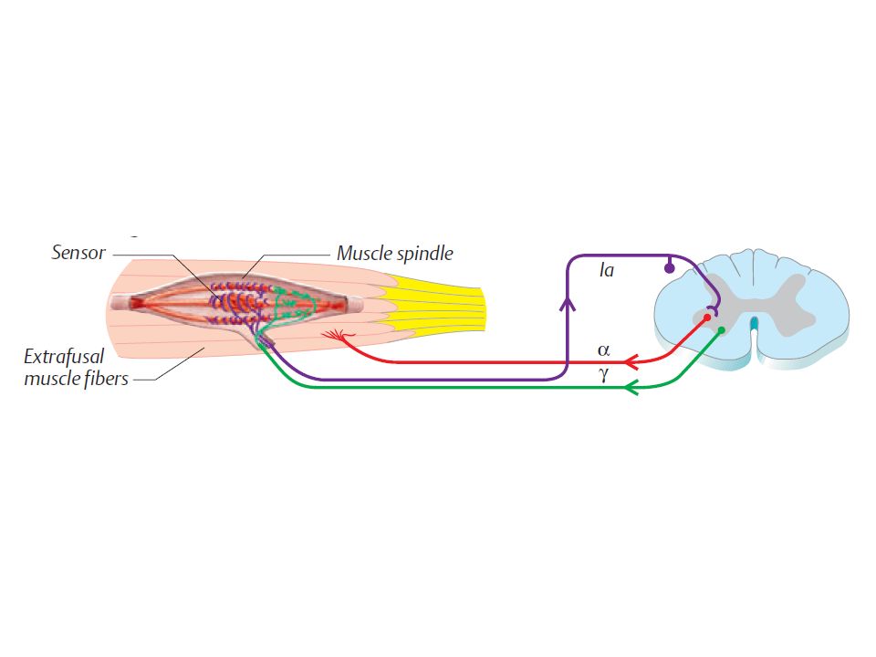
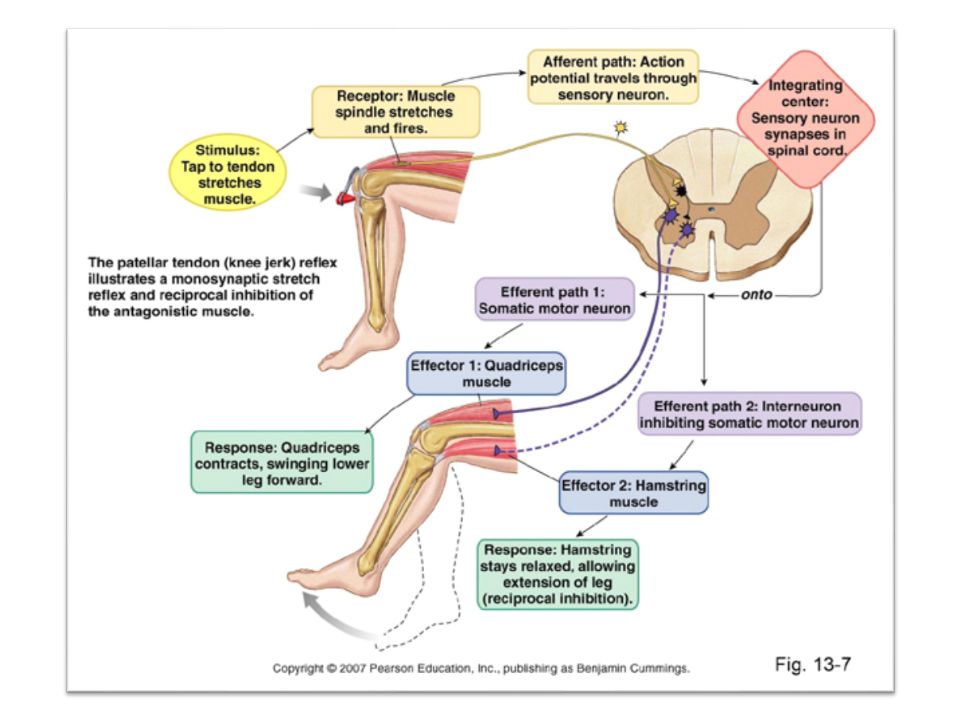
Stimulation of -MNs afferent impulses along -afferent fibers contraction of the peripheral contractile parts of the intrafusal muscle fibers stretch of the central (receptor) part depolarization of the central part action potential & impulse discharge along 1ry & 2ry endings stimulation of α-MNs innervating the stretched muscle muscle contraction (stretch reflex). impulses along the α- efferent fibers N.B.: contraction of the peripheral contractile part causes stretch of the central part as: a) the intrafusal muscle fiber is not too strong to shorten the muscle. b) the ends of the intrafusal muscle fiber is fixed to the membranes of the extrafusal muscle fibers.
 the intrafusal muscle fiber is not too strong to shorten the muscle. b) the ends of the intrafusal muscle fiber is fixed to the membranes of the extrafusal muscle fibers..")
20
- Control of - MNs discharge: I- Supraspinal control (mainly). II- Reflex control.
. II- Reflex control.")
21
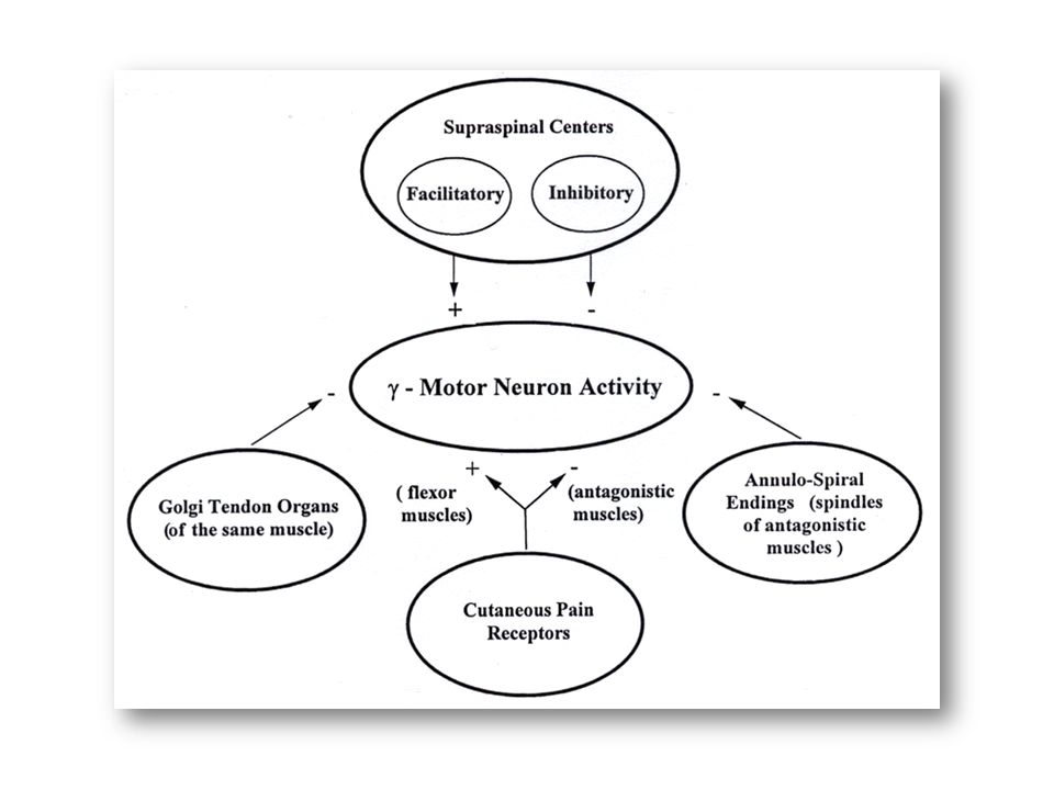
I- Supraspinal control (mainly): - Presynaptic impulses from higher centers in the brain & brainstem reach the - MNs through the descending motor pathways. - These impulses may: - Stimulate (facilitate) - MNs ----> ↑discharge along efferents ------> ↑the magnitude of the existing stretch reflex. - Inhibit - MNs ----> ↓ discharge along efferents ------> ↓ the magnitude of the existing stretch reflex. - The centers which send these impulses are called "Facilitatory centers" & they include: 1)1ry motor area (area 4). 2)Neocerebellum. 3)Facilitatory reticular formation. 4)Vestibular nuclei. - The centers which send these impulses are called "inhibitory centers" & they include: 1)Premotor area (area 6). 2)Paleocerebellum. 3)Inhibitory reticular formation. 4) Red nucleus. 4) Red nucleus.
 - MNs ----> ↑discharge along efferents > ↑the magnitude of the existing stretch reflex. - Inhibit - MNs ----> ↓ discharge along efferents > ↓ the magnitude of the existing stretch reflex. - The centers which send these impulses are called Facilitatory centers & they include: 1)1ry motor area (area 4). 2)Neocerebellum. 3)Facilitatory reticular formation. 4)Vestibular nuclei. - The centers which send these impulses are called inhibitory centers & they include: 1)Premotor area (area 6). 2)Paleocerebellum. 3)Inhibitory reticular formation. 4) Red nucleus. 4) Red nucleus..")
22
II- Reflex Control: By impulses along a presynaptic terminals arising from peripheral receptors such as: Golgi tendon organ of the same muscle ------> inhibition of -MNs (this is called inverse stretch reflex). 1ry sensory endings of the antagonistic muscles -------> inhibition of -MNs (this is called reciprocal innervation). Other nearby contracted muscles ------> facilitation of -MNs (this is called Jendrassik maneuver). Cutaneous receptors (especially pain receptors) ------> facilitation of -MNs of the flexor muscles & inhibition of -MNs of the antagonistic muscles.
. Other nearby contracted muscles > facilitation of -MNs (this is called Jendrassik maneuver). Cutaneous receptors (especially pain receptors) > facilitation of -MNs of the flexor muscles & inhibition of -MNs of the antagonistic muscles..")
24
- Significance (Importance) of - MNs innervation: Stimulation of - MNs ------> stimulation of the muscle spindle -----> ↑ magnitude of the already existing stretch reflex & vice versa. 1) Adjust muscle spindle sensitivity: The sensitivity of the muscle spindle is controlled by the level of activity of the - MNs as follows:
 Adjust muscle spindle sensitivity: The sensitivity of the muscle spindle is controlled by the level of activity of the - MNs as follows:.")
25
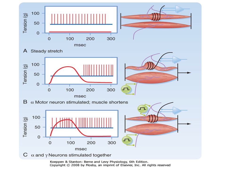
↑ activity of - MNs (↑the level of motor impulses discharge along - efferents) ↓ activity of - MNs (↓ the level of motor impulses discharge along - efferents) more contraction of the intrafusal muscle fibers -------> slight stretch of the whole muscle is needed to stretch the spindle to the adequate level needed for sensory discharge -----> production of the required level of the stretch reflex. less contraction of the intrafusal muscle fibers -------> greater stretch of the whole muscle is needed to stretch the spindle to the adequate level needed for sensory discharge & production of the required level of the stretch reflex. i.e., the spindle becomes more sensitive.i.e., the spindle becomes less sensitive. So, continuous discharge along the efferent fibers keeps the muscle spindle sensitive to the slightest change in the muscle length.

26
2) Adjusting Muscle Length: - The higher centers determine the proper length of the muscle needed to achieve certain posture. - This is done through adjustment of the magnitude of the discharge of -MNs by impulses from the higher centers as follow: If the actual length of the muscle > the desired length (i.e., the contraction of the muscle is not enough) If the actual length of the muscle < the desired length (i.e., the contraction of the muscle is not much) Facilitatory higher centers -----> ↑discharge along -MNs ----> ↑stretch of the muscle spindles ---> ↑magnitude of the stretch reflex -----> more contraction --- -> shortening of the muscle to the desired length. inhibitory higher centers ----> ↓ discharge along -MNs ----> ↓ stretch of the muscle spindles ---> ↓ magnitude of the stretch reflex -----> less contraction ----> lengthening of the muscle to the desired length.
 If the actual length of the muscle < the desired length (i.e., the contraction of the muscle is not much) Facilitatory higher centers -----> ↑discharge along -MNs ----> ↑stretch of the muscle spindles ---> ↑magnitude of the stretch reflex -----> more contraction --- -> shortening of the muscle to the desired length. inhibitory higher centers ----> ↓ discharge along -MNs ----> ↓ stretch of the muscle spindles ---> ↓ magnitude of the stretch reflex -----> less contraction ----> lengthening of the muscle to the desired length..")
27
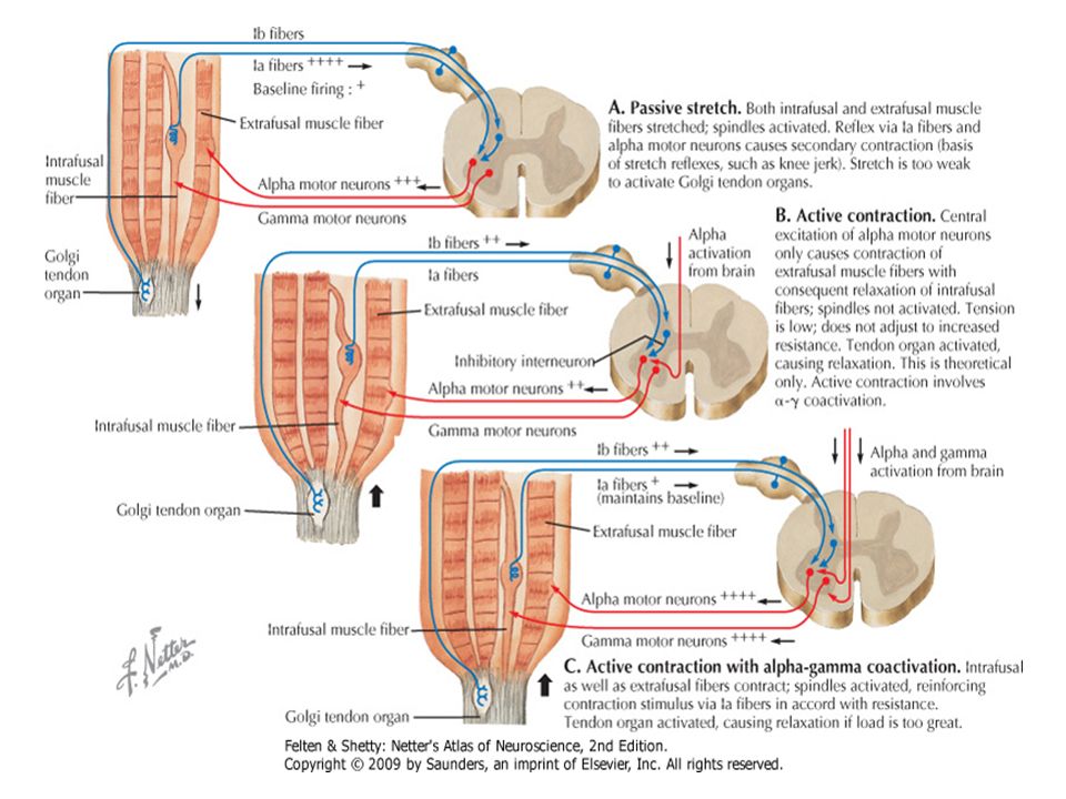
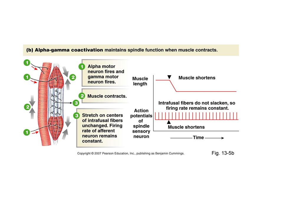
3) Co- activation of and -MNs: - When the higher motor centers in the brain direct a particular muscle to contract, there is almost always an associated ↑ of -MNs along with the delivered -MNs discharge that initiate the movement. -This is called co-activation of -and -MNs. - This co –activation of -MNs helps to: a) Prevent laxation of muscle spindles during active muscle contraction. This maintains the spindle continuously sensitive & maintains continuous information of the higher centers especially the cerebellum about the changes in the muscle length & body posture during contraction. b) maintain adequate excitatory impulses to α-MNs needed to perform the desired motor performance (see before).
 Prevent laxation of muscle spindles during active muscle contraction. This maintains the spindle continuously sensitive & maintains continuous information of the higher centers especially the cerebellum about the changes in the muscle length & body posture during contraction. b) maintain adequate excitatory impulses to α-MNs needed to perform the desired motor performance (see before)..")
34
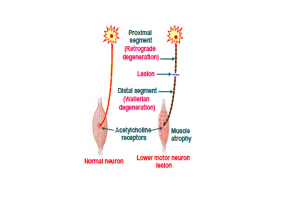
Lower Motor Neuron Lesions Injury or damage of the lower motor neurons in the spinal ventral horns (or the corresponding neurons in the motor nuclei of cranial nerves) innervating a given muscle impairs the ability of the muscle to contract and thereby results in its paralysis. Effects of LMN Lesions 1) Flaccid Paralysis The denervated muscle is completely paralyzed, since there is loss of all types of movements, including voluntary movements, postural movements, and reflex movements
 Flaccid Paralysis The denervated muscle is completely paralyzed, since there is loss of all types of movements, including voluntary movements, postural movements, and reflex movements.")
36
The paralyzed muscle is also hypotonic (flaccid) due to interruption of the motor component of the stretch reflex pathway, and as a result there is loss of the tendon jerks in the affected muscle. The extent of paralysis is often limited in LMN lesions, because the lower motor neurons are widely spread in the brain stem and spinal cord, and their axons proceed to innervate skeletal muscles via a large number of peripheral somatic nerves. Thus, it is not common to have a lesion which would cause damage of a large proportion of these neurons simultaneously.

37
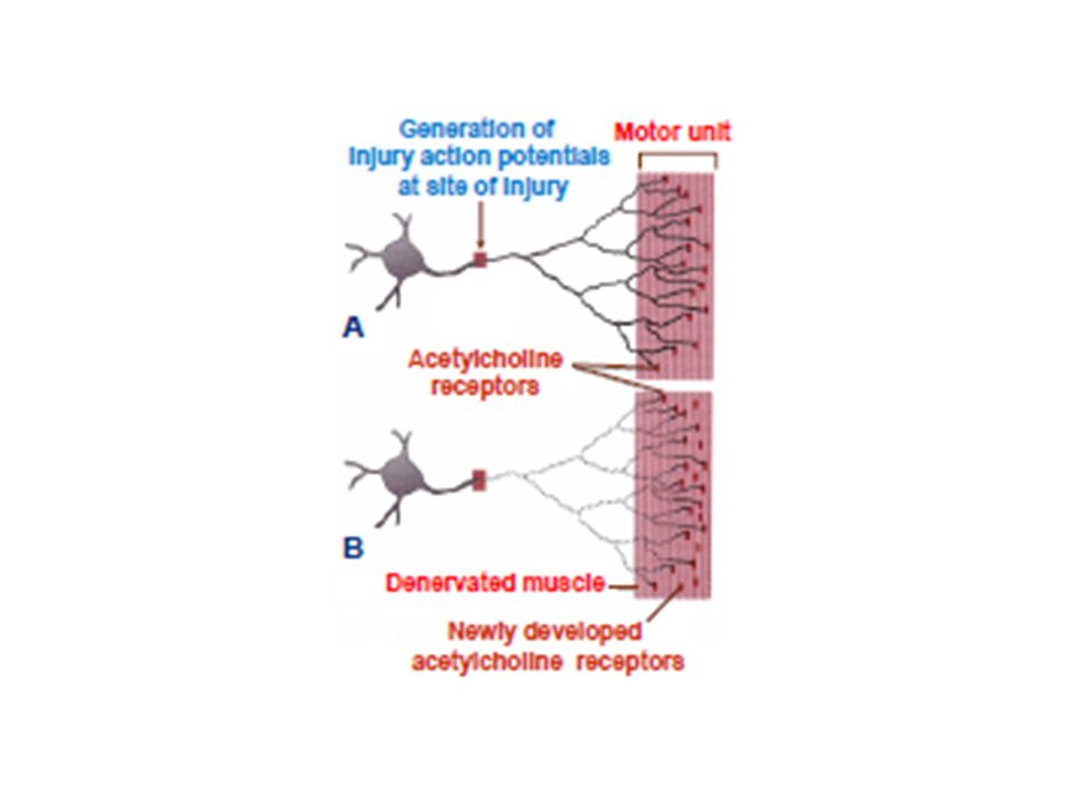
2) Fasiculations and Fibrillations These are abnormal contractions that occur in the denervated muscle. Fasiculations These begin to appear few days after muscle denervation. As a part of the axon degenerates, injury potentials produced by depolarization at the sites of injury of the neuronal membrane cause spontaneous action potentials to travel along the intact distal portions of the axon and stimulate all the muscle fibers innervated by the injured axon (a complete motor unit).
..")
38
This elicits synchronous contractions of all the muscle fibers of the unit, which are thus visible to the observer. These contractions are called “fasiculations” and persist as long the distal segment of the axon is still intact.

40
Fibrillations As the axon degenerates further, its terminal branches become isolated from each other, but the injury potentials are still generated in these branches and still cause muscular contractions. Since the muscle fibers of the affected motor unit are no longer joined by a functioning main axon, the different muscle fibers no longer contract simultaneously as a unit.

41
The resulting contractions are very small and are not visible on the body surface. These contractions are called “fibrillations”, and can be detected electrically by means of an electromyogram. Fibrillations start few weeks after the occurrence of motor neuron injury and persist untill the motor nerve terminals degenerate completely, or until the regenerating proximal segment of the axon successfully re-innervates the muscle fibers.

42
3) Denervation Supersensitivity When the motor innervation to a skeletal muscle is damaged, we find that, instead of being restricted only to the region of the motor end plate, progressively more and more of the muscle cell’s surface becomes sensitive to acetylcholine; a phenomenon called “denervation supersensitivity” Denervation supersensitivity is due to spread out of acetylcholine receptors which ultimately cover the entire surface of the muscle cell’s membrane
 Denervation Supersensitivity When the motor innervation to a skeletal muscle is damaged, we find that, instead of being restricted only to the region of the motor end plate, progressively more and more of the muscle cell’s surface becomes sensitive to acetylcholine; a phenomenon called denervation supersensitivity Denervation supersensitivity is due to spread out of acetylcholine receptors which ultimately cover the entire surface of the muscle cell’s membrane")
43
4) Abnormal Response to Electrical Stimulation : Reaction of Degeneration Response of the Normal Muscle (i) Faradic currents: These currents produce clonic or tetanic contraction, depending on the frequency of the alternating cycles of the current (ii) Galvanic currents: Galvanic currents produce contraction only at: (1) the beginning of current flow (closing the electric circuit) giving rise to closing contraction, and (2) at the termination of the current flow (opening of the circuit) giving rise to opening contraction. No contraction is produced when the galvanic current is flowing in the muscle at a steady level.

44
Also, the muscle responds to the galvanic current at these two instances whether the stimulating electrode is the “cathode” of the electric circuit producing a cathodal contraction, or is the “anode” producing an anodal contraction. When we start stimulation of the muscle by the least effective galvanic current then gradually increase its strength, we will notice that the first contraction to appear is the cathodal closing contraction (CCC). A stronger current is needed to evoke the anodal closing contraction (ACC), then the anodal opening contraction (AOC) comes next, while the cathodal opening contraction (COC) is the last one to appear. These responses can be expressed by the following shortened formula : CCC > ACC > AOC > COC.
. A stronger current is needed to evoke the anodal closing contraction (ACC), then the anodal opening contraction (AOC) comes next, while the cathodal opening contraction (COC) is the last one to appear. These responses can be expressed by the following shortened formula : CCC > ACC > AOC > COC..")
45
Response of the Denervated Muscles (i) Failure of response to faradic currents, because of decreased excitability of the denervated muscle, therefore, the duration of any faradic current cycle becomes too short to be effective. (ii) The muscle still responds to galvanic current (because of its longer duration), but the contractions produced are weak and slow, and the anodal closing current becomes more effective than the cathodal closing current, thus the “ACC” becomes greater than the “CCC”; ACC > CCC.
 The muscle still responds to galvanic current (because of its longer duration), but the contractions produced are weak and slow, and the anodal closing current becomes more effective than the cathodal closing current, thus the ACC becomes greater than the CCC ; ACC > CCC..")
46
These characteristic changes in the response of the denervated muscle to electric stimulation are known as the “reaction of degeneration”. This reaction can be elicited from the muscle only during the period that passes between loss of excitability of the nerve and loss of excitability of the muscle. But, it disappears if re-innervation fails with consequent complete degeneration of the muscle, or when the muscle is successfully re- innervated by the regrowing motor neuron axons.

Similar presentations