Download presentation
Presentation is loading. Please wait.

1
KCP 774 경북대학교병원 병리과 전공의 박보은

2
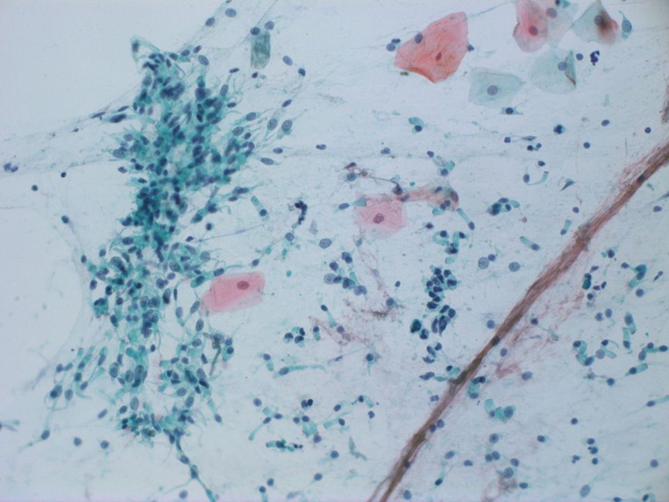
M/28 좌측 전 종격동에 약 5.6 cm 크기의 종괴 좌측 폐문부와 하엽, 중엽에도 결절 두꺼워진 엽간격막 좌측 두정엽에 부종과 출혈을 동반한 병변 α fetoprotein 20,051.0 ng/ml, CEA 7.5 ng/ml, CA19-9 155.7 U/mL, CRP 8.79 mg/dl, LD 475 U/L, β-HCG 2217.0 mIU/ml 기관지 세척 세포 검체

13
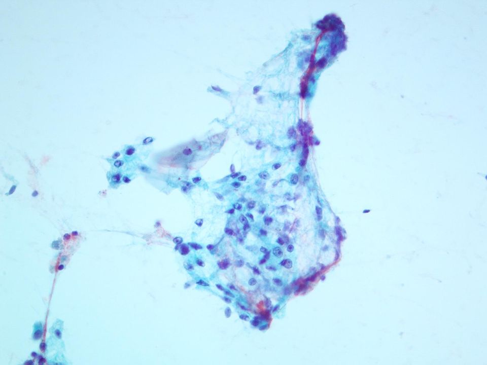
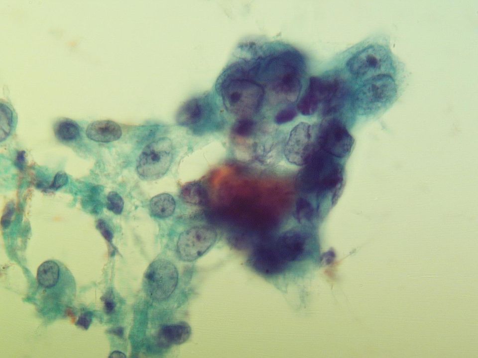
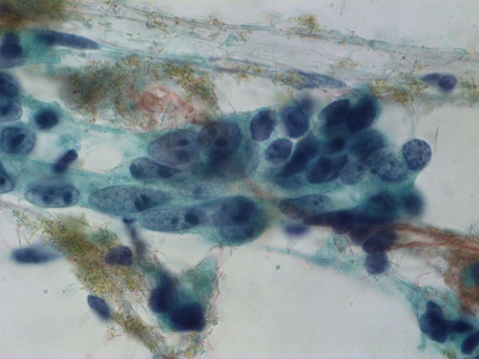
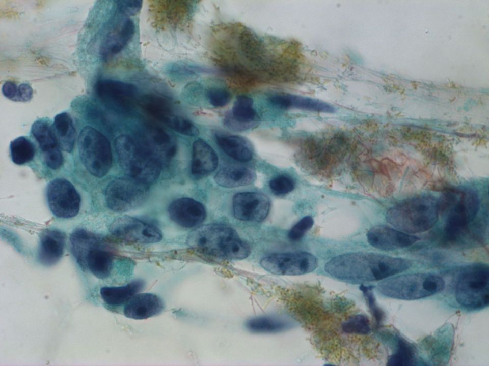
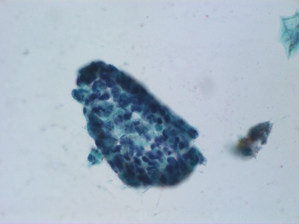
Clean backgroud, rare lymphocytes Loosely cohesive cell groups or clusters Poorly-defined cell borders Multinucleated giant cells Oval, spindle, and pleomorphic neclei Vesicular nuclei, coarsely granular chromatin Multiple necleoli No mitosis, no necrosis

14
Differential diagnosis Thymic carcinoma Poorly differentiated adenocarcinoma Embryonal carcinoma choriocarcinoma Endodermal sinus tumor (Yolk sac tumor)
")
15
Thymic carcinoma Very rare Significant diagnostic dilemma since they resemble other types of carcinomas Cytologically and histologically, thymic carcinomas cannot be differentiated from other malignant neoplasm with similar histomorphology.

16
Lung-Poorly differentiated adenocarcinoma malignant cells isolated, in loosely cohesive groups and in syncytial tissue fragments Pleomorphic in size High N/C ratio Multiple micronucleoli/ macronucleoli Variable cytoplasm with vacuoles +/- AFP and HCG producing lung cancer

17
choriocarcinoma Usually seen as a component of other germ cell tumors Both malignant syncytiotrophoblasts and cytotrophoblasts –Syncytiotrophoblast : large, multinucleated cells with eosinophilic cytoplasm –Cytotrophoblast : round or polygonal Large giant form with bizarre nuclei

18
Endodermal sinus tumor (Yolk sac tumor) Malignant cells in loosely cohesive groups or in syncytial tissue fragments With or without acinar and papillary pattern Cell borders poorly- difined Pleomorphic irregular nuclei Intra and extracellular hyaline droplets Schiller-Duval bodies
 Malignant cells in loosely cohesive groups or in syncytial tissue fragments With or without acinar and papillary pattern Cell borders poorly- difined Pleomorphic irregular nuclei Intra and extracellular hyaline droplets Schiller-Duval bodies")
19
Embryonal carcinoma Highly malignant tumor Histologic pattern typically is variable Scant cytoplasm, poorly-defined cell borders Nuclei : irregular, pleomorphic in size Coarse chromatin Multiple nucleoli Mitosis +/ necrosis +

20
Diagnosis Bronchial washing, conventional cytology: Malignancy. possibly, embryonal carcinoma, metastatic. NOTE) ancillary immunohistochemistry is needed for definite diagnosis.
 ancillary immunohistochemistry is needed for definite diagnosis..")
Similar presentations


>")

>")


 School of Diagnostic Cytology Health Sciences Centre.>")
>")


 Chapter: Neoplasia Definitions Nomenclature Characteristics of benign and malignant neoplasms Epidemiology.>")

