Download presentation
Presentation is loading. Please wait.

1
Chapter 8 The Nervous system Department of Histology & embryology Lecturer: Ph. D xu Short number: 630397 Office:7A210

2
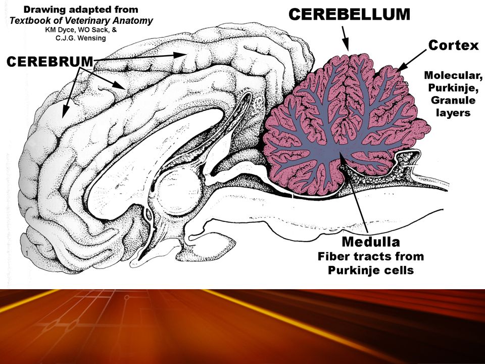
introduction The central nervous system consists of the cerebrum, cerebellum, and spinal cord. When sectioned, the cerebrum, cerebellum, and spinal cord show regions that are white (white matter) and that are gray (gray matter). Gray matter at the periphery of the cerebral hemispheres is folded into many gyri and sulci called the cerebral cortex
 and that are gray (gray matter). Gray matter at the periphery of the cerebral hemispheres is folded into many gyri and sulci called the cerebral cortex.")
6
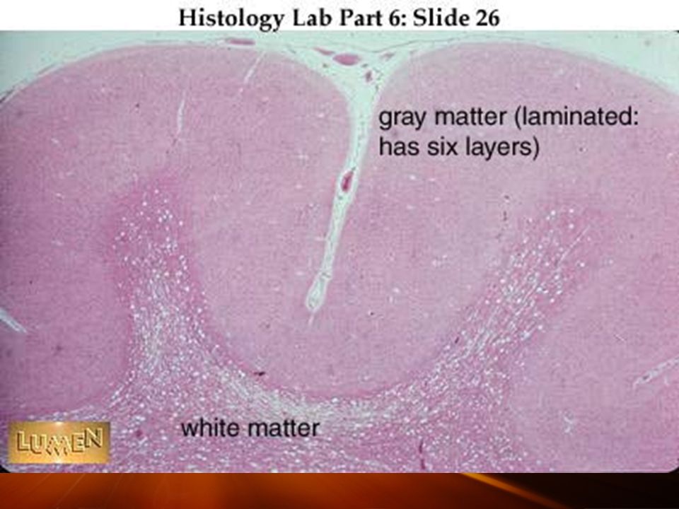
Cross section of the spinal cord in the transition between gray matter (below) and white matter (above).
 and white matter (above).")
7
Cerebral cortex function The cerebral cortex is responsible for learning, memory, sensory integration, information analysis, and initiation of motor responses.

11
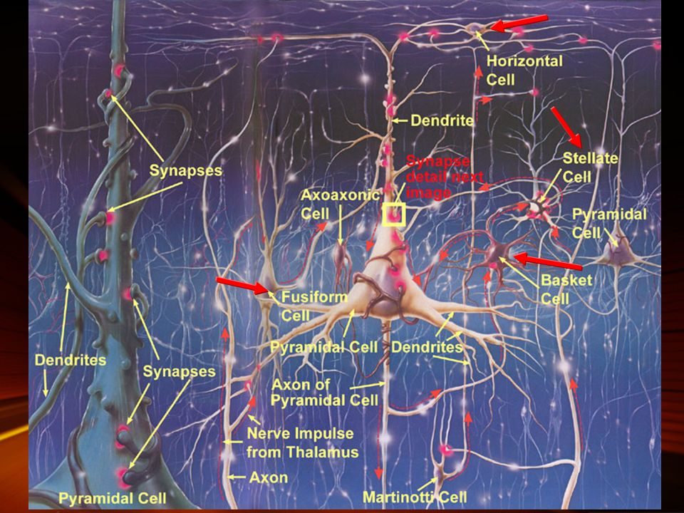
Neurons types in the Cerebral cortex

12
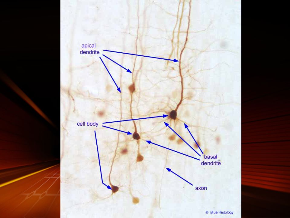
Pyramidal cells Morphology: Axon: Dendrite:

14
Granular cells Morphology: Classification: stellate cells; horizontal cells; basket cells; ascending axons cells. Function:

16
Fusiform cells Morphology: Location: Axon:

17
A, from spinal ganglion; B, from ventral horn of spinal cord; C, pyramidal cell from cerebral cortex; D, Purkinje cell from cerebellar cortex; E, Golgi cell of type II from spinal cord; F, fusiform cell from cerebral cortex;

18
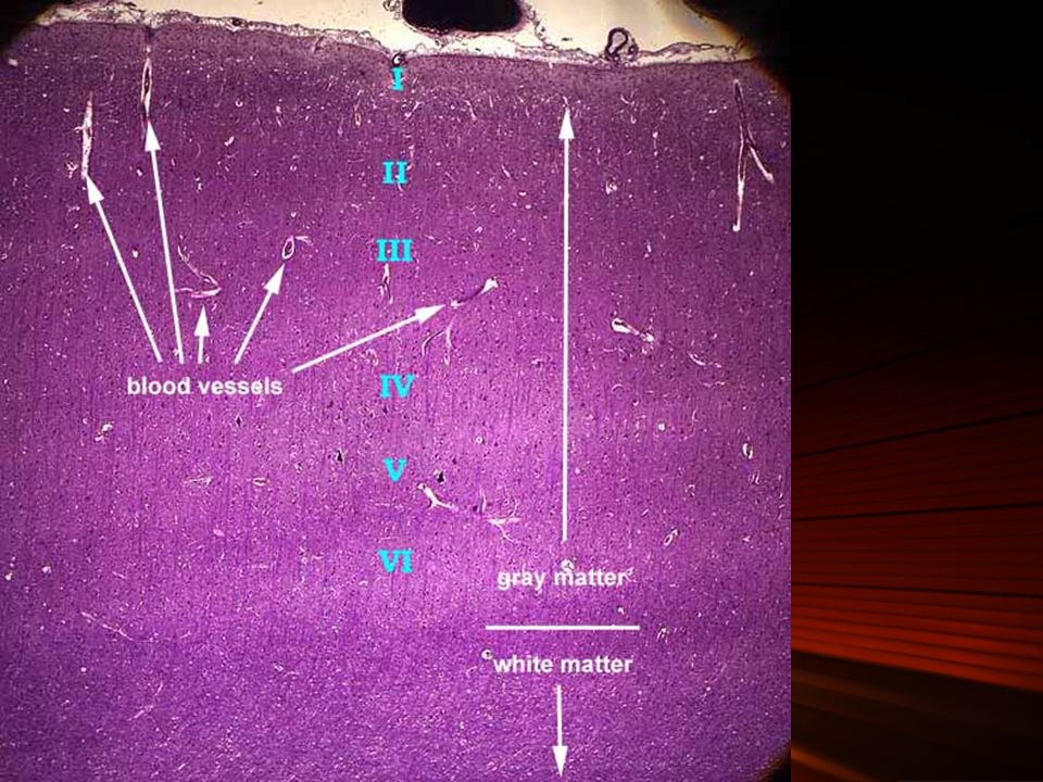
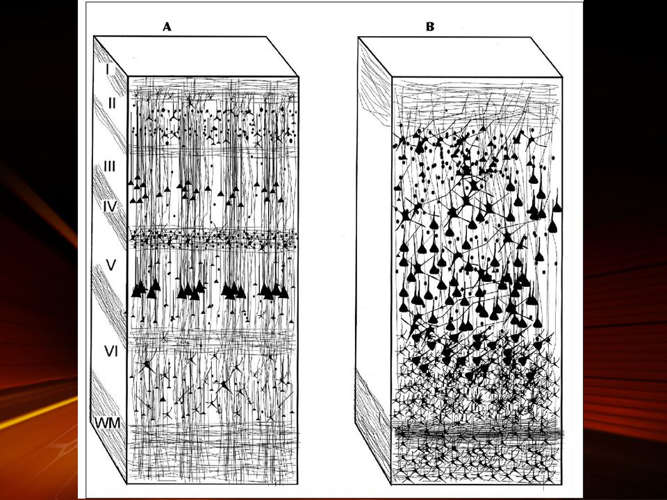
Layers of cerebral cortex

19
molecular layer (1) Composition: mostly of nerve terminals originating in other areas of the brain, horizontal cells, and neuroglia. Location:

21
Outer granular cells (2) Composition: granule (stellate) cells and neuroglial cells; small pyramidal cells. Location:

23
Outer pyramidal cells(3) Composition: medium and small sized pyramidal cells Dendrites extend to the molecular layers and their axon enter the white matter to form association fibers.
 Composition: medium and small sized pyramidal cells Dendrites extend to the molecular layers and their axon enter the white matter to form association fibers.")
24
Inner granular layers(4) Composition: small granule cells (stellate cells), pyramidal cells, and neuroglia. Characteristics: This layer has the greatest cell density of the cerebral cortex.

26
Inner pyramidal cell layers(5) contains the largest pyramidal cells and neuroglia This layer has the lowest cell density of the cerebral cortex.

27
Polymorphic layers (6) Contains small pyramidal cells, granular cells, fusiform cells. Characteristics:

28
The cerebellum has a cortex and a medulla ; o that if you could follow the outermost surface you would dip down between the leaf-like folds (the foliae, F) and back up again. The medulla consists of nerve fibers (NF) leading out of the cortex.
 leading out of the cortex..")
29
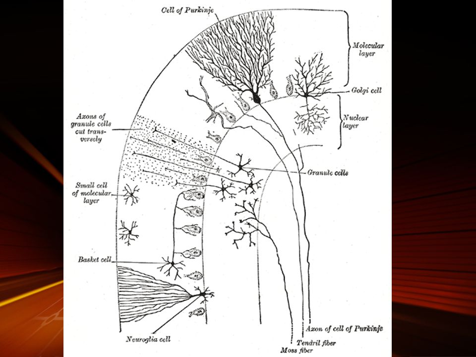
Cerebellar Cortex The cerebellar cortex has three layers: an outer molecular layer, a central layer of large Purkinje cells, and an inner granule layer.

30
The molecular layer Location: lies directly below the pia mater and contains superficially located stellate cells, dendrites of Purkinje cells, basket cells, and unmyelinated axons from the granular layer.

32
The Purkinje cell layer contains the large, flask-shaped Purkinje cells, which are present only in the cerebellum. axon: Dendrite:

34
The granular layer (the deepest layer) consists of small granule cells and golgi cells.
 consists of small granule cells and golgi cells.")
38
Spinal cord In the transverse section of the spinal cord, the gray matter has the shape of a butterfly or an H

41
Spinal cord white matter

42
Ganglia Ganglia are ovoid structures containing neuronal cell bodies and glial cells supported by connective tissue. Because they serve as relay stations to transmit nerve impulses, one nerve enters and another exits from each ganglion. The direction of the nerve impulse determines whether the ganglion will be a sensory or an autonomic ganglion.

43
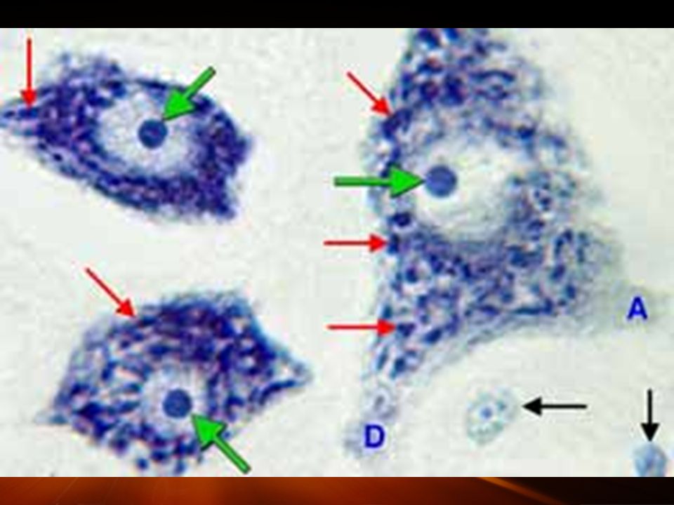
Sensory Ganglia Sensory ganglia receive afferent impulses that go to the central nervous system. cranial ganglia spinal ganglia. The latter comprise large neuronal cell bodies with prominent fine Nissl bodies surrounded by abundant small glial cells called satellite cells.

45
Autonomic Ganglia Autonomic ganglia appear as bulbous dilatations in autonomic nerves. Some are located within certain organs, especially in the walls of the digestive tract. Autonomic ganglia usually have multipolar neurons. As with craniospinal ganglia, autonomic ganglia have neuronal perikaryons with fine Nissl bodies.

47
Meninges The outermost layer of the meninges is the dura mater, the intermediat e layer is the arachnoid, and the innermost or intimate layer of the meninges is the pia mater

51
Dura Mater The dura mater covering the brain is a dense, collagenous connective tissue.

52
Arachnoid The arachnoid is the intermediate layer of the meninges.The arachnoid layer of the meninges is avascular, although blood vessels course through it.

53
Pia Mater Pia mater, the innermost highly vascular layer of the meninges, is in close contact with the brain.

54
Blood–brain Barrier The blood–brain barrier is a functional barrier that prevents the passage of some substances, such as antibiotics and chemical and bacterial toxic matter, from the blood to nerve tissue.

55
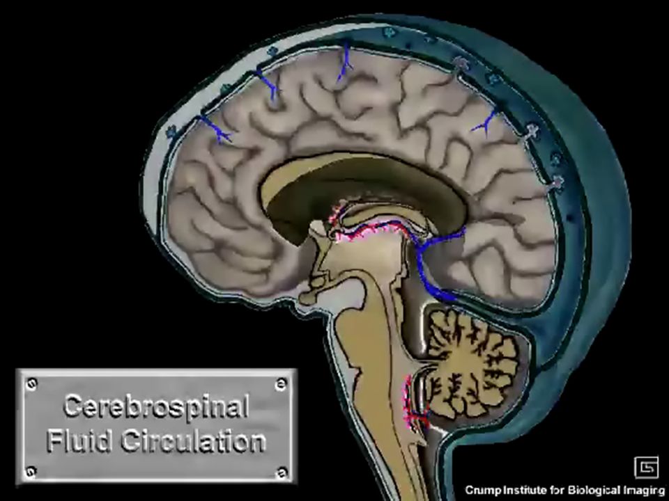
Choroid Plexus & Cerebrospinal Fluid The choroid plexus consists of invaginated folds of pia mater, rich in dilated fenestrated capillaries.

56
Each plexus consists of a tortuous loop or tuft of capillaries, overlain by a neatly cuboidal epithelial covering. The plexi protrude into the fluid-filled lumen of the ventricle

57
Small cells closely associated with neurons in peripheral ganglia may be called: A satellite cells B pyramidal cells C postganglionic cells D autonomic cells Cell bodies of the peripheral receptor neurons associated with spinal sensory nerve roots are located: A near the peripheral receptor organ (in skin or muscle). B in spinal gray matter. C in dorsal root ganglia. D two of the above. White matter appears white because of: A the absence of blood vessels. B the absence of glial cells. C the presence of many collagen fibers. D the presence of many myelinated axons. Epineurium and perineurium are names for the connective tissue which ensheaths peripheral nerves and axon bundles within nerves. True. False. Myelin is an extracellular, secretory product of Schwann cells. True. False. Myelin is the cell membrane of the Schwann cell. Quiz

58
A typical peripheral mixed nerve includes all of the following EXCEPT: A motor axons. B connective tissue of the perineurium and/or endoneurium. C Schwann cells. D sensory axons. E astrocytes. Dura mater consists of: A loose connective tissue with CSF as ground substance. B dense fibrous connective tissue. C white matter. D gray matter. The arachnoid layer of the meninges consists of: loose connective tissue with CSF as ground substance. A dense fibrous connective tissue. B white matter. C gray matter.

60
Optical test

Similar presentations


>")


 It is phylogenetically newest and structurally most complex. Neocortex is.>")


 Central Nervous System Communication and coordination system of the body Seat of intellect and reasoning Consists of the.>")
 Brain and spinal cord Peripheral Nervous System (PNS) ◦ nerves.>")

