Download presentation
Presentation is loading. Please wait.

1
Vasculitis CVS 7 Hisham Alkhalidi

2
Lecture 7 Vasculitis – pathology of polyarteritis nodosa, giant cell arteritis and Buerger’s disease. ANCA mediated vasculitis (Wegener’s granulomatosis and hypersensitivity/ leukocytoclastic vasculitis)
")
3
Vascular inflammatory injury,
Vasculitis Vascular inflammatory injury, often with necrosis

4
Vasculitis Causes immune-mediated :
Immune complex deposition Antineutrophil cytoplasmic antibodies (ANCAs) Anti-endothelial cell antibodies invasion of vascular walls by infectious pathogens Physical and chemical injury Immun complex: Hepatitib-B & AB in PAN, Drugs like penicillin (foreign material or durg modified self protiens) Circulating immune (antigen-antibody) complexes may also be seen-for example, DNA-anti-DNA complexes in systemic lupus erythematosus (SLE)-associated vasculitis Infections can also indirectly induce a noninfectious vasculitis, for example, by generating immune complexes or triggering cross-reactivity Antiendothelial : Kawasaki
 Anti-endothelial cell antibodies. invasion of vascular walls by infectious pathogens. Physical and chemical injury. Immun complex: Hepatitib-B & AB in PAN, Drugs like penicillin (foreign material or durg modified self protiens) Circulating immune (antigen-antibody) complexes may also be seen-for example, DNA-anti-DNA complexes in systemic lupus erythematosus (SLE)-associated vasculitis. Infections can also indirectly induce a noninfectious vasculitis, for example, by generating immune complexes or triggering cross-reactivity. Antiendothelial : Kawasaki.")
6
Giant-Cell (Temporal) Arteritis
The most common Chronic, typically granulomatous inflammation of large to small-sized arteries Principally affects the arteries in the head-especially the temporal arteries Rarely the aorta (giant-cell aortitis) -but also the vertebral and ophthalmic arteries
 -but also the vertebral and ophthalmic arteries.")
7
Giant-Cell (Temporal) Arteritis
Unknown cause Likely immune origin, T cell-mediated An immune origin is supported by the characteristic granulomatous response with associated helper T cells, a correlation with certain major histocompatibility complex (MHC) class II haplotypes, and a therapeutic response to steroids. The extraordinary predilection for a single vascular site (temporal artery) remains unexplained
 class II haplotypes, and a therapeutic response to steroids. The extraordinary predilection for a single vascular site (temporal artery) remains unexplained.")
8
Nodularity, thickness and firm vessel
Can be segmental process

9
The granuloma is centered on the interanl elastic lamina

10
Nodular intimal thickness
Can be healed (remember ATCHscle) , you don’t have to see granuloma
 , you don’t have to see granuloma.")
11
Giant-Cell (Temporal) Arteritis Clinical features
> 50 years of age Vague symptoms: Fever, fatigue and weight loss May involve facial pain or headache Most intense along the course of the superficial temporal artery, which is painful to palpation Ocular symptoms (associated with involvement of the ophthalmic artery) abruptly appear in about 50% of patients Female predominance these range from diplopia to complete vision loss. Plymyalgaia rheumatica association: myalgia, arhtaralgia and fever
 abruptly appear in about 50% of patients. Female predominance. these range from diplopia to complete vision loss. Plymyalgaia rheumatica association: myalgia, arhtaralgia and fever.")
12
Giant-Cell (Temporal) Arteritis
- Definite diagnosis depends on: biopsy of an adequate segment and histological confirmation - Treatment: corticosteroids However, because temporal arteritis is extremely segmental, adequate biopsy requires at least a 2- to 3-cm length of artery; even then, a negative biopsy result does not exclude the diagnosis

14
Polyarteritis Nodosa Systemic Small or medium-sized muscular arteries
But not arterioles, capillaries, or venules Typically involving renal and visceral vessels but sparing the pulmonary circulation

15
Biopsy is needed for Dx with a mixed infiltrate of neutrophils, eosinophils, and mononuclear cells, frequently accompanied by fibrinoid necrosis Thrombosis, transmural arrow NOT involved Sharply demarcated

16
Polyarteritis Nodosa all stages of activity (from early to late) may coexist in different vessels or even within the same vessel Later, the acute inflammatory infiltrate is replaced by fibrous (occasionally nodular) thickening of the vessel wall that can extend into the adventitia Can lead to an aneurysm suggesting ongoing and recurrent pathogenic insults
 thickening of the vessel wall that can extend into the adventitia. Can lead to an aneurysm. suggesting ongoing and recurrent pathogenic insults.")
17
Polyarteritis Nodosa Clinical picture
Largely young adults Typically episodic, with long symptom-free intervals Because the vascular involvement is widely scattered, the clinical findings may be varied and puzzling The course can vary from acute to chronic

18
Polyarteritis Nodosa Clinical picture
Fever and weight loss Examples on systemic involvement: Renal (arterial) involvement is common and a major cause of death Hypertension, usually developing rapidly Abdominal pain and melena (bloody stool) Diffuse muscular aches and pains Peripheral neuritis Biopsy is often necessary to confirm the diagnosis caused by vascular GI lesions
 involvement is common and a major cause of death. Hypertension, usually developing rapidly. Abdominal pain and melena (bloody stool) Diffuse muscular aches and pains. Peripheral neuritis. Biopsy is often necessary to confirm the diagnosis. caused by vascular GI lesions.")
19
Polyarteritis Nodosa No association with ANCA
Some 30% of patients with PAN have hepatitis B antigenemia If untreated, the disease is fatal in most cases Therapy with corticosteroids and other immunosuppressive therapy results in remissions or cures in 90%

20
Polyarteritis Nodosa Complications
Vessel rupture Impaired perfusion: Ulcerations Infarcts Ischemic atrophy (not infarction) Haemorrhages in the distribution of affected vessels may be the first sign of disease Like other vasculitis
 Haemorrhages in the distribution of affected vessels may be the first sign of disease. Like other vasculitis.")
22
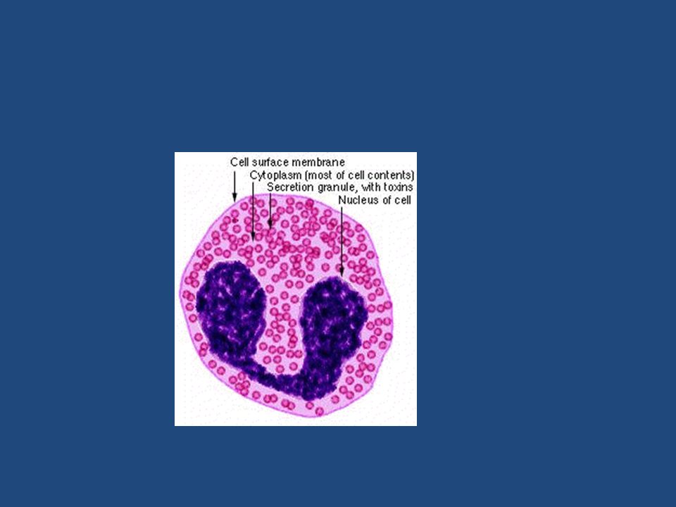
c-ANCA

23
p-ANCA

24
Antineutrophil Cytoplasmic Antibodies
Cytoplasmic localization (c-ANCA) -> the most common target antigen is proteinase-3 (PR3) typical of Wegener granulomatosis Perinuclear localization (p-ANCA) -> most of the autoantibodies are specific for myeloperoxidase (MPO) microscopic polyangiitis and Churg-Strauss syndrome ANCAs serve as useful diagnostic markers for the ANCA-associated vasculitides Their levels can reflect the degree of inflammatory activity A plausible mechanism for ANCneutrophil A vasculitis is: Neutrophil release of PR3 and MPO (e.g., in the setting of infections) incites ANCA formation in a susceptible host. Some underlying disorder (e.g., infection, endotoxin exposure, etc.) elicits inflammatory cytokines, such as TNF, that result in surface expression of PR3 and MPO on neutrophils and other cell types. ANCAs react with these cytokine-primed cells and either cause direct injury (e.g., to endothelium) or induce activation (e.g., in neutrophils). ANCA-activated neutrophils degranulate and also cause injury by the release of reactive oxygen species, engendering EC toxicity and other direct tissue injury.
 -> the most common target antigen is proteinase-3 (PR3) typical of Wegener granulomatosis. Perinuclear localization (p-ANCA) -> most of the autoantibodies are specific for myeloperoxidase (MPO) microscopic polyangiitis and Churg-Strauss syndrome. ANCAs serve as useful diagnostic markers for the ANCA-associated vasculitides. Their levels can reflect the degree of inflammatory activity. A plausible mechanism for ANCneutrophil A vasculitis is: Neutrophil release of PR3 and MPO (e.g., in the setting of infections) incites ANCA formation in a susceptible host. Some underlying disorder (e.g., infection, endotoxin exposure, etc.) elicits inflammatory cytokines, such as TNF, that result in surface expression of PR3 and MPO on neutrophils and other cell types. ANCAs react with these cytokine-primed cells and either cause direct injury (e.g., to endothelium) or induce activation (e.g., in neutrophils). ANCA-activated neutrophils degranulate and also cause injury by the release of reactive oxygen species, engendering EC toxicity and other direct tissue injury.")
26
Microscopic Polyangiitis
Necrotizing vasculitis that generally affects capillaries as well as arterioles and venules of a size smaller than those involved in PAN Rarely, larger arteries may be involved All lesions of microscopic polyangiitis tend to be of the same age in any given patient Necrotizing glomerulonephritis (90% of patients) and pulmonary capillaritis are particularly common skin, mucous membranes, lungs, brain, heart, GI tract, kidneys, and muscle can all be involved Disseminated vascular lesions of hypersensitivity angiitis can also occur as a presentation of other disorders (e.g., Henoch-Schönlein purpura, essential mixed cryoglobulinemia, and vasculitis associated with connective tissue disorders).
 and pulmonary capillaritis are particularly common. skin, mucous membranes, lungs, brain, heart, GI tract, kidneys, and muscle can all be involved. Disseminated vascular lesions of hypersensitivity angiitis can also occur as a presentation of other disorders (e.g., Henoch-Schönlein purpura, essential mixed cryoglobulinemia, and vasculitis associated with connective tissue disorders).")
27
Microscopic Polyangiitis Pathogenesis
In many cases, an antibody response to antigens such as drugs (e.g., penicillin), microorganisms (e.g., streptococci), heterologous proteins, or tumor proteins is the presumed cause This can result in immune complex deposition, or it may trigger secondary immune responses p-ANCAs are present in more than 70% of patients . Recruitment and activation of neutrophils within a particular vascular bed are probably responsible for the manifestations of the disease.
, microorganisms (e.g., streptococci), heterologous proteins, or tumor proteins is the presumed cause. This can result in immune complex deposition, or it may trigger secondary immune responses. p-ANCAs are present in more than 70% of patients. . Recruitment and activation of neutrophils within a particular vascular bed are probably responsible for the manifestations of the disease.")
28
granulomatous inflammation is absent

29
Microscopic Polyangiitis
Depending on the organ involved, major clinical features include: Hemoptysis Hematuria and proteinuria Bowel pain or bleeding Muscle pain or weakness Palpable cutaneous purpura

31
Wegener Granulomatosis
Triad: Acute necrotizing granulomas of the upper and lower respiratory tract (lung), or both Necrotizing or granulomatous vasculitis affecting small to medium-sized vessels (most prominent in the lungs and upper airways) Focal necrotizing, often crescentic, glomerulitis No causative agent, although immune complexs occasionally seen clinically, this resembles PAN except that there is also respiratory involvement. Kidney severity is variable
, or both. Necrotizing or granulomatous vasculitis affecting small to medium-sized vessels (most prominent in the lungs and upper airways) Focal necrotizing, often crescentic, glomerulitis. No causative agent, although immune complexs occasionally seen. clinically, this resembles PAN except that there is also respiratory involvement. Kidney severity is variable.")
33
Wegener Granulomatosis
40-50 years Without Rx -> 80% die With Rx > 90% live (not cured) The Rx -> immunosuppression M>F
 The Rx -> immunosuppression. M>F.")
36
Churg-Strauss syndrome
Eosinophil-rich and granulomatous inflammation involving the respiratory tract and necrotizing vasculitis affecting small vessels Associated with asthma and blood eosinophilia Associated with p-ANCAs. Lung and splenic vessels

37
Churg-Strauss syndrome
strong association with Allergic rhinitis Bronchial asthma Eosinophilia Vessels in the lung, heart, spleen … are frequently involved by: intravascular and extravascular granulomas infiltration of vessels and perivascular tissues by eosinophils is striking . However, an early, prevasculitic phase marked by tissue infiltration by eosinophils without overt vasculitis may be present in some cases. Severe renal disease is infrequent. Coronary arteritis and myocarditis are the principal causes of morbidity and mortality. The disorder is thought to result from hyperresponsiveness to an allergic stimulus

39
Thromboangiitis obliterans (Buerger disease)
")
40
Thromboangiitis obliterans (Buerger disease)
Segmental, thrombosing acute and chronic inflammation of medium-sized and small arteries Tibial and radial arteries, with occasional secondary extension into extremity veins and nerves Unknown cause GENETIC OR TOBACco The strong relationship to cigarette smoking is thought to involve direct toxicity to endothelium by some tobacco products, or an idiosyncratic immune response to the same agents. Most Buerger patients have hypersensitivity to intradermally injected tobacco extracts, and their vessels show impaired endothelium-dependent vasodilation when challenged with acetylcholine. Genetic influences are suggested by an increased prevalence in certain ethnic groups (Israeli, Indian subcontinent, Japanese) and an association with certain MHC haplotypes.
 and an association with certain MHC haplotypes.")
41
sharply segmental acute and chronic vasculitis
Thrombus Abscess microabscess composed of neutrophils surrounded by granulomatous inflammation Large lesions organize and recanalize

42
Thromboangiitis obliterans Clinical picture
Heavy smoker Begins before the age of 35 years Superficial nodular phlebitis Cold sensitivity of the Raynaud type in the hands (see figure below) Pain in the instep of the foot induced by exercise (so-called instep claudication)
 Pain in the instep of the foot induced by exercise (so-called instep claudication)")
43
Thromboangiitis obliterans
In contrast to the vascular insufficiency caused by atherosclerosis, in Buerger disease the insufficiency tends to be accompanied by severe pain, even at rest, related undoubtedly to the neural involvement Chronic ulcerations of the toes, feet, or fingers may appear, perhaps followed in time by frank gangrene Abstinence from cigarette smoking in the early stages of the disease often brings dramatic relief from further attacks

Similar presentations


 Dr. Raid Jastania. Vasculitis Inflammation of the walls of the vessels Causes of inflammation: –Infectious, physical, chemical,>")


 Aneurysms & Dissections Veins & Lymphatics Tumors.>")


