Download presentation
Presentation is loading. Please wait.

1
Objectives: 1.diagram, explain and/or define terms on page 86 2.layers of tear film 3.components of the eye, its three main layers, and the mechanisms of focusing 4.the origin, function, flow and fate of aqueous humor

2
dura mater subarachnoid space optic nerve blind spot lens

3
dura mater subarachnoid space optic nerve blind spot lens iris

4
dura mater subarachnoid space optic nerve blind spot pupil cornea sclera lens ciliary body ciliary processes limbus

5
dura mater subarachnoid space optic nerve lens anterior chamber posterior chamber vitreous chamber

6
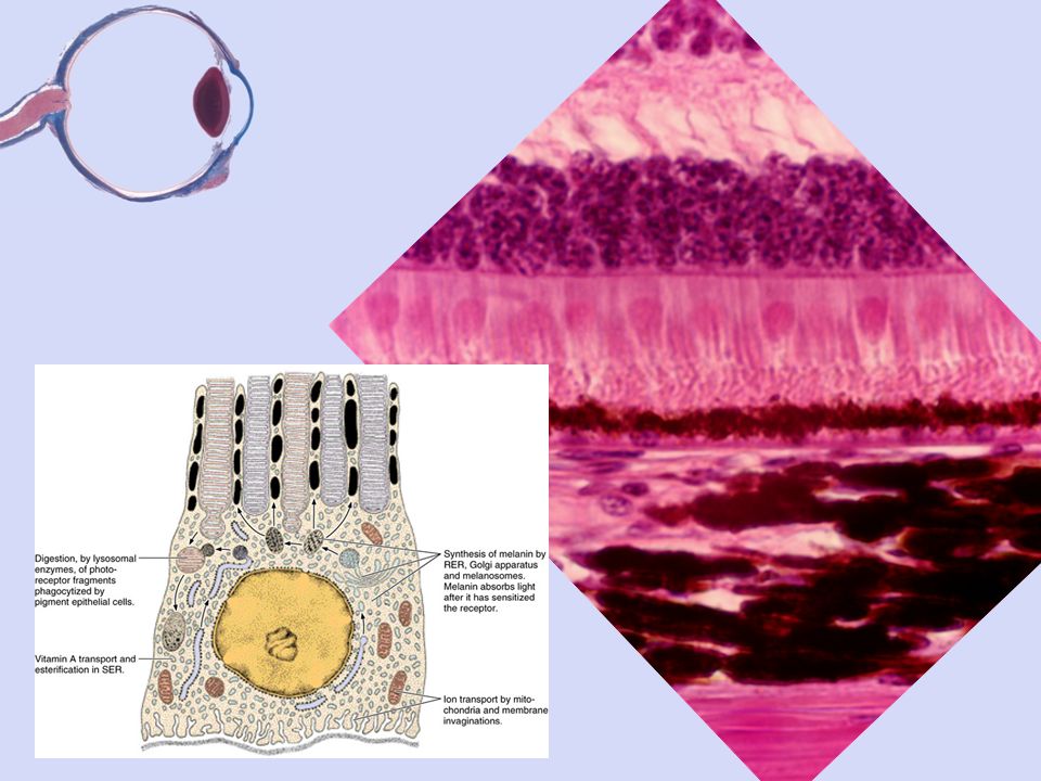
Three tunics or layers of the eye … -outer fibrous tunic or connective tissue layer -cornea -limbus -sclera - middle vascular tunic -choroid layer beneath the retina -ciliary body -ciliary processes -most of iris - neural layer -sensory layer (visual retina) -pigmented layer of retina -ora serrata -pigmented and non-pigmented layers of the ciliary processes -pigmented layer on the posterior surface of the iris
 -pigmented layer of retina -ora serrata -pigmented and non-pigmented layers of the ciliary processes -pigmented layer on the posterior surface of the iris")
7
Three tunics or layers of the eye … -outer fibrous tunic or connective tissue layer -cornea -limbus -sclera - middle vascular tunic -choroid layer beneath the retina -ciliary body -ciliary processes -most of iris - neural layer -sensory layer (visual retina) -pigmented layer of retina -ora serrata -pigmented and non-pigmented layers of the ciliary processes -pigmented layer on the posterior surface of the iris Concepts: 1.The posterior retina is a bi-layer consisting of the visual retina and the pigmented layer of the retina. They have only a loose connection. Separation (by trauma or degeneration of connective tissues) leads to detached retina. 2.A combination of blood vessels from the vascular tunic and the non-pigmented cells of the retina produce a tissue similar to the choroid plexus of the brain, called the ciliary processes. These produce aqueous humor, a CSF-like fluid in the eye. 3.At the time of death of an animal, blood pressure drops off, intraocular pressure remains, so blood is forced from the choroid layer, producing an artifactual thinness to its appearance. 4.Eye is composed of both dense and soft, porous tissues. During processing for histology, separation and breakage of tissues is common. Lenses usually shatter. 5.Excess aqueous humor in the eye may increase intra-ocular pressure, occluding blood flow and creating a condition known as glaucoma.
 leads to detached retina. 2.A combination of blood vessels from the vascular tunic and the non-pigmented cells of the retina produce a tissue similar to the choroid plexus of the brain, called the ciliary processes. These produce aqueous humor, a CSF-like fluid in the eye. 3.At the time of death of an animal, blood pressure drops off, intraocular pressure remains, so blood is forced from the choroid layer, producing an artifactual thinness to its appearance. 4.Eye is composed of both dense and soft, porous tissues. During processing for histology, separation and breakage of tissues is common. Lenses usually shatter. 5.Excess aqueous humor in the eye may increase intra-ocular pressure, occluding blood flow and creating a condition known as glaucoma..")
9
3 components of tear film …

10
cornea limbus third eyelid eyelid hyaline cartilage

11
cornea limbus third eyelid eyelid hyaline cartilage

12
cornea limbus third eyelid eyelid hyaline cartilage

13
lymphocyte infiltration mucous cells stratified columnar epithelium

14
cornea stroma endothelium epithelium active pump avascular rich with free nerve endings

15
bacterial pink eye

16
lens ciliary body ciliary processes zonule fibers sclera limbus cornea iris conjunctiva

19
lens zonule fibers iris PC AC

20
sclera choroid visual retina pigmented layer of the retina

21
nuclei of the rod and cone cells layer with rods and cones pigmented layer of the retina choroid sclera

23
ora serrata macula or fovea

25
tapetum

26
In conclusion … -how do we focus an image? -how do we adjust light level? -how do we nourish the avascular cornea? -how do we maintain the overall shape of the eye? -how do birds of prey see so amazingly well?

Similar presentations


 1. Cornea 2. Sclera Middle Tunic (pg. 470-474) 3. Choroid Coat 4. Ciliary Body 5. Lens & Accommodation 6. Aqueous.>")


>")


. 2. *The sclera is the.>")


>")