Download presentation
Presentation is loading. Please wait.

1
The Circulatory System Part II

2
2 Heartbeat Systole: contraction Diastole: filling Normal rate: 60-100 Slow: bradycardia Fast: tachycardia ***Note: blood goes to RA, then RV, then lungs, then LA, then LV, then body; but the fact that a given drop of blood passes through the heart chambers sequentially does not mean that the four chambers contract in that order; the 2 atria always contract together, followed by the simultaneous contraction of the 2 ventricles Definition: a single sequence of atrial contraction followed by ventricular contraction See http://www.geocities.com/Athens/Forum/6100/1heart.html

3
#1. I can name the components of the conduction system of the heart, and trace the conduction pathway.

4
4 Electrical conduction system: (Explanation in next slides) specialized cardiac muscle cells that carry impulses throughout the heart musculature, signaling the chambers to contract in the proper sequence
 specialized cardiac muscle cells that carry impulses throughout the heart musculature, signaling the chambers to contract in the proper sequence")
5
5 Conduction system SA node (sinoatrial) In wall of RA Sets basic rate: 70-80 Is the normal pacemaker Impulse from SA to atria Impulse also to AV node via internodal pathway AV node In interatrial septum
 In wall of RA Sets basic rate: Is the normal pacemaker Impulse from SA to atria Impulse also to AV node via internodal pathway AV node In interatrial septum")
6
6 Conduction continued SA node through AV bundle (bundle of His) Into interventricular septum Divides R and L bundle branches become subendocardial branches (“Purkinje fibers”) Contraction begins at apex
 Into interventricular septum Divides R and L bundle branches become subendocardial branches ( Purkinje fibers ) Contraction begins at apex")
7
#2. I can draw a diagram of a normal electrocardiogram tracing; name the individual waves and intervals, and indicate what each represents.

8
8

9
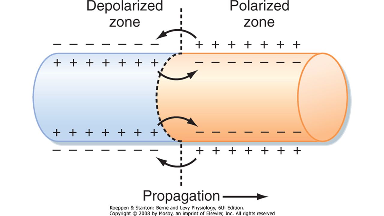
9 “EKG” ( or ECG, electrocardiogram) Electrical depolarization is recorded on the body surface by up to 12 leads Pattern analyzed in each lead P wave=atrial depolarization QRS=ventricular depolarization T wave=ventricular repolarization
 Electrical depolarization is recorded on the body surface by up to 12 leads Pattern analyzed in each lead P wave=atrial depolarization QRS=ventricular depolarization T wave=ventricular repolarization")
10
10 12 lead EKG

11
#3. I can name some abnormalities that can be detected on an ECG tracing.

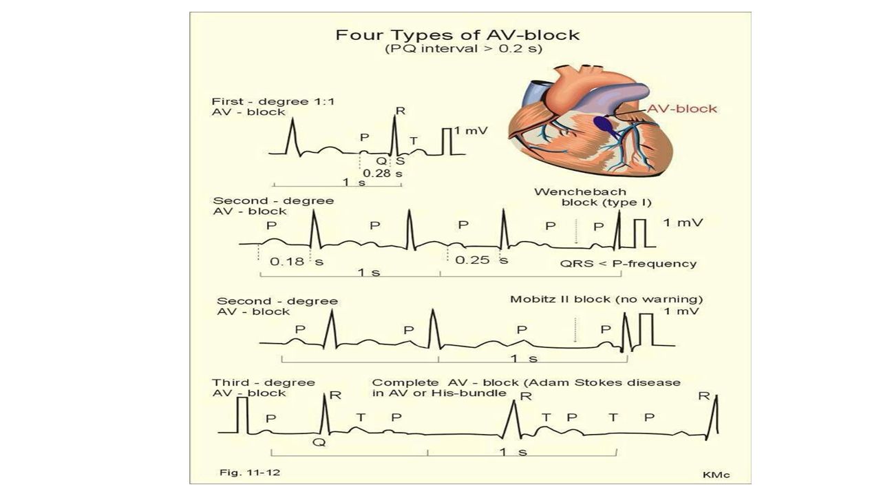
13
Heart Blocks Second Degree:Signals from AV node to ventricles are blocked/bradycardia. Third Degree:Signals from SA node to AV node and ventricles are blocked/severe bradycardia.

15
Ventricular Tachycardia Fast heartbeat originating in the ventricles.

16
Atrial Fibrillation Irregular, fast heartbeat originating in the atria.

17
Ventricular Fibrillation Most serious. Ventricles quiver and can not pump blood.

18
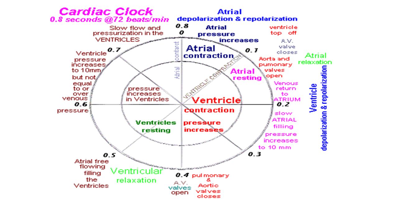
#4. I can describe the timing and events of the cardiac cycle.

20
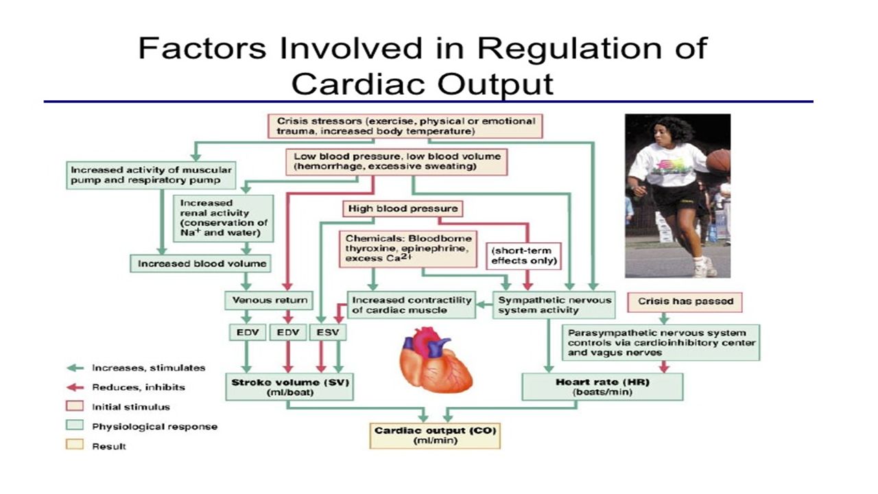
#5. I can name and explain the effects of the various factors involved in the regulation of heart rate.

22
#6. I can explain the role of the autonomic nervous system in regulating cardiac output.

23
23 Autonomic innervation Sympathetic Increases rate and force of contractions Parasympathetic (branches of Vagus n.) Slows the heart rate http://education.med.nyu.edu/courses/old/physiology/courseware/ekg_pt1/EKGseq.html For a show on depolarization:
 Slows the heart rate For a show on depolarization:")
25
25 Artificial Pacemaker

Similar presentations


, the atrioventricular node.>")

 node Atrioventricular (AV) node.>")

 – is found in the right atrium and initiates.>")


