Download presentation
Presentation is loading. Please wait.

1
بسم الله الرحمن الرحيم

2
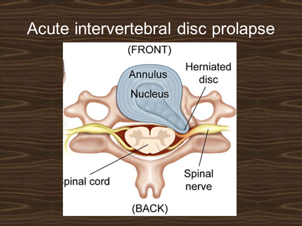
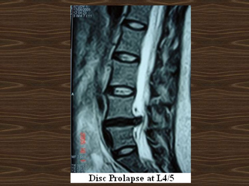
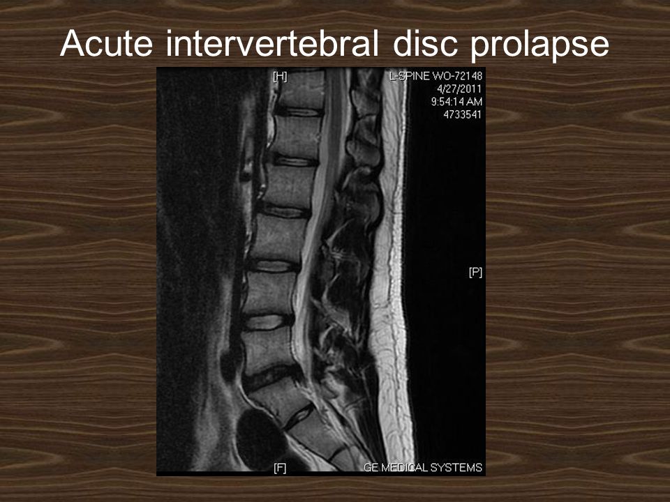
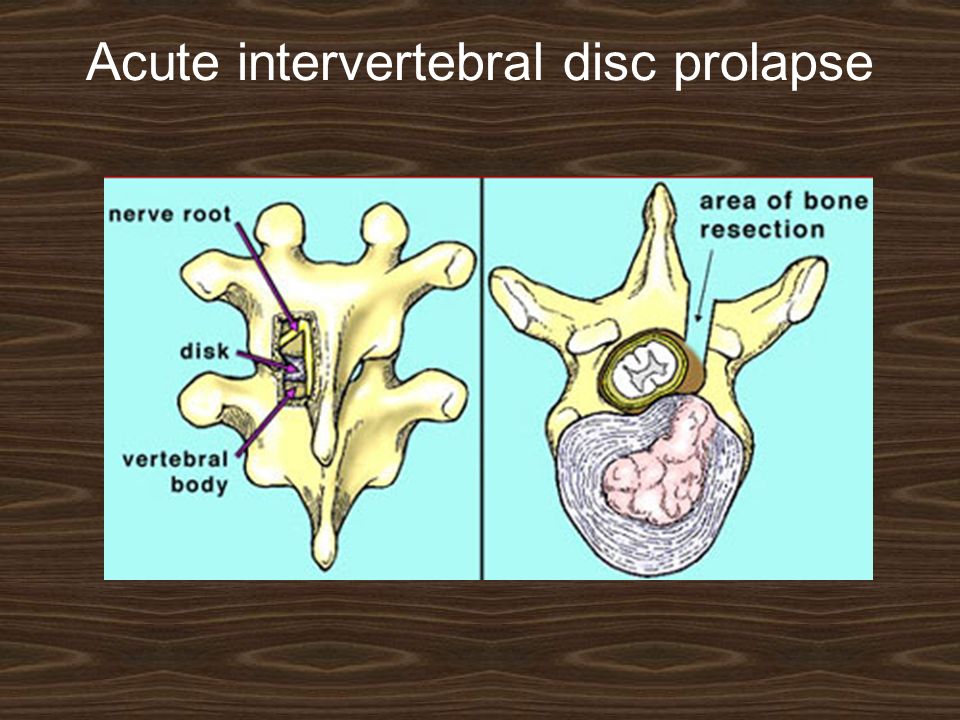
Acute intervertebral disc prolapse

4
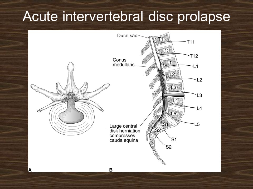
more common in adults 20–45 years. If the pain is felt in the buttock and lower limb, this is called sciatica. Cauda equina compression is rare but may cause urinary retention and perineal numbness (saddle anaesthesia). The patient has a list to one side (sciatic scoliosis). All back movements are restricted.
. The patient has a list to one side (sciatic scoliosis). All back movements are restricted..")
5
Acute intervertebral disc prolapse Sciatica

6
Acute intervertebral disc prolapse There is often tenderness in the midline of the low back, and para-vertebral muscle spasm. Straight leg rising is restricted and painful on the affected side. Sometimes raising the unaffected leg causes acute sciatic tension on the painful side (crossed sciatic tension). Neurological examination may show muscle weakness (and, later, wasting), diminished reflexes and sensory loss related to the affected level. Cauda equina compression causes urinary retention and sensory loss over the sacrum.
. Neurological examination may show muscle weakness (and, later, wasting), diminished reflexes and sensory loss related to the affected level. Cauda equina compression causes urinary retention and sensory loss over the sacrum..")
7
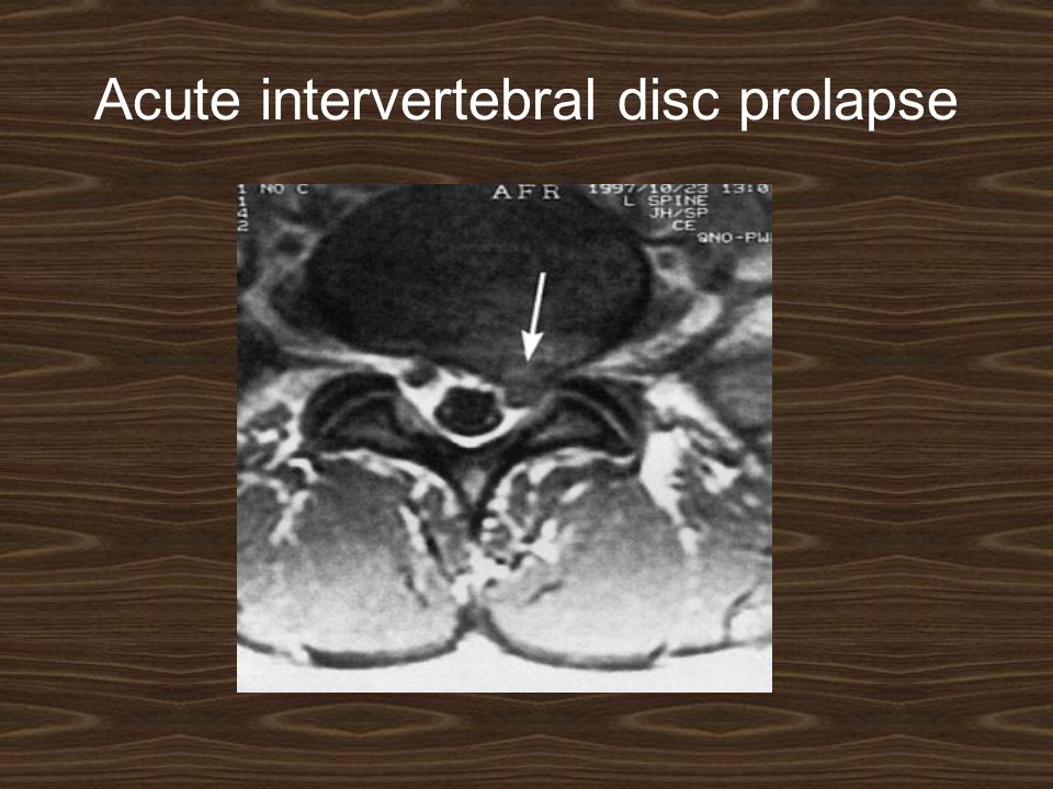
Acute intervertebral disc prolapse

10
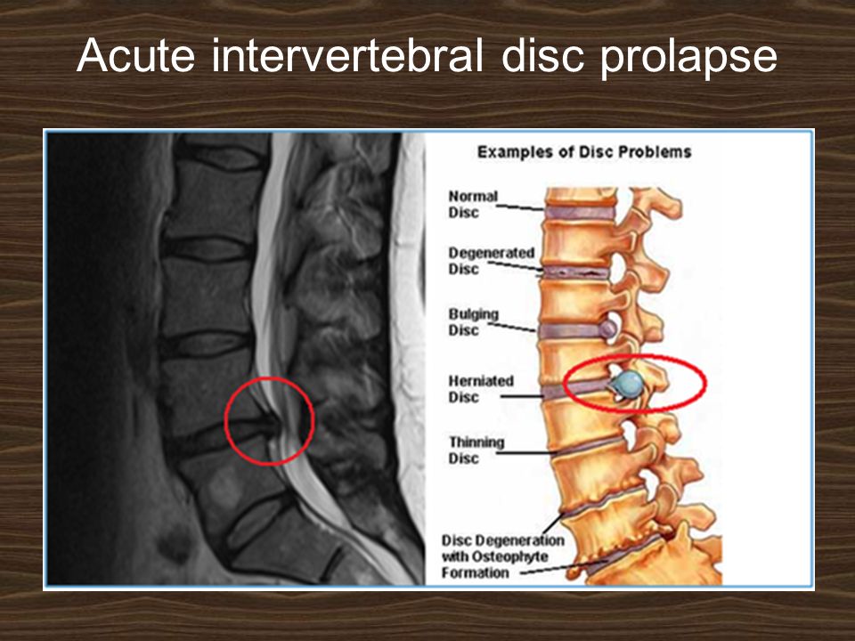
X-rays: show an abnormal narrowed disc space and to exclude bone diseases. After several attacks the disc space may be narrowed and small osteophytes appear. CT and MRI are more reliable than myelography.

11
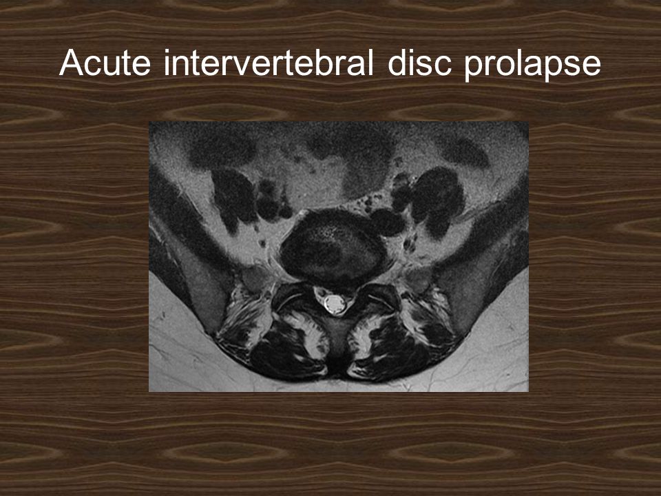
Acute intervertebral disc prolapse

19
Differential diagnosis: 1. Inflammatory disorders such as infection or ankylosing spondylitis, cause severe stiffness, a raised ESR and erosive changes on x-ray. 2. Vertebral tumours cause severe pain, marked muscle spasm and pain through the night. With metastases the patient is ill, the ESR is raised and the x-rays show bone destruction or sclerosis. 3. Nerve tumours such as a neurofibroma of the cauda equina May cause sciatica but pain is continuous. Advanced imaging will confirm the diagnosis.

20
Acute intervertebral disc prolapse

21
FEATURES OF CAUDA EQUINA SYNDROME: 1. Bladder and bowel incontinence 2. Perineal numbness (saddle paresthesia). 3. Bilateral sciatica 4. Lower limb weakness 5. Crossed straight-leg raising sign
. 3. Bilateral sciatica 4. Lower limb weakness 5. Crossed straight-leg raising sign.")
22
Acute intervertebral disc prolapse Treatment : 3 Rs: Rest. Removal. Rehabilitation.

23
Acute intervertebral disc prolapse Rest: With an acute attack the patient should be kept in bed. A non-steroidal anti-inflammatory drug and heat is useful. If the symptoms and signs do not improve, an epidural injection of corticosteroid and local anesthetic may help.

24
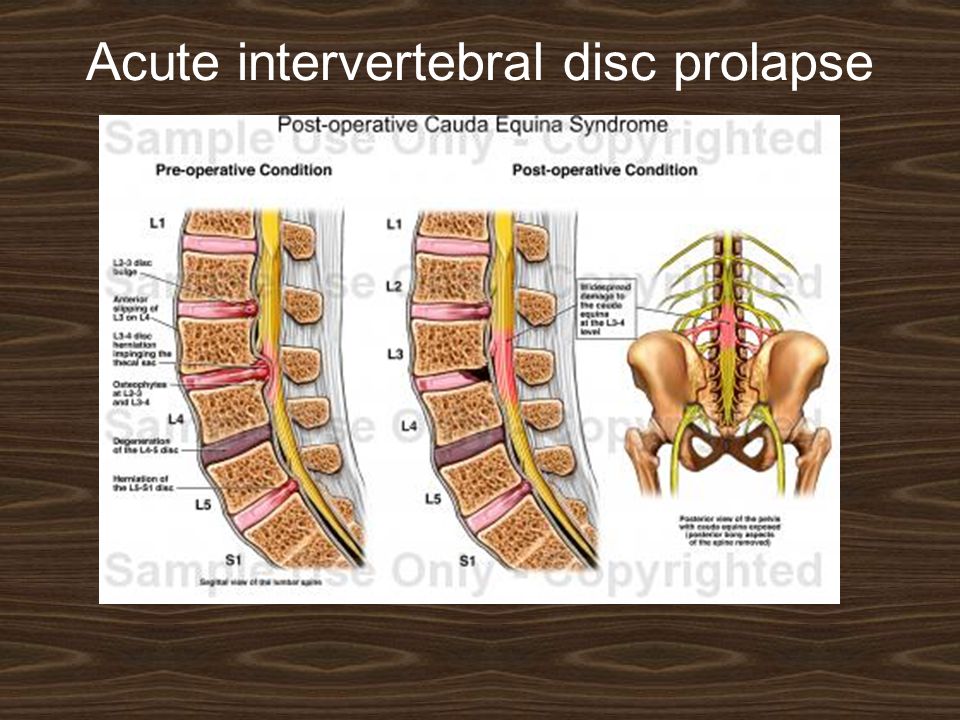
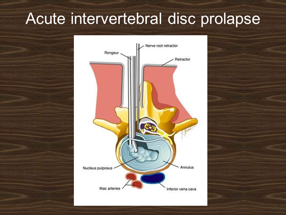
Acute intervertebral disc prolapse RemovalRemoval: The indications for operative removal of a prolapse are: (1) a cauda equina compression syndrome (emergency). (2) neurological deterioration while under conservative treatment. (3) persistent pain and signs of sciatic tension (especially crossed sciatic tension) after 2–3 weeks of conservative treatment.
 neurological deterioration while under conservative treatment. (3) persistent pain and signs of sciatic tension (especially crossed sciatic tension) after 2–3 weeks of conservative treatment..")
25
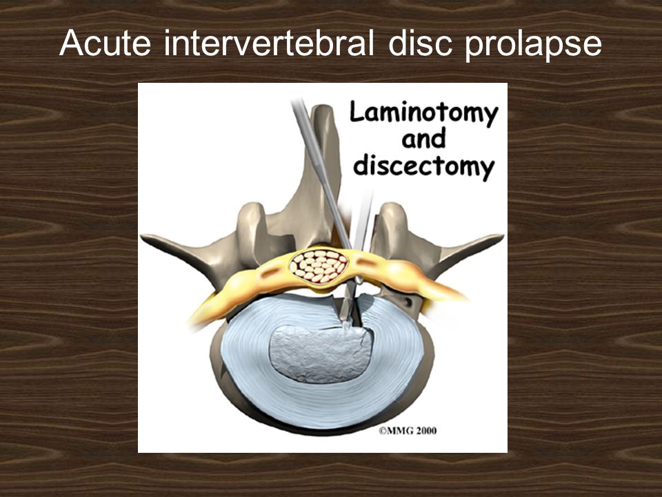
Acute intervertebral disc prolapse Laminotomy and micro-discectomy. Laminotomy: Ligamentum flavum on the relevant side and at the relevant level is removed, if necessary with some margin of the bordering laminae and medial third of the facet joint. The dura and nerve root are then gently retracted towards the midline and disc is excised. Micro-discectomy is essentially similar to the standard posterior operation, except that the exposure is very limited and the procedure is carried out with the aid of an operating microscope. The major postoperative complication is (disciitis). Recurrent prolapse with sciatica is more common and may require revision surgery.
. Recurrent prolapse with sciatica is more common and may require revision surgery..")
26
Acute intervertebral disc prolapse

30
Rehabilitation: After recovery from an acute disc rupture, or disc removal, isometric exercises and physiotherapy are started.

31
PERSISTENT POSTOPERATIVE BACKACHE AND SCIATICA Persistent symptoms after operation may be due to: (1) Residual disc material in the spinal canal. (2) Disc prolapse at another level. (3) Nerve root pressure by a hypertrophic facet joint or a narrow lateral recess (root canal stenosis). Treated by re-operation (revision).
 Disc prolapse at another level. (3) Nerve root pressure by a hypertrophic facet joint or a narrow lateral recess (root canal stenosis). Treated by re-operation (revision)..")
32
CHRONIC INTERVERTEBRAL DISC DEGENERATION This is an age related phenomenon that occurs in over 80 per cent of people who live for more than 50 years and in most cases it is asymptomatic.

33
CHRONIC INTERVERTEBRAL DISC DEGENERATION Pathology: With normal ageing the disc gradually dehydrated. The annulus fibrosus develops fissures. The discs flatten down and bulge slightly beyond the margins of the vertebral bodies. Where they protrude against the ligaments, reactive new bone formation produces bony ridges osteophytic projections which lead to spinal stenosis. The picture as a whole is referred to as spondylosis. Then displacement of the facet joints and this lead to segmental spinal instability.

34
CHRONIC INTERVERTEBRAL DISC DEGENERATION

35
CHRONIC INTERVERTEBRAL DISC DEGENERATION Clinical features: Disc degeneration of itself is usually asymptomatic. Sometimes chronic backache or low-back pain on activity may appear. Clinical features: Disc degeneration of itself is usually asymptomatic. Sometimes chronic backache or low-back pain on activity may appear.

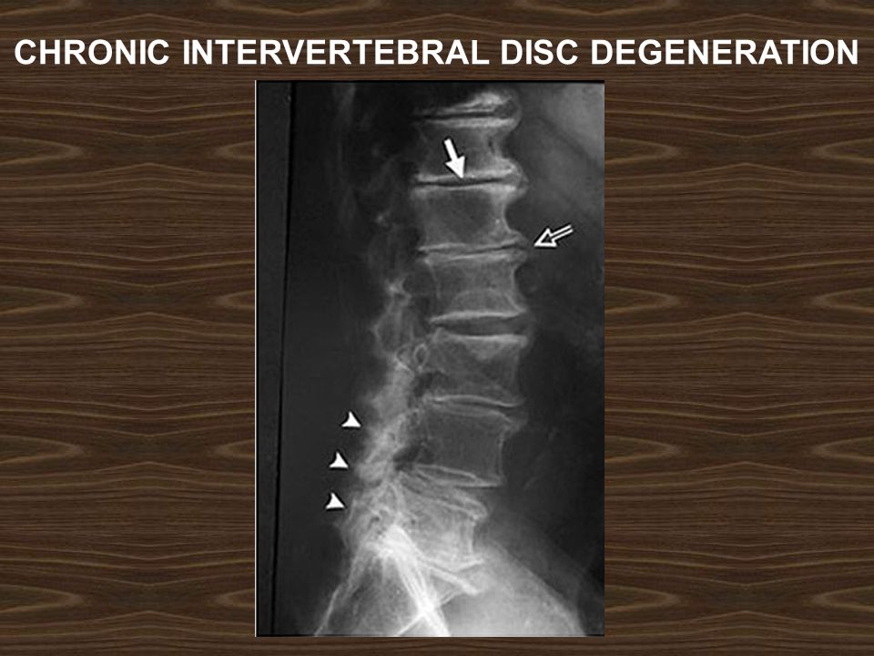
36
CHRONIC INTERVERTEBRAL DISC DEGENERATION X-Rays: Flattening of the disc space and marginal osteophyte formation appear later.

37
CHRONIC INTERVERTEBRAL DISC DEGENERATION

39
MRI: Bulging of the annulus fibrosus in both sagittal and axial projections and diminished thickness and reduced signal intensity (dehydration) of the degenerating disc.
 of the degenerating disc.")
40
CHRONIC INTERVERTEBRAL DISC DEGENERATION

41
Treatment: Asymptomatic lumbar disc degeneration does not need any treatment. Secondary features of disc degeneration, such as spinal stenosis, spinal instability, or spondylolisthesis may needs management, usually operative treatment.

42
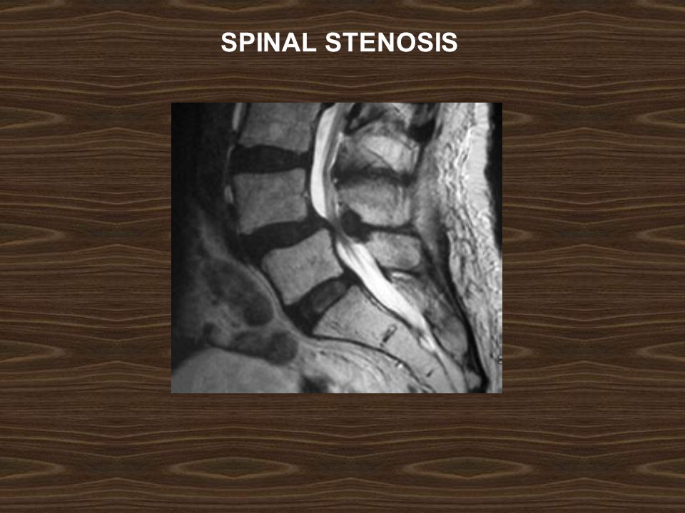
SPINAL STENOSIS Abnormal narrowing of the central canal, the lateral recesses, or the intervertebral foramina to the point where the neural elements are compromised and the patient develops neurological symptoms and signs in the lower limbs.

43
SPINAL STENOSIS

44
The causes of spinal stenosis are: (1) congenital vertebral dysplasia (achondroplasia). (2) chronic disc degeneration. (3) displacement of facet joints. (4) hypertrophy of ligamentum flavum. (5) bone thickening due to Paget's disease. (6) spondylolisthesis. Unilateral narrowing of the intervertebral foramen (root canal stenosis) may result from lateral disc herniation.
 chronic disc degeneration. (3) displacement of facet joints. (4) hypertrophy of ligamentum flavum. (5) bone thickening due to Paget s disease. (6) spondylolisthesis. Unilateral narrowing of the intervertebral foramen (root canal stenosis) may result from lateral disc herniation..")
45
SPINAL STENOSIS Two measurements are used: the mid- sagittal (anteroposterior) diameter and the inter-pedicular (transverse) diameter of the spinal canal. Normally, the diameters are 15 mm for the anteroposterior and 20 mm for the transverse. Anything less than 11 mm for the anteroposterior diameter and 16 mm for the transverse diameter is considered abnormal.

46
SPINAL STENOSIS

49
Clinical features: The patient, usually over 50 years, complains of aching, heaviness, numbness and paresthesia in the thighs and legs; it comes on after standing upright or walking for 5–10 minutes, and is relieved by sitting, squatting or leaning to flex the spine (spinal claudication) and should be differentiated form arterial claudication which occur in atherosclerosis. The patient sometimes has a previous history of disc prolapse, chronic backache or spinal operation.

50
SPINAL STENOSIS Imaging: X-rays will show features of disc degeneration or spondylolisthesis. Measurement of the spinal canal can be carried out on CT and MRI.

51
SPINAL STENOSIS Treatment: Conservative measures, including instruction in spinal posture, may enough. Operative decompression is almost always successful (laminectomy with spinal fusion).
..")
52
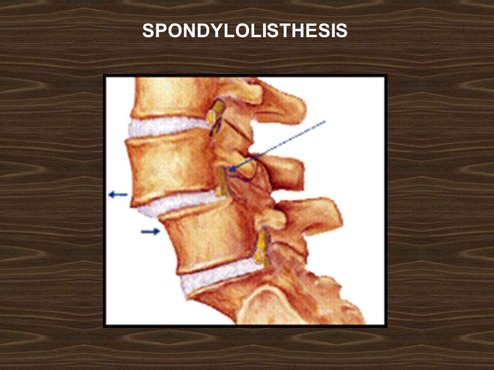
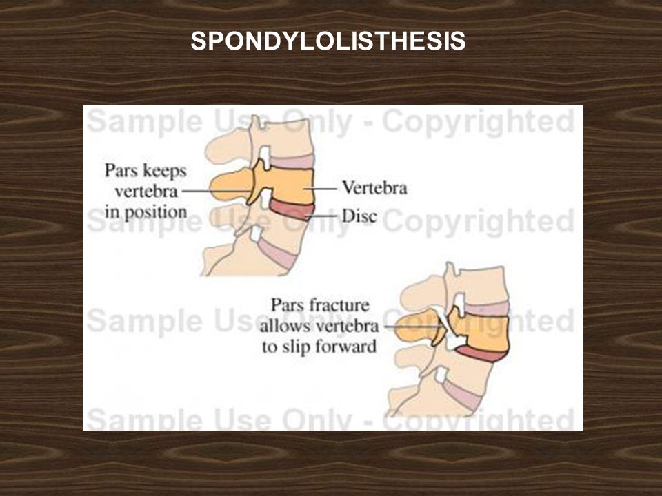
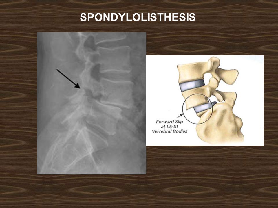
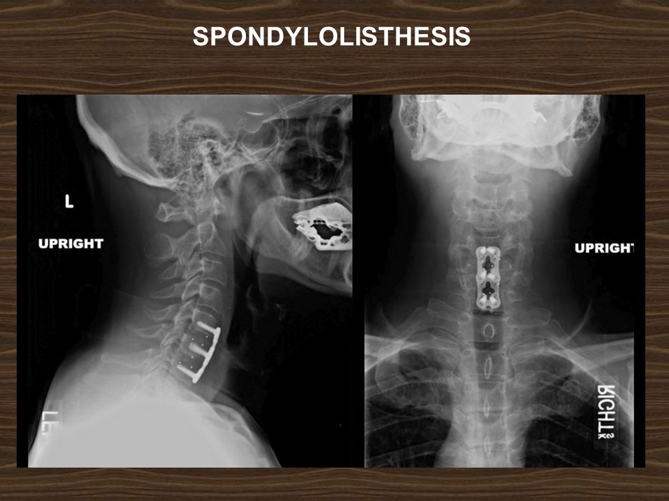
SPONDYLOLISTHESIS Spondylolisthesis means forward translation of one segment of the spine upon another. The shift is nearly always between L4 and L5, or between L5 and the sacrum. Backward translation is called retrolisthesis.

54
RETROLISTHESIS

55
SPONDYLOLISTHESIS Classification: Basically there are six types of spondylolisthesis: 1. Dysplastic (20 %): The superior sacral facets are congenitally defective. Associated anomalies (usually spina bifida occulta) are common. 2. Lytic or isthmic (50 %): In this, there is a defect in the pars interarticularis (spondylolysis). It may be a genetic factor and more common in adolescents.
: The superior sacral facets are congenitally defective. Associated anomalies (usually spina bifida occulta) are common. 2. Lytic or isthmic (50 %): In this, there is a defect in the pars interarticularis (spondylolysis). It may be a genetic factor and more common in adolescents..")
56
SPONDYLOLISTHESIS

57
3. Degenerative (25%): Degenerative changes in the facet joints and the discs permit forward slip (always at L4/L5). 4. Post-traumatic: Unusual fractures may result in destabilization of the lumbar spine.
: Degenerative changes in the facet joints and the discs permit forward slip (always at L4/L5). 4. Post-traumatic: Unusual fractures may result in destabilization of the lumbar spine..")
58
SPONDYLOLISTHESIS 5. Pathological: Bone destruction (e.g. due to tuberculosis or neoplasm) may lead to vertebral slipping. 6. Postoperative (iatrogenic): Occasionally, excessive operative removal of bone like in laminectomy.
 may lead to vertebral slipping. 6. Postoperative (iatrogenic): Occasionally, excessive operative removal of bone like in laminectomy..")
59
SPONDYLOLISTHESIS Pathology: In the common lytic type, the pars interarticularis on both sides is disrupted (spondylolysis), leaving the posterior arch separated from the vertebral body anteriorly; the gap is occupied by fibrous tissue. With stress, spondylolisthesis developed. The degree of slip is measured by the amount of overlap of adjacent vertebral bodies and is usually expressed as a percentage. With slipping there will be spinal stenosis.

62
Clinical features: Spondylolisthesis, may be discovered incidentally during routine x-ray examination which is usually asymptomatic. In children: the condition is usually painless but there is protruding abdomen and peculiar gait.

63
SPONDYLOLISTHESIS Clinical features: In adolescents and adults: intermittent backache is the usual presenting symptom. Patients over 50: are usually women with degenerative spondylolisthesis. They always have backache; some have claudication due to spinal stenosis. On examination the buttocks look flat, a step can often be felt when the fingers are run down the spine.

64
SPONDYLOLISTHESIS X-ray: Lateral views show the forward shift of the vertebra above on the vertebra below. The gap in the pars interarticularis is best seen in the oblique views. CT-scan and MRI are helpful.

65
SPONDYLOLISTHESIS Spondylolysis

66
SPONDYLOLISTHESIS

67
Treatment: Conservative treatment, similar to that for disc prolapse, is helpful. Operative treatment is indicated: (1) if the symptoms are interfere significantly with work; (2) if the slip is more than 50 % and progressing; (3) if neurological signs are significant. The surgery is posterior or anterior intervertebral body fusion.
 if the symptoms are interfere significantly with work; (2) if the slip is more than 50 % and progressing; (3) if neurological signs are significant. The surgery is posterior or anterior intervertebral body fusion..")
68
SPONDYLOLISTHESIS

70
APPROACH TO DIAGNOSIS IN PATIENTS WITH LOW BACK PAIN Careful history taking and examination will uncover one of five pain patterns: 1. Transient backache following muscular activity: This suggests a simple back strain that will respond to a short period of rest followed by gradually increasing exercise.

71
APPROACH TO DIAGNOSIS IN PATIENTS WITH LOW BACK PAIN 2. Sudden, acute pain and sciatica: --- In young people (those under the age of 20) it is important to exclude infection and spondylolisthesis; both produce recognizable x-ray changes. --- Patients aged 20–40 years are more likely to have an acute disc prolapse: diagnostic features are: (1) a history of a lifting strain, (2) sciatica; (3) neurological symptoms and signs. --- Elderly patients may have osteoporotic compression fractures, but metastatic disease and myeloma must be excluded.
 it is important to exclude infection and spondylolisthesis; both produce recognizable x-ray changes. --- Patients aged 20–40 years are more likely to have an acute disc prolapse: diagnostic features are: (1) a history of a lifting strain, (2) sciatica; (3) neurological symptoms and signs. --- Elderly patients may have osteoporotic compression fractures, but metastatic disease and myeloma must be excluded..")
72
APPROACH TO DIAGNOSIS IN PATIENTS WITH LOW BACK PAIN 3. Intermittent low back pain after exertion: Patients of almost any age may complain of recurrent backache following exertion or lifting activities and this is relieved by rest. In early cases x-rays usually show no abnormality; later there may be signs of lumbar spondylosis in those over 50 years and osteoarthritis of the facet joints is common, disorders such as ankylosing spondylitis, chronic infection (TB or Brucellosis), myelomatosis and other bone diseases must be excluded.
, myelomatosis and other bone diseases must be excluded..")
73
APPROACH TO DIAGNOSIS IN PATIENTS WITH LOW BACK PAIN 4. Back pain + claudication with walking: These patients are usually aged over 50 and may give a history of previous, longstanding back problem. The diagnosis of spinal stenosis should be confirmed by CT and/or MRI.

74
APPROACH TO DIAGNOSIS IN PATIENTS WITH LOW BACK PAIN 5. Severe and constant pain localized to a particular site: This suggests local bone pathology, such as a compression fracture, Paget's disease, a tumour or infection.

Similar presentations


 - Lumbar>")


