Download presentation
Presentation is loading. Please wait.

1
The structure of the kidney

2
There are two kidneys located on the dorsal (back) wall of the abdomen. Each of the kidneys is supplied with blood by a renal artery and is drained by a renal vein. Leading from each kidney is a tube known as the ureter which carries urine from the kidney to the bladder. In section each kidney consists of an outer layer known as the cortex and an inner layer known as the medulla. Each kidney contains around one million kidney tubules or nephrons. These are the functional units of the kidney.

3
The kidney in section The next slide shows the structure of a kidney that has been cut longitudinally Line of cut Side view Line of cut End view Two halves produced

4
Go to next slide for labels

5
CORTEX MEDULLA URETER RENAL ARTERY RENAL VEIN PELVIS OF THE KIDNEY Pyramid of medulla The next slide shows the location of one nephron

6
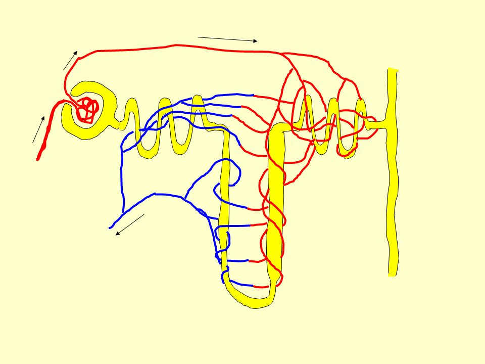
A single nephron in the kidney

7
To test your knowledge of kidney structure………………. ClickClick To test your knowledge of the labels…………Click simply move the labels to the correct position and check your answer with the originalClick

8
The next five slides shows a single nephron in isolation and the relevant labels. In addition the blood supply from a branch of the renal artery is shown and the collection of blood to return back to the renal vein.

10
Cortex Medulla

11
BOWMAN’S CAPSULE PCT LOOP OF HENLE DCT COLLECTING DUCT DESCENDING LIMB ASCENDING LIMB (THICK SECTION) ASCENDING LIMB (THIN SECTION)
 ASCENDING LIMB (THIN SECTION)")
13
AFFERENT VESSEL (FROM RENAL ARTERY) EFFERENT VESSEL TO RENAL VEIN TO THE PELVIS OF THE KIDNEY
 EFFERENT VESSEL TO RENAL VEIN TO THE PELVIS OF THE KIDNEY")
14
Bowman’s capsule This structure is like a hollow sphere with part of one side pushed in……..

15
Surface view View in section

16
Inside the Bowman’s capsule is a knot of blood capillaries known as the glomerulus. The vessel supplying the glomerulus is referred to as the afferent vessel and the one draining it is referred to as the efferent vessel

17
AFFERENT VESSEL EFFERENT VESSEL GLOMERULUS BOWMAN’S CAPSULE PODOCYTES These are the cells that line the Bowman’s capsule

18
Podocytes These are a single layer of cells that line the Bowman’s capsule. What makes these cells special is the fact that they don’t tessellate. In other words they don’t fit tightly together, there are gaps in between the cells.

19
A podocyte Gaps produced because the cells don’t tessellate

20
Cells of the PCT These cells are involved in the re- absorption of material from the nephron.

22
Epithelial cell Microvilli Blood Mitochondria Nucleus Lumen containing filtrate

23
Glucose and sodium pumped in actively through symport Glucose moved into blood by facilitated diffusion Sodium actively pumped into the blood in exchange for potassium

24
To see a range of animations regarding the kidney………………..clickclick

25
This is the end of this section

Similar presentations