Download presentation
Presentation is loading. Please wait.

1
Plasma Cell Disorders Kanwar Kahlon M.D. October 2014

2
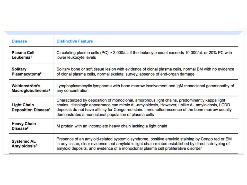
Subtypes of Plasma Cell Disorders
Increased Plasma Cells Monoclonal Gammopathy Myeloma Macroglobulinemia (IgM) Increased / Altered Products of Plasma Cells Light Chain Amyloidosis Light Chain Deposition Disease
 Increased / Altered Products of Plasma Cells. Light Chain Amyloidosis. Light Chain Deposition Disease.")
3
B cell Development As a Framework for Malignancies
Lymphoplasmacyte IgM Lymph node Germinal Center SOMATIC Lymphoblast HYPERMUTATION Plasmablast SWITCH RECOMBINATION In order to characterize the translocations in MM we need first to determine when they are occuring, during VDJ recombination, or isotype switching. This slide shows a summary of B cell differentiation. In the BM a pre-B cell undergoes VDJ recombination to make a functional Ig gene. An error resulting in a translocation to bcl-1 can result in mantle zone lymphoma, to bcl-2 in indolent lymphoma, and to c-myc to Burkitt’s lymphoma. This cell can enter the bloodstream, and the secondary lymphoid tissue where if it encounters antigen directly as part of the primary immun response, it can differentiate into a short lived plasma cell, that live about 3 days. Alternatively if it encounters antigen in the context of an antigen presenting cell it can enter a germinal center where it undergoes multiple rounds of division and somatic hypermutation of its immunoglobulin genes. The cells who Ig genes have retained or increased their affinity for antigen exit the germinal center, and they may undergo isotype switching and home to the BM where they can differentiate into long lived plasma cells with a lifespan of about 30 days. It is this cell that is the counterpart of the malignant plasma cell in myeloma. It has switched isotype, and its Ig genes have undergone extensive somatic hypermutation indicaing passage throuh the germinal center. Based on this analysis we postulated that the translocations in MM would occur at this stage, after passing through the germinal, during the process of isotype switch recombination Virgin B cell V(D)J RECOMBINATION Bone Marrow Plasma cell G,A,E Pre-B cell
J. RECOMBINATION. Bone. Marrow. Plasma cell. G,A,E. Pre-B cell.")
4
B cell Development As a Framework for Malignancies
Lymphoplasmacyte IgM WALDENSTROM’S Lymph node Germinal Center SOMATIC Lymphoblast HYPERMUTATION BURKITT’S LYMPHOMA Plasmablast FOLLICULAR LYMPHOMA In order to characterize the translocations in MM we need first to determine when they are occuring, during VDJ recombination, or isotype switching. This slide shows a summary of B cell differentiation. In the BM a pre-B cell undergoes VDJ recombination to make a functional Ig gene. An error resulting in a translocation to bcl-1 can result in mantle zone lymphoma, to bcl-2 in indolent lymphoma, and to c-myc to Burkitt’s lymphoma. This cell can enter the bloodstream, and the secondary lymphoid tissue where if it encounters antigen directly as part of the primary immun response, it can differentiate into a short lived plasma cell, that live about 3 days. Alternatively if it encounters antigen in the context of an antigen presenting cell it can enter a germinal center where it undergoes multiple rounds of division and somatic hypermutation of its immunoglobulin genes. The cells who Ig genes have retained or increased their affinity for antigen exit the germinal center, and they may undergo isotype switching and home to the BM where they can differentiate into long lived plasma cells with a lifespan of about 30 days. It is this cell that is the counterpart of the malignant plasma cell in myeloma. It has switched isotype, and its Ig genes have undergone extensive somatic hypermutation indicaing passage throuh the germinal center. Based on this analysis we postulated that the translocations in MM would occur at this stage, after passing through the germinal, during the process of isotype switch recombination Virgin B cell CLL Bone Marrow MULTIPLE MYELOMA Plasma cell ALL G,A,E Pre-B cell

6
Case Presentation 48 yo Jamaican male
Admitted with 2 wk history of fatigue and diffuse bone pain Evaluation: Increased serum creatinine 5.5 mg/dl (normal < 1.2 mg/dl) Increased serum calcium 12 mg/dl (normal < 10.5 mg/dl) Increased serum globulin 5.0 g/dl (normal < 3 g/dl)
 Increased serum calcium 12 mg/dl (normal < 10.5 mg/dl) Increased serum globulin 5.0 g/dl (normal < 3 g/dl)")
7
Electrophoresis (S/UPEP) Electrophoresis (IFE)
Evaluation of Abnormal Serum Globulins Serum Urine Serum/Urine Protein Electrophoresis (S/UPEP) Immunofixation Electrophoresis (IFE) Serum M spike 4 g/dl; typed as IgG kappa monoclonal Ig
 Immunofixation. Electrophoresis (IFE) Serum M spike 4 g/dl; typed as IgG kappa monoclonal Ig.")
8
Case Presentation…cont’d
Bone Marrow biopsy- Increased plasma cells

9
Skeletal survey—Lytic bone disease

10
Clinical spectrum of clonal expansions of
transformed plasma cells in patients Stable intramedullary expansion Asymptomatic. MGUS (premalignant) Normal cell Transformed Cell Progressive intramedullary expansion. Anemia, bone pain, infections Lytic bone disease. Incurable, limited survival. 13000 deaths/yr in USA. Multiple myeloma (malignant)
 Normal. cell. Transformed. Cell. Progressive intramedullary. expansion. Anemia, bone pain, infections. Lytic bone disease. Incurable, limited survival deaths/yr in USA. Multiple myeloma. (malignant)")
12
Diagnostic Criteria in Monoclonal Gammopathies
MGUS < 10% bone marrow plasma cells and M spike < 3 g/dl Monoclonal protein / clonal plasma cell population No End organ damage Myeloma > 10% marrow plasma cells End Organ Damage Indolent / Smoldering Myeloma > 10% marrow plasma cells or M spike > 3 g/dl

13
Criteria for End-Organ Damage in Monoclonal Gammopathies
CRAB Calcium > 1 mg/dl above ULN Renal Insufficiency (> 2 mg/dl) Anemia (< 10 g/dl) Bone Lesions (lytic lesions or osteopenia)
 Anemia (< 10 g/dl) Bone Lesions (lytic lesions or osteopenia)")
14
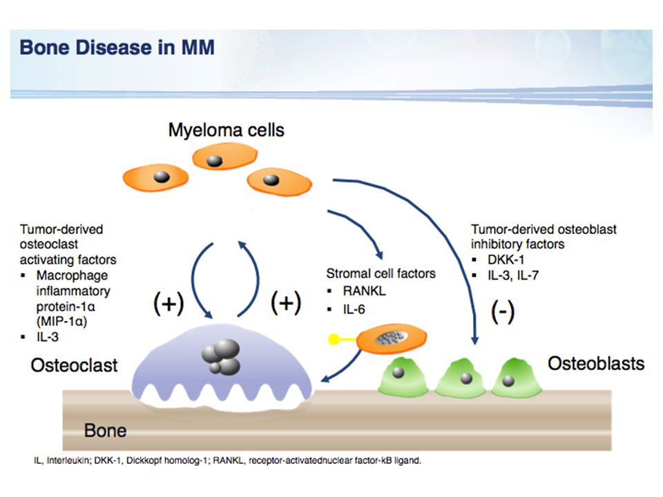
A Model for Pathogenesis of Myeloma

16
Monoclonal Gammopathy of Undetermined Significance (MGUS)
Common, age-related Prevalence: 3.2% in persons over 50 yrs old (Minnesota) ~5% in age >70 Higher prevalence in African populations. ? Association with inflammatory states: obesity, Gaucher’s disease Increased risk for thrombosis and fractures Risk of progression in entire population: 1% /yr Risk factors for progression: %PC, level M spike, Free light chain, IgA protein, ?Decline in uninvolved Ig’s
 ~5% in age >70. Higher prevalence in African populations. Association with inflammatory states: obesity, Gaucher’s disease. Increased risk for thrombosis and fractures. Risk of progression in entire population: 1% /yr. Risk factors for progression: %PC, level M spike, Free light chain, IgA protein, Decline in uninvolved Ig’s.")
17
Smoldering Myeloma (SMM)
Patients with PC > 10% or M spike > 3 g/dl, but lacking CRAB symptoms. 10% per yr progression to overt MM Most eventually require therapy. Current recommendation is observation until progressive disease.

18
Disease Progression in MGUS and SMM
Kyle et al. NEJM 356: 2582, 2007

19
Risk Groups in Asymptomatic Myeloma
G1: BMPC >10% M > 3 g/dl; G2: BMPC >10% M < 3 g/dl G3: BMPC <10% M > 3 g/dl Kyle et al. NEJM 356: 2582, 2007

20
Multiple myeloma Uncontrolled proliferation of Ig secreting plasma cells most commonly IgG (57%), IgA (21%) or light chain only (18%) Twice as frequent in men as women, and in blacks as whites 1% of all cancers 2% in African Americans Incurable Median survival 4-6 years

21
5 year survival (SEER)
")
22
Work-up in suspected myeloma
Assessment of serum/urine protein SPEP/IF, 24 hr urine for UPEP/IF Free light chains (kappa, lambda) CBC, sCr, Calcium, Albumin, LDH, Serum beta 2 microglobulin (B2M) Skeletal survey Bone marrow aspirate and biopsy Cytogenetics (including FISH) Under investigation: MRI spine PET scans Bone densitometry, Urine n-telopeptide
 CBC, sCr, Calcium, Albumin, LDH, Serum beta 2 microglobulin (B2M) Skeletal survey. Bone marrow aspirate and biopsy. Cytogenetics (including FISH) Under investigation: MRI spine. PET scans. Bone densitometry, Urine n-telopeptide.")
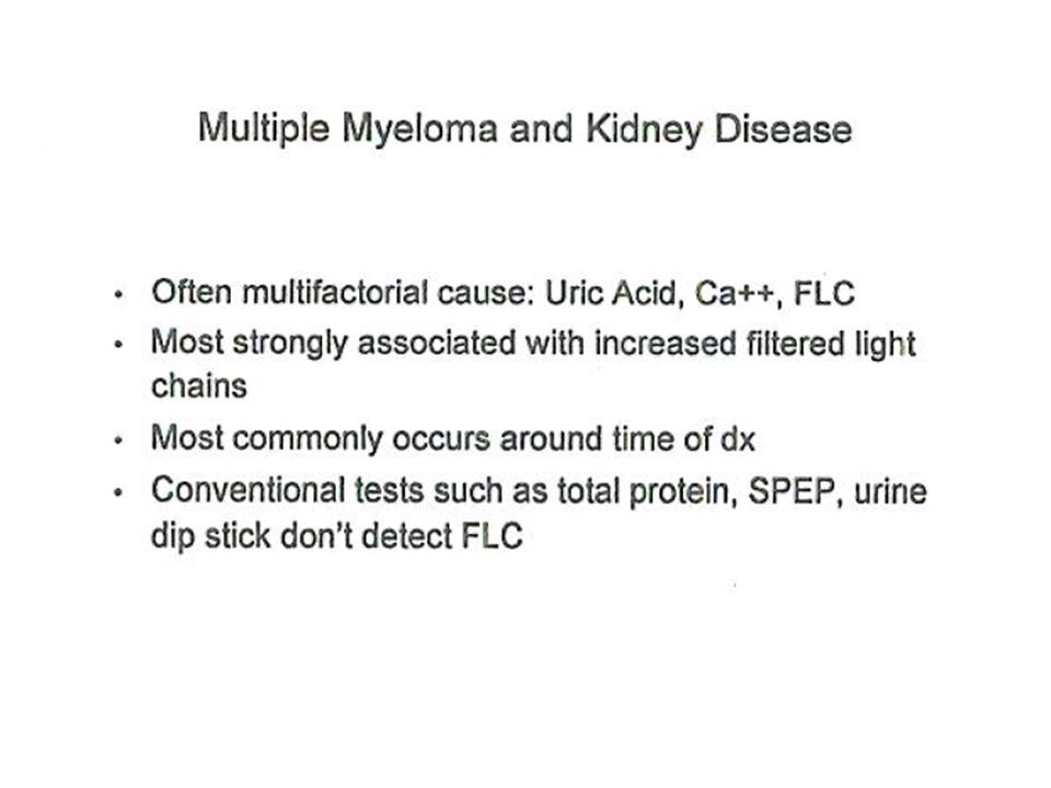
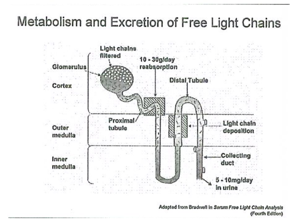
23
* Kappa and Lambda FLC’s are both increased in renal failure/CKD

24
Key clinical aspects of myeloma
Predominantly intra-medullary growth. Absence of clinical LN or spleen involvement. Low proliferative fraction. Long periods of stability in MGUS. Osteoclast activation, osteoblast inhibition, and bone loss. Multi-focal growth of tumor cells.

25
Clinical presentation

26
Manifestations of Clonal Plasma Cell Proliferation
Ig deposition Cast nephropathy RENAL FAILURE Osteoclast Osteoblast LYTIC BONE DZ HYPERCALCEMIA Immune-paresis Hypogamm INFECTION Erythropoiesis ANEMIA

27
Historical aside… At age 39, developed fatigue and bone pain from several fractures. She died 4 years later; autopsy showed that her marrow was replaced by a red, gelatinous substance

28
Multiple Myeloma: Skeletal Complications
Examples of fractures and spinal cord compression in multiple myeloma patients. Bone pain is the most frequent symptom at presentation; 68%-80% of patients present with destructive osteolytic lesions at time of diagnosis. Sheridan CA, Serrano M: Multiple myeloma, in Yarbro CH, Frogge MH, Goodman M et al (eds). Cancer Nursing: Principles and Practice (5th ed) Jones and Bartlett, Boston, pp ; Sheridan CA et al. Semin Oncol Nurs. 1996;12:1-12; Wilkes GM, Ingwersen K, Barton-Burke M Oncology Nursing Drug Handbook, Boston, Jones and Bartlett. 2003; Tariman J et al. Clin J Oncol Nurs. 2003;7: ; Annas GJ et al. Am J Pub Health. 1999;89:98-101; van de Poel, Pasman Schoutnen et al. Netherlands J Med. 2001;59:45-49.
. Cancer Nursing: Principles and Practice (5th ed) Jones and Bartlett, Boston, pp ; Sheridan CA et al. Semin Oncol Nurs. 1996;12:1-12; Wilkes GM, Ingwersen K, Barton-Burke M Oncology Nursing Drug Handbook, Boston, Jones and Bartlett. 2003; Tariman J et al. Clin J Oncol Nurs. 2003;7: ; Annas GJ et al. Am J Pub Health. 1999;89:98-101; van de Poel, Pasman Schoutnen et al. Netherlands J Med. 2001;59:")
30
Original Article Romosozumab in Postmenopausal Women with Low Bone Mineral Density
Michael R. McClung, M.D., Andreas Grauer, M.D., Steven Boonen, M.D., Ph.D., Michael A. Bolognese, M.D., Jacques P. Brown, M.D., Adolfo Diez-Perez, M.D., Ph.D., Bente L. Langdahl, Ph.D., D.M.Sc., Jean-Yves Reginster, M.D., Ph.D., Jose R. Zanchetta, M.D., Scott M. Wasserman, M.D., Leonid Katz, M.D., Judy Maddox, D.O., Yu-Ching Yang, Ph.D., Cesar Libanati, M.D., and Henry G. Bone, M.D. N Engl J Med Volume 370(5): January 30, 2014
: January 30,")
32
Light Chain Deposition Disease Light Chain Cast Nephropathy
Renal Pathology in MM Light Chain Deposition Disease Light Chain Cast Nephropathy AL Amyloid Fig 1. The characteristic appearance of light chain deposition disease by light microscopy is that of a nodular glomerulosclerosis, which strongly resembles diabetic nephropathy, as in this case (periodic acid-Schiff stain; original magnification x400). Fig 5. Glomerular capillary loop, mesangial staining, and linear tubular staining are characteristic of light chain deposition disease. Either kappa or lambda light chain paraprotein may cause light chain deposition disease, although kappa more commonly is the culprit (antibody to kappa light chain, immunofluorescence; original magnification x200). Fig 3. Close-up of intratubular refractile casts with surrounding syncytial giant cell reaction with chronic tubulointerstitial nephritis and fibrosis, characteristic of myeloma cast nephropathy (PAS stain; original magnification x400). Fig 7. When the Congo red stain is viewed under polarized light, areas of amyloid show apple green birefringence. In this case, amyloid deposits are seen in the mesangial areas, the capillary loops, and in vessels (Congo red stain; original magnification x100).
. Fig 5. Glomerular capillary loop, mesangial staining, and linear tubular staining are characteristic of light chain deposition disease. Either kappa or lambda light chain paraprotein may cause light chain deposition disease, although kappa more commonly is the culprit (antibody to kappa light chain, immunofluorescence; original magnification x200). Fig 3. Close-up of intratubular refractile casts with surrounding syncytial giant cell reaction with chronic tubulointerstitial nephritis and fibrosis, characteristic of myeloma cast nephropathy (PAS stain; original magnification x400). Fig 7. When the Congo red stain is viewed under polarized light, areas of amyloid show apple green birefringence. In this case, amyloid deposits are seen in the mesangial areas, the capillary loops, and in vessels (Congo red stain; original magnification x100).")
35
International Staging System
Stage I (B2M <3.5 mg/l; Albumin >3.5 g/dl) Median OS 62 months Stage III (B2M >5.5 mg/l) Median OS 29 months Stage II (Neither Stage I or III) Median OS 44 months
 Median OS 62 months. Stage III (B2M >5.5 mg/l) Median OS 29 months. Stage II (Neither Stage I or III) Median OS 44 months.")
36
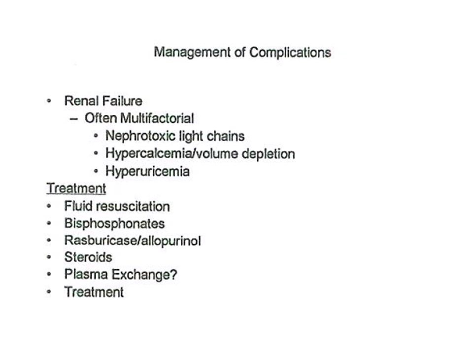
Principles Of Treatment
No evidence (yet) that early treatment prolongs survival Wait for symptoms, or evidence of disease progression, to start treatment Supportive measures are critically important drink plenty of fluids daily treat infections promptly prophylactic bisphosphonates reduce skeletal complications in patients with osteopenia and lytic bone disease anemia often responds to erythropoietin.
 that early treatment prolongs survival. Wait for symptoms, or evidence of disease progression, to start treatment. Supportive measures are critically important. drink plenty of fluids daily. treat infections promptly. prophylactic bisphosphonates reduce skeletal complications in patients with osteopenia and lytic bone disease. anemia often responds to erythropoietin.")
37
“Myeloma treatment is a marathon, not a sprint”

41
JCO, 30(4), Feb 2012
, Feb 2012")
44
Major drugs in myeloma Alkylators - 1962 Melphalan, cyclophosphamide
High dose melphalan and ASCT Glucocorticoids Prednisone, dexamethasone IMiDs Thalidomide Revlimid Pomalidomide Proteasome Inhibitors Bortezomib Carfilzomib

45
Treatment course Asymptomatic Symptomatic Acute MGUS Stable MM
Pancytopenia Plasma cell leukemia M protein Treatments Years Months Days

46
Cytogenetic Profiles of Myeloma
Carrasco et al Cancer Cell, Sawyer et al CGG, 2011

47
Percentage of patients
IMWG Molecular Classification of Myeloma Percentage of patients Clinical and laboratory features Hyperdiploid 45 More favorable, IgG-κ, older patients. Non-hyperdiploid 40 Aggressive, IgA-λ, younger individuals Cyclin D translocation 18 t(11;14)(q13;q32) 16 Upregulation of CCND1; favorable prognosis; bone lesions. Two subtypes by GEP; CD20+ in one subset t(6;14q)(p21;32) 2 Probably same as CCND1 t(12;14)(p13;q32) <1 Rare MMSET translocation 15 t(4;14)(p16;q32) Upregulation of MMSET; upregulation of FGFR3 in 75% unfavorable prognosis with conventional therapy; bone lesions less frequent MAF translocation 8 Aggressive t(14;16)(q32;q23) 5 Confirmed as aggressive by at least two series t(14;20)(q32;q11) One series shows more aggressive disease. t(8;14)(q24;q32) 1 Unknown effect on outcome but presumed aggressive. Unclassified (other) Various subtypes and some with overlap IMWG. Leukemia 2010
(q13;q32) 16. Upregulation of CCND1; favorable prognosis; bone lesions. Two subtypes by GEP; CD20+ in one subset. t(6;14q)(p21;32) 2. Probably same as CCND1. t(12;14)(p13;q32) <1. Rare. MMSET translocation. 15. t(4;14)(p16;q32) Upregulation of MMSET; upregulation of FGFR3 in 75% unfavorable prognosis with conventional therapy; bone lesions less frequent. MAF translocation. 8. Aggressive. t(14;16)(q32;q23) 5. Confirmed as aggressive by at least two series. t(14;20)(q32;q11) One series shows more aggressive disease. t(8;14)(q24;q32) 1. Unknown effect on outcome but presumed aggressive. Unclassified (other) Various subtypes and some with overlap. IMWG. Leukemia")
48
Factors Associated with Increased Disease Risk in MM
Gene expression profile (GEP) 70 (or GEP15) high risk signature FISH: t(4:14); t(14:16) Del 17p 1q amp; hypodiploidy Abnormal cytogenetics by metaphase, including del chr 13 ISS Stage 3 (increased beta 2 m) High LDH > 10 focal lesions on MR
 70 (or GEP15) high risk signature. FISH: t(4:14); t(14:16) Del 17p. 1q amp; hypodiploidy. Abnormal cytogenetics by metaphase, including del chr 13. ISS Stage 3 (increased beta 2 m) High LDH. > 10 focal lesions on MR.")
49
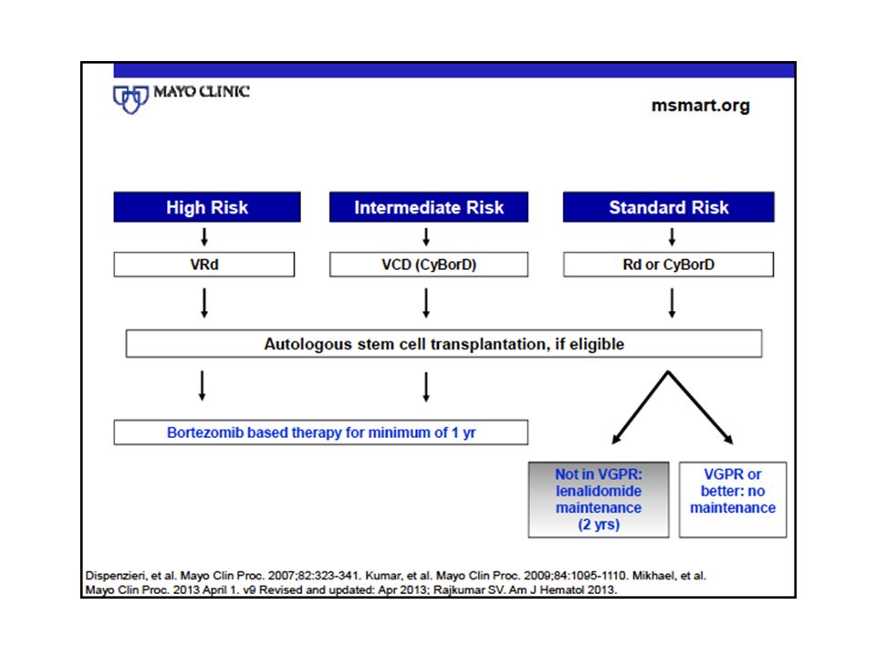
Mayo Clin Proc, Apr 2013

50
Initial therapy in myeloma
Current approach based on risk status and potential candidacy for stem cell transplantation. Combination therapy superior to single agents.

52
High Response Rates to Induction Therapies in MM
Combinations in the Upfront Treatment of MM [Blood. Copyright Reproduced with permission of Blood in the format Journal via Copyright Clearance Center]. ORR, objective response rate; VGPR, very good partial response; CR, complete response; nCR, near CR; VAD, vincristine, adriamycin, dexamethasone; TD, thalidomide and dexamethasone; RD, lenalidomide and dexamethasone; PAD, bortezomib, doxorubicin, dexamethasone; VTD, bortezomib, thalidomide and dexamethasone; CVD, cyclophosphamide, velcade, dexamethasone; RVD, lenalidomide, bortezomib and dexamethasone; CVRD, bortezomib, dexamethasone, cyclophosphamide, lenalidomide. © This slide is made available for non-commercial use only. Please note that permission may be required for re-use of images in which the copyright is owned by a third party.

56
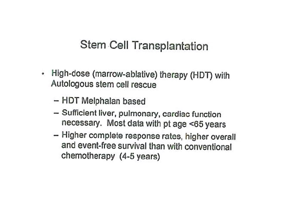
High dose therapy in myeloma
Small randomized trials showing superiority to conventional therapy without ASCT (two with mature data). Early ASCT not superior to late ASCT. No conditioning regimen superior to melphalan alone. No benefit from CD34+ selected grafts. Superiority of SCT is being tested in the setting of new drugs.
. Early ASCT not superior to late ASCT. No conditioning regimen superior to melphalan alone. No benefit from CD34+ selected grafts. Superiority of SCT is being tested in the setting of new drugs.")
57
Initial Therapy: Transplant Ineligible
Melphalan + Prednisone was once the standard approach. RCTs show superiority of addition of thalidomide (MPT) or Velcade (MPV) to MP. Lenalidomide and dexamethasone is also active.
 or Velcade (MPV) to MP. Lenalidomide and dexamethasone is also active.")
58
The future… JCO, 30(4), Feb 2012
, Feb 2012")
59
Waldenström Macroglobulinemia
Uncontrolled proliferation of lymphoplasmacytes producing IgM Median age 63 years Presents with weakness, fatigue, epistaxis, blurred vision Bone pain and lytic bone lesions are uncommon (<5%) 25% have hepatomegaly, splenomegaly and lymphadenopathy Hyperviscosity is common
 25% have hepatomegaly, splenomegaly and lymphadenopathy. Hyperviscosity is common.")
60
Mayo Clin Proc, Sept 2010

61
Hyperviscosity syndrome
bleeding (nasal and gums) blurred vision dizziness, headaches, ataxia congestive heart failure retinal vein engorgement, and papilledema rarely occurs with serum viscosity <4 centipoises (cp) (normal 1.8 cp) IgM pentamer
 blurred vision. dizziness, headaches, ataxia. congestive heart failure. retinal vein engorgement, and papilledema. rarely occurs with serum viscosity <4 centipoises (cp) (normal 1.8 cp) IgM pentamer.")
62
Macroglobulinemia: Principles of Therapy
Observation in patients with asymptomatic disease. Active drugs for therapy Alkylating agents: Chlorambucil, Cytoxan MAbs: Rituxan Purine analogues: Fludarabine, Cladribine Bendamustine Steroids Bortezomib Thalidomide analogues

63
Plasma Cell Disorders Manifest Due to Clonal Immunoglobulin
AL Amyloid Light chain deposition dz Neuropathy Cryoglobulinemia Acquired vWD

64
Clinical Settings Where Ig Deposition Diseases (Including AL Amyloid) Should Be Suspected In Pts With Monoclonal Ig Congestive Heart Failure Neuropathy (including autonomic neuropathy) Nephrotic syndrome, Renal Failure Malabsorption Hepatosplenomegaly Carpal tunnel syndrome Macroglossia Unexplained constitutional symptoms
 Nephrotic syndrome, Renal Failure. Malabsorption. Hepatosplenomegaly. Carpal tunnel syndrome. Macroglossia. Unexplained constitutional symptoms.")
65
Diagnostic Approach in Suspected AL Amyloid

66
Principles of Management in AL Amyloid
Therapeutic approach guided by age, organ involvement. Cardiac involvement and dysfunction as a major predictor. Therapy directed at the underlying clonal plasma cells. Melphalan Steroids Proteasome Inhibitors (Velcade) Thalidomide/lenalidomide
 Thalidomide/lenalidomide.")
67
Conclusion Plasma cell dyscrasias are a heterogeneous group of disorders. Clinical presentation may be due to the clone itself or the properties of the secreted Ig. Therapy largely directed (if indicated) at reducing the underlying clone.
 at reducing the underlying clone.")
Similar presentations


 Family Medicine Review Course 2011 Christian Cable, MD, FACP.>")


 Idiopathic Associated with other diseases (autoimmune, infectious, non-heme.>")


>")
 DEFINITION CLL is a neoplastic disease characterized by proliferation and accumulation (blood, marrow and lymphoid.>")

.>")