Download presentation
Presentation is loading. Please wait.

1
Radiology

2
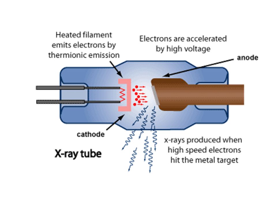
The Cathode… Provides a source of electrons and directs the electrons towards to anode. The cathode has a coiled wire filament that emits electrons when heated. This filament is similar to one found in a light bulb. When the filament is heated electrons are held less tightly and become excited. They can now travel to the anode

3
The anode: A beveled target placed on a cylindrical base. The base is usually made of copper which acts as a conductor of heat. It draws the heat away from the tungsten target.

5
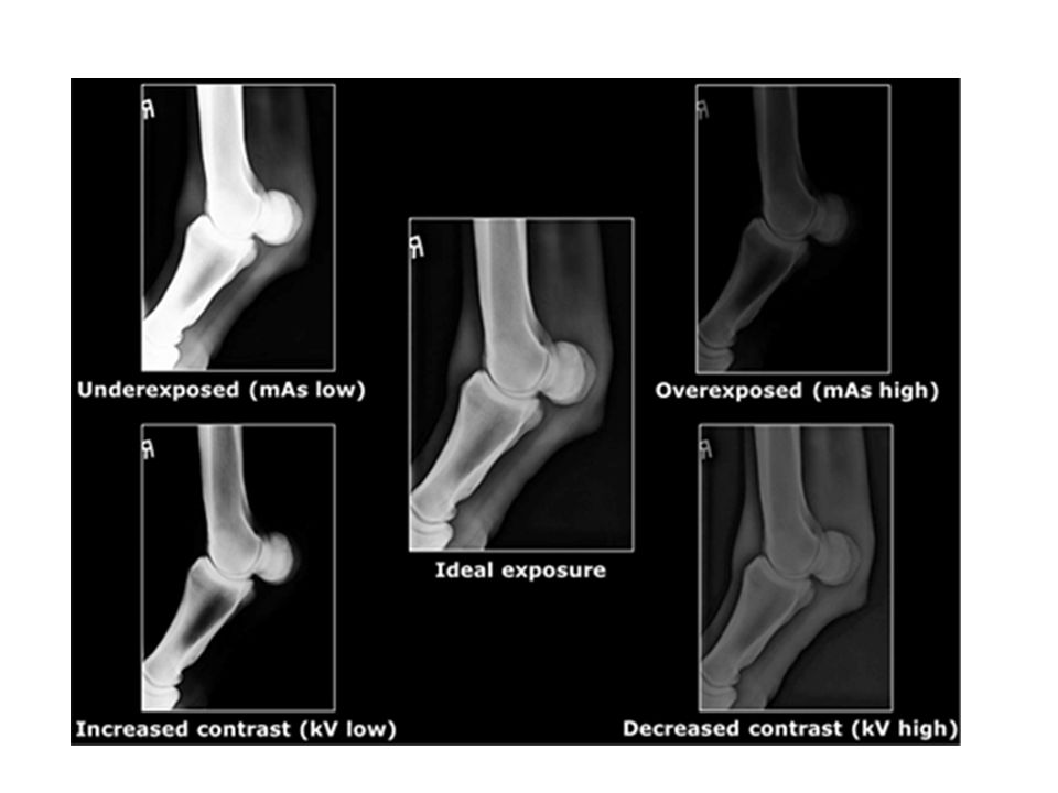
Amperage, Milliamperage and Kilovoltage Amperage = unit of current Milliampere = 1/1000 ampere Radiology uses mA to regulate the number of electrons used to produce x-ray photons The amount of energy in the circuit is referred to as milliamperage (mA) This controls the heat of the filament which in turn controls the number of electrons. Acceleration of the electrons is controlled by the kilovoltage.

8
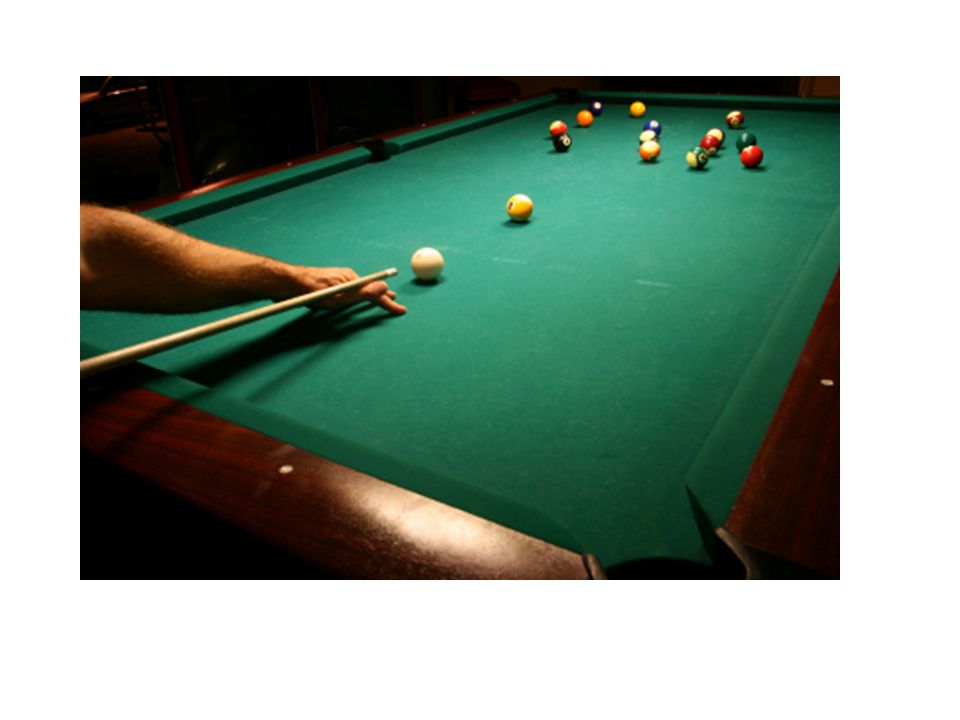
Several things can influence the primary signal which in turn will change the outcome of the radiograph. Energy (how hard did you throw the ball) Frequency (how many balls did you throw) Wavelength (What path did the balls take) Number of photons produced (How many balls do you have left to throw) Penetrability (how hard is the object you are throwing the balls at)
 Frequency (how many balls did you throw) Wavelength (What path did the balls take) Number of photons produced (How many balls do you have left to throw) Penetrability (how hard is the object you are throwing the balls at).")
9
The Inverse Square Law : source to image distance. The intensity of the x-ray beam is inversely proportional to the square of the distance from the source. Distance increases, intensity decreases.

11
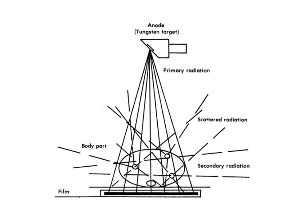
Using the grid helps to reduce scatter. You should use the grid for larger body parts (greater than 10 cm).
..")
12
Technique chart gives settings based on the thickness of the body part.

13
Machine settings: Use the fastest time Use the highest kvp (wrong) Use a constant distance (40 inches is standard) Measure accurately.
 Use a constant distance (40 inches is standard) Measure accurately.")
14
Calipers are used to measure the body part.

15
Most radiographic evals are made with 2 views. Anterior/Posterior Dorsal/Ventral Lateral Proximal/Distal

16
Patient Motion

17
Film Cassette/Intensifying Screens Purposes: Reduces the amount of radiation required to produce a diagnostic radiograph Enhances contrast The screens in an x-ray cassette sandwich the film. Radiation causes screen to glow. Screens are rated by how fast the x-rays are converted to light by the phosphors in its layers.

19
Digital Xray Machine

20
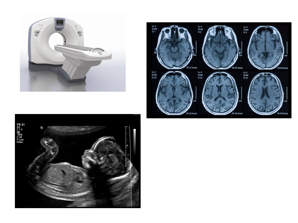
Alternative Modalities Computerized Tomography Fluoroscopy Ultrasound Nuclear medicine Magnetic resonance imaging

22
Tissue density: The less dense the tissue: the darker is becomes on the radiograph

Similar presentations