Download presentation
Presentation is loading. Please wait.

1
Insulin Improves Myocardial Blood Flow in Patients with Type 2 Diabetes and Coronary Artery Disease Riikka Lautamäki, K.E. Juhani Airaksinen, Marko Seppänen, Jyri Toikka, Risto Härkönen, Matti Luotolahti, Ronald Borra, Jan Sundell, Juhani Knuuti R2 박지나 Diabetes, Vol 55 2006 Feb

2
The main aim of this study … to determine whether insulin is able to increase myocardial blood flow in the regions of compromised myocardial perfusion in subjects with type 2 diabetes and ischemic CAD. compare the flow values between ischemic and nonischemic regions. Introduction Insulin infusion improves myocardial blood flow (MBF) in healthy subjects. Until now, the effect of insulin on myocardial perfusion in type 2 diabetic subjects with coronary artery disease (CAD) has been unknown.
 in healthy subjects. Until now, the effect of insulin on myocardial perfusion in type 2 diabetic subjects with coronary artery disease (CAD) has been unknown..")
3
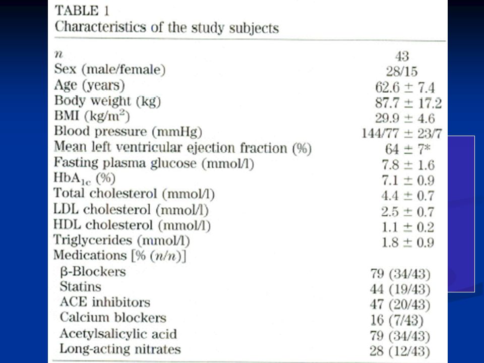
Research disign and Methods Inclusion criteria past or present angina pectoris symptoms under stress, type 2 diabetes treated with diet or metformin and/or a sulfonylurea, and good or moderate glycemic control (HbA1c <8.5%) Exclusion criteria unstable angina pectoris, symptomatic tachy- or bradyarrhythmias, history of percutaneous transluminal coronary angioplasty during the preceding 6 months, asthma, chronic use of insulin, or clinical signs of heart failure.
 Exclusion criteria unstable angina pectoris, symptomatic tachy- or bradyarrhythmias, history of percutaneous transluminal coronary angioplasty during the preceding 6 months, asthma, chronic use of insulin, or clinical signs of heart failure.")
5
SPECT perfusion imaging 99m-Myoview-tetrofosmin was used in the SPECT studies. Subjects refrained from taking β-blockers, calcium blockers, and long acting nitrates for 48 h before the study Both rest and stress imaging studies Coronary angiography PET study Fig 1 Echocardiographic examination To evaluate the global left ventricular function, all subjects underwent rest echocardiographic examination after the PET imaging during Insulin stimulation

7
The SPECT analyses, echocardiography, and coronary angiography were performed by different independent experienced observers; afterwards, the results were matched together. The purpose was to match the ischemia in SPECT with the stenotic lesion found in the angiography and with the possible wall motion abnormality in echocardiography. The ischemic region a completely or partially reversible perfusion deficit in SPECT, coronary artery stenosis in the respective coronary artery in the angiogram, and a possible wall motion abnormality in the echocardiogram. A nonischemic region normal wall motion in the echocardiogram, had no reversible perfusion deficit in the SPECT, and was associated with nonstenotic coronary artery.

8
Results

11
0.043(13%) 0.018(20%)0.045(18%) 0.003(22%)
 0.018(20%)0.045(18%) 0.003(22%)")
12
Conclusion This study demonstrates… insulin improves myocardial blood flow similarly in ischemic and nonischemic regions both at rest and during adenosine-induced hyperemia in subjects with type 2 diabetes and CAD. insulin-induced vasodilation, in addition to its metabolic effect, improves endothelial function and thus increases the threshold for ischemia in these subjects.

13
Discussion Data from many previous studies have suggested reduced coronary vasodilation is present in diabetic subjects. The level of myocardial blood flow during pharmacologically applied stress conditions is significantly decreased in subjects with uncomplicated type 2 diabetes and in type 2 diabetic subjects with CAD However, in type 2 diabetic subjects, myocardial blood flow at rest does not significantly differ from that in healthy control subjects or from that in type 2 diabetic subjects with CAD In another study, resting myocardial blood flow was higher in type 2 diabetic subjects as compared with in healthy volunteers In many studies, comparisons of the flow values are made between healthy control subjects and patient groups. In the present study we measured myocardial blood flow in the same region in the fasting state and during hyperinsulinemia; thus we were able to compare the values within each patient and exclude between-subject variability.

14
Discussion In the present study the subjects were on several medications, which is typical for this patient population. To avoid the confounding effects of other medications, the subjects refrained from all their medications for 12 h before the PET study, but otherwise the subjects were on stable medical therapy throughout the study. It is known that some medications may affect absolute myocardial blood flow. ex) the statin and ACE inhibitor,β-adrenergic blockade However, the results of our study indicate that insulin has additive effects over the concomitant medication on the absolute myocardial blood flow.
 the statin and ACE inhibitor,β-adrenergic blockade However, the results of our study indicate that insulin has additive effects over the concomitant medication on the absolute myocardial blood flow..")
Similar presentations


 Inclusion criteria.>")


