Download presentation
Presentation is loading. Please wait.

1
Introduction to Pulmonary Function Tests By Shaimaa Ahmed Attia

2
Indications Evaluate symptoms Cough, wheezing, dyspnea Cough, wheezing, dyspnea Evaluate effects of work exposure Dusts, chemical Dusts, chemical Assess patients at risk for lung disease Screen smokers >45 y/o for COPD Screen smokers >45 y/o for COPD >25% will have abnormal results >25% will have abnormal results Risk assessment prior to surgery Objective assessment of disability Follow progression of disease Lung transplant patients Lung transplant patients Response to treatment Response to treatment

3
Pulmonary function VentilationDiffusionPerfusion

4
Pulmonary Function Test Spirometry Lung volumes Diffusing Capacity of Carbon Monoxide (DLCO) PO2,PCO2, PH (non- specific) Pulmonary angiography Bronchodilator tests and challenge tests
 PO2,PCO2, PH (non- specific) Pulmonary angiography Bronchodilator tests and challenge tests")
5
Spirometry Measurement of airflow rate and expired volume over time Reference range depends on height, weight, sex and race

6
Spirometry FVC- Forced vital capacity Volume of air that can be exhaled during a maximal forced expiration effort. Volume of air that can be exhaled during a maximal forced expiration effort. FEV1- Forced expiratory volume in one second Volume of air exhaled in the first second under force after a maximal inhalation. Volume of air exhaled in the first second under force after a maximal inhalation. FEV1/FVC ratio- Percentage of the FVC expired in one second.

7
PEF (Peak expiratory flow rate) FEF25-75%- Forced expiratory flow over the middle one half of the FVC Average flow from the point at which 25% of the FVC has been exhaled to the point at which 75% of the FVC has been exhaled
 FEF25-75%- Forced expiratory flow over the middle one half of the FVC Average flow from the point at which 25% of the FVC has been exhaled to the point at which 75% of the FVC has been exhaled")
8
Lung Volumes 4 ways to measure Helium dilution Helium dilution Nitrogen washout Nitrogen washout Body plethysmography (body box) Body plethysmography (body box) Gold standard CXR measurements CXR measurements
 Body plethysmography (body box) Gold standard CXR measurements CXR measurements")
9
Lung Volumes Maximum exp effort Maximum insp. effort

10
Lung Volumes ERV-Expiratory reserve volume Maximal volume of air exhaled from end-expiration. Maximal volume of air exhaled from end-expiration. IRV-Inspiratory reserve volume Maximal volume of air inhaled from end-inspiration. Maximal volume of air inhaled from end-inspiration. RV-Residual volume Volume of air remaining in the lungs after a maximal exhalation. Volume of air remaining in the lungs after a maximal exhalation. VT-Tidal volume Volume of air inhaled or exhaled during each respiratory cycle. Volume of air inhaled or exhaled during each respiratory cycle.

11
Lung Capacities FRC-Functional residual capacity Volume of air in the lungs at resting end-expiration. Volume of air in the lungs at resting end-expiration. IC-Inspiratory capacity Maximal volume of air that can be inhaled from the resting expiratory level. Maximal volume of air that can be inhaled from the resting expiratory level. TLC-Total lung capacity Volume of air in the lungs at maximal inflation. Volume of air in the lungs at maximal inflation. VC-Vital capacity Largest volume measured on complete exhalation after full inspiration. Largest volume measured on complete exhalation after full inspiration.

12
Obstruction FEV1/FVC is reduced Lung volumes can not differentiate between asthma and COPD Normal reduction of FEV1 is 30mL/yr Smokers: 90-150 mL/ yr reduction

13
Restrictive Reduction of lung volumes Intrinsic Interstitial (parenchymal) lung disease Interstitial (parenchymal) lung diseaseExtrinsic Chest wall disorders Chest wall disorders Pleural disease Pleural disease Neuromuscular disease Normal DLCO Normal DLCO
 lung disease Interstitial (parenchymal) lung diseaseExtrinsic Chest wall disorders Chest wall disorders Pleural disease Pleural disease Neuromuscular disease Normal DLCO Normal DLCO")
14
Lung Volumes Obstructive lung disease If FVC is reduced, RV is increased proportionally (FVC usually normal), FEV1 decreased, FEV1/FVC ratio decreased If FVC is reduced, RV is increased proportionally (FVC usually normal), FEV1 decreased, FEV1/FVC ratio decreased TLC remains normal TLC remains normal FRC remains normal FRC remains normal Restrictive lung disease Decrease in TLC Decrease in TLC Mainly decrease in lung volumes, FVC is reduced, FEV1 normal, FEV1/FVC ratio normal or increased Mainly decrease in lung volumes, FVC is reduced, FEV1 normal, FEV1/FVC ratio normal or increased
, FEV1 decreased, FEV1/FVC ratio decreased If FVC is reduced, RV is increased proportionally (FVC usually normal), FEV1 decreased, FEV1/FVC ratio decreased TLC remains normal TLC remains normal FRC remains normal FRC remains normal Restrictive lung disease Decrease in TLC Decrease in TLC Mainly decrease in lung volumes, FVC is reduced, FEV1 normal, FEV1/FVC ratio normal or increased Mainly decrease in lung volumes, FVC is reduced, FEV1 normal, FEV1/FVC ratio normal or increased")
15
Lung Volumes Combination of Restrictive and Obstructive Normal lung volumes Normal lung volumes Normal expiratory flow rate Normal expiratory flow rate DLCO is reduced DLCO is reduced Example: Pulmonary fibrosis Example: Pulmonary fibrosis

16
Forced inspiratory maneuvers Flow-volume loops Flow-volume loops Upper airway obstruction can not always be detected by PFTs (Pharynx, larynx, trachea) in stridor Indication of median to small airway obstruction like asthma and emphysema, the shape of the expiratory limb from 75% to RV depend on elastic recoil so never be malingering
 in stridor Indication of median to small airway obstruction like asthma and emphysema, the shape of the expiratory limb from 75% to RV depend on elastic recoil so never be malingering")
17
Flow Volume Loop

18
Restrictive Restrictive Normal Normal Obstructive Obstructive Flow Volume Curves

19
Diffusing Capacity (DLCO) Indirect measurement of lung’s ability to transfer an inhaled gas From distal airspaces across pulmonary capillary walls to circulating blood From distal airspaces across pulmonary capillary walls to circulating blood Can determine the nature and extent of interstitial or pulmonary vascular disease Pulmonary fibrosis – low Pulmonary fibrosis – low Emphysema – low Emphysema – low Bronchitis – normal Bronchitis – normal Asthma – normal to high Asthma – normal to high
 Indirect measurement of lung’s ability to transfer an inhaled gas From distal airspaces across pulmonary capillary walls to circulating blood From distal airspaces across pulmonary capillary walls to circulating blood Can determine the nature and extent of interstitial or pulmonary vascular disease Pulmonary fibrosis – low Pulmonary fibrosis – low Emphysema – low Emphysema – low Bronchitis – normal Bronchitis – normal Asthma – normal to high Asthma – normal to high")
20
Interpreting PFTs Look at FEV1/FVC first <70% = obstructive lung disease <70% = obstructive lung disease Then FEV1 Grade severity of obstruction by this Grade severity of obstruction by this Used in management of pts Used in management of pts Then FVC If ratio is normal and FVC is low = restrictive lung disease If ratio is normal and FVC is low = restrictive lung disease Take a quick look at the time (FET100%) Take a quick look at the time (FET100%)
 Take a quick look at the time (FET100%)")
21
Post bronchodilators If baseline tests are normal– no need to check post measurements Pts are given albuterol, repeat test in 10 minutes Response is clinically important if FEV1, FVC or PEF increases by >12% or >200mL

22
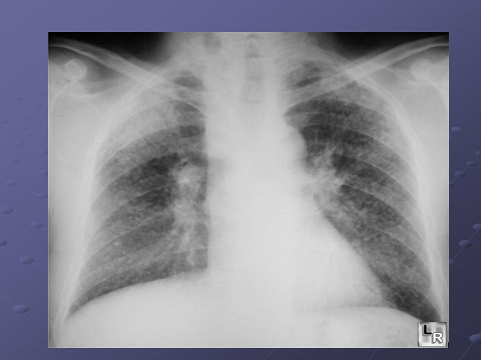
Case 1 65 y/o male DM, HTN 50+ pack history of smoking SOB with exertion CXR: Increased reticular markings at the bilateral bases, which appear chronic

23
Case 1 FEV1/FVC: 48% FVC: 3.24L (86%) FEV1: 1.55L (48%) FEF25-75%: 0.64L (28%)
 FEV1: 1.55L (48%) FEF25-75%: 0.64L (28%)")
24
Case 1 FEV1/FVC: 48% Severely ↓ FVC: 3.24L (86%) Normal FEV1: 1.55L (48%) Severely ↓ FEF25-75%: 0.64L (28%) Severely ↓
 Normal FEV1: 1.55L (48%) Severely ↓ FEF25-75%: 0.64L (28%) Severely ↓")
25
Case 1 FEV1/FVC: 48% Severely ↓ FVC: 3.24L (86%) Normal FEV1: 1.55L (48%) Severely ↓ FEF25-75%: 0.64L (28%) Severely ↓ Interpretation: Moderate obstruction Diagnosis: COPD
 Normal FEV1: 1.55L (48%) Severely ↓ FEF25-75%: 0.64L (28%) Severely ↓ Interpretation: Moderate obstruction Diagnosis: COPD")
26
Case 2 31 y/o female h/o chronic sinusitis, persistent cough with daily sputum production, situs inversus and infertility

27
Case 2 FEV1/FVC: 59% FVC: 2.27L (71%) FEV1: 1.34L (49%) FEF25-75%: 0.95L (22%)
 FEV1: 1.34L (49%) FEF25-75%: 0.95L (22%)")
28
Case 2 FEV1/FVC: 59% Moderately ↓ FVC: 2.27L (71%) Mildly ↓ FEV1: 1.34L (49%) Severely ↓ FEF25-75%: 0.95L (22%) Severely ↓
 Mildly ↓ FEV1: 1.34L (49%) Severely ↓ FEF25-75%: 0.95L (22%) Severely ↓")
29
Case 2 FEV1/FVC: 59% Moderately ↓ FVC: 2.27L (71%) Mildly ↓ FEV1: 1.34L (49%) Severely ↓ FEF25-75%: 0.95L (22%) Severely ↓ Interpretation: Moderate obstructive impairment, FVC mildly reduced, can not rule out restrictive impairment
 Mildly ↓ FEV1: 1.34L (49%) Severely ↓ FEF25-75%: 0.95L (22%) Severely ↓ Interpretation: Moderate obstructive impairment, FVC mildly reduced, can not rule out restrictive impairment")
30
Case 2 FEV1/FVC: 59% Moderately ↓ FVC: 2.27L (71%) Mildly ↓ FEV1: 1.34L (49%) Severely ↓ FEF25-75%: 0.95L (22%) Severely ↓ Interpretation: Moderate obstructive impairment, FVC mildly reduced, can not rule out restrictive impairment Diagnosis: Kartagener’s Syndrome (PCD)
 Mildly ↓ FEV1: 1.34L (49%) Severely ↓ FEF25-75%: 0.95L (22%) Severely ↓ Interpretation: Moderate obstructive impairment, FVC mildly reduced, can not rule out restrictive impairment Diagnosis: Kartagener’s Syndrome (PCD)")
31
Role of PFT in Occupational lung diseases Diagnostic(early detection) restrictive and obstructive impairment restrictive and obstructive impairment small airway disease small airway disease Bronchodilators to diagnose reversibility Bronchodilators to diagnose reversibility Stop resume test(pre and post shift( Stop resume test(pre and post shift( Disability evaluation Pre employment and periodic exam Follow up
 restrictive and obstructive impairment restrictive and obstructive impairment small airway disease small airway disease Bronchodilators to diagnose reversibility Bronchodilators to diagnose reversibility Stop resume test(pre and post shift( Stop resume test(pre and post shift( Disability evaluation Pre employment and periodic exam Follow up")
32
Early detection of occupational lung diseases Most lung diseases take many years of exposure to develop. Symptoms of lung disease such as shortness of breath, wheezing, and coughing usually develop gradually. Lung function tests and an awareness of pulmonary symptoms are useful in early detection of lung disease, beside they are non-invasive techniques. PFTs can help detect illness at an early stage before symptoms are apparent to the worker.

33
Screening and Monitoring Serial measurement of lung function (monitoring) may be useful in tracking pulmonary expressions of diseases, quantifying responses to therapy, and making early diagnoses of lung injury after occupational exposures and drug or radiation therapy
 may be useful in tracking pulmonary expressions of diseases, quantifying responses to therapy, and making early diagnoses of lung injury after occupational exposures and drug or radiation therapy")
34
diagnosis, prognosis,follow up & treatment of different types of occupational lung diseases: Occupational lung diseases have three common characteristics: Caused or aggravated by a workplace exposure. Preventable Potentially compensable.

35
Silicosis Calcified lymph nodes Upper lobe nodules

36
Diffuse alveolar Damage Non cardiogenic pulmonary edema=ARDS can be delayed

37
Imaging findings in silicosis Multiple small rounded opacities 1-10 mm in size Usually in upper lobes Mostly in apical and posterior regions of upper lobes and apical portion of lower lobes

39
May occasionally calcify centrally (20%) May occasionally calcify centrally (20%) Lymph node enlargement common Eggshell calcification of hilar nodes (5%) DDx: Sarcoidosis Large opacities are conglomerations of small opacities
 May occasionally calcify centrally (20%) Lymph node enlargement common Eggshell calcification of hilar nodes (5%) DDx: Sarcoidosis Large opacities are conglomerations of small opacities")
40
Complicated Silicosis (Progressive Massive Fibrosis—PMF) Massive fibrosis and conglomerate nodule formation in upper lobes with scarring and retraction of hila upwards Conglomerate nodules are >1 cm in size Conglomerate nodules are >1 cm in size Usually in mid-zone or periphery of upper lobes Usually in mid-zone or periphery of upper lobes Compensatory emphysema occurs in lower lung fields Nodules tend to disappear from rest of lung when PMF develops
 Massive fibrosis and conglomerate nodule formation in upper lobes with scarring and retraction of hila upwards Conglomerate nodules are >1 cm in size Conglomerate nodules are >1 cm in size Usually in mid-zone or periphery of upper lobes Usually in mid-zone or periphery of upper lobes Compensatory emphysema occurs in lower lung fields Nodules tend to disappear from rest of lung when PMF develops")
Similar presentations


>")


