Download presentation
Presentation is loading. Please wait.

1
Lecturer in Biochemistry
HEME SYNTHESIS Dr. S. Annie Jeyachristy Lecturer in Biochemistry Faculty of Medicine AIMST University

2
to provide an overview of synthesis of heme and porphyrias.
OBJECTIVES to provide an overview of synthesis of heme and porphyrias.

3
LEARNING OUTCOMES List the heme proteins and their functions.
List the site, subcellular location and explain the steps in pathway of heme synthesis. Explain with examples and clinical significance the inhibition of heme synthesis. Describe the mechanisms by which heme biosynthesis is regulated and how heme synthesis regulates globin synthesis. Define and classify porphyrias. List the enzyme deficiencies and key clinical features associated with each porphyria. Briefly describe the utility of laboratory tests in analysis of porphyrias.

4
Heme proteins and their functions
Group of specialized proteins that contain heme as prosthetic group Heme is metalloporphyrin Contains metal iron in the centre of the porphyrin ring Heme proteins are referred to as metalloporphyrinoproteins

5
Examples of heme proteins are
Cytochromes and cytochrome oxidase Components of respiratory chain and involved in electron transport Myoglobin , & Hemoglobin Involved in oxygen transport in animals and vertebrates

6
Cytochrome P450 Involved in detoxification of drugs Tryptophan dioxygenase Involved in metabolism – tryptophan catabolism Cyclooxygenase Involved in metabolism – prostaglandin synthesis Catalase & Peroxidase Involved in the removal of H2O2

7
Myoglobin Hemoglobin Made up of single chain Tetramer of four subunits- each subunit structurally similar to myoglobin Found in Muscles Found in Red Blood Cells In invertebrates Erythrocruorins – responsible for oxygen transport

8
Structure of Hemoglobin
Found exclusively in red blood cells Conjugated protein nonprotein part – heme protein part – globulin Example of the four levels of organization of protein structure – primary, secondary, tertiary and quartenary

9
Globulin is a tetramer - four noncovalently linked chains
Two identical dimers (αβ), one α- and one β- chain These chains make up the adult, normal hemoglobin (Hb A) Heme consists of a cyclic tetrapyrrole ring (porphyrin IX) with Fe2+ in the centre.
, one α- and one β- chain. These chains make up the adult, normal hemoglobin (Hb A) Heme consists of a cyclic tetrapyrrole ring (porphyrin IX) with Fe2+ in the centre.")
10
Structure of Heme Pyrrole ring is the fundamental unit Four pyrrole rings (tetrapyrrole) are linked to one another by =CH- bridges to create porphyrin Side chains are added to the porphyrin ring to yield protoporphyrin Ferrous iron binds to the four nitrogen atoms of protoporphyrin at the centre to yield heme
 are linked to one another by =CH- bridges to create porphyrin. Side chains are added to the porphyrin ring to yield protoporphyrin. Ferrous iron binds to the four nitrogen atoms of protoporphyrin at the centre to yield heme.")
11
Structure of pyrrole Four pyrrole rings shown by Roman numerals Positions at which substituents attached shown by Arabic numerals Methylene bridges shown by Greek letters Schematic representation of porphyrin

12
Forms of Hemoglobin Hemoglobin A – adult hemoglobin 98% made up of α2β2 and minor portion made of α2δ2 Subunit N-terminal C-terminal α (141 residues) Val Arg β (146 residues) His δ (146 residues) Gly
 Val. Arg. β (146 residues) His. δ (146 residues) Gly.")
13
Forms of Hemoglobin Hemoglobin F – fetal hemoglobin 80-90% made up of α2γ2 in newborns Subunit N-terminal C-terminal α (141 residues) Val Arg γ (146 residues) His Embryonic hemoglobin α2ε2
 Val. Arg. γ (146 residues) His. Embryonic hemoglobin. α2ε2.")
14
Subunits – primary structure
Subunit consisting of seven helical structures alternating with some short, nonhelical ones – secondary structure Weak secondary forces – hydrogen bonds and nonpolar interaction – tertiary structure Globular tetrameric molecule made up of two pairs of peptide chains – folded and coiled secondary and tertiary structures – own prosthetic group – stabilized by hydrophobic, hydrogen and electrostatic bonds – quartenary structure Thus, it is the tetramer that forms the functional unit of hemoglobin

15
Functions of Hemoglobin
Transports oxygen from lungs to tissues (oxygen carrying capacity of Hb is 20%, 1g Hb – 4O2 / 1.34ml of O2; 20ml of O2 is carried by 100ml of blood) Also transports CO2 from the tissues to lungs. Major buffer of blood which removes free H+ from blood
 Also transports CO2 from the tissues to lungs. Major buffer of blood which removes free H+ from blood.")
16
Site, Subcellular location and the Steps in heme synthesis
Site and subcellular location of heme synthesis Synthesized by most of the cells except mature erythrocytes Bone marrow (80%) and liver (20%) are the chief organs involved in heme production. Liver is the nonerythrocyte source of heme Heme synthesis takes place in mitochondria and cytosol.
 and liver (20%) are the chief organs involved in heme production. Liver is the nonerythrocyte source of heme. Heme synthesis takes place in mitochondria and cytosol.")
17
5- δ Aminolevulinate (ALA) Aminolevulinate (ALA) synthase
Mitochondrion Succinyl CoA + Glycine 5- δ Aminolevulinate (ALA) Aminolevulinate (ALA) synthase Pyridoxal phosphate CO2 CoASH Condensation reaction Rate limiting step
 Aminolevulinate (ALA) synthase. Pyridoxal phosphate. CO2. CoASH. Condensation reaction. Rate limiting step.")
18
Cytosol 5- δ Aminolevulinate (ALA) PBG synthase or
Aminolevulinate (ALA) dehydratase Condensation reaction of two ALA molecules Porphobilinogen Four porphobilinogen combine to form linear tetrapyrrole chain Porphobilinogen deaminase Uroporphyrinogen I Linear tetrapyrrole chain cyclizes to yield uroporphobilinogen III Uroporphyrinogen cosynthase Uroporphyrinogen III (pyrrole) Acetate chains decarboxylated to methyl groups. Decarboxylase CO2 Only coproporphyrinogen enters mitochondria Coproporphyrinogen III
 dehydratase. Condensation reaction of two ALA molecules. Porphobilinogen. Four porphobilinogen combine to form linear tetrapyrrole chain. Porphobilinogen deaminase. Uroporphyrinogen I. Linear tetrapyrrole chain cyclizes to yield uroporphobilinogen III. Uroporphyrinogen cosynthase. Uroporphyrinogen III (pyrrole) Acetate chains decarboxylated to methyl groups. Decarboxylase. CO2. Only coproporphyrinogen enters mitochondria. Coproporphyrinogen III.")
19
Mitochondrion Coproporphyrinogen III Coproporphobilinogen Oxidase
Decarboxylation and oxidation of propionic acid side chains in I and II pyrrole H+ +e- CO2 Protoporphyrinogen IX Protoporphyrinogen Oxidase Oxidation Protoporphyrin IX Incorporation of Fe2+ into protoporphyrin IX. Fe2+ occupies the center of the planar structure. Ferrochelatase (Heme synthase) Fe2+ Heme
 Fe2+ Heme.")
20
Overview of Heme Synthesis
Uroporphyrinogen I Coproporphyrinogen I Overview of Heme Synthesis Heme synthesis occurs in all cells due to the requirement for heme as a prosthetic group on enzymes and electron transport chain. By weight, the major locations of heme synthesis are the liver and the erythroid progenitor cells of the bone marrow. Succinyl CoA + Glycine -aminolevulinic acid Porphobilinogen Uroporphyrinogen III Coproporphyrinogen III Protoporphyrinogen IX Protoporphyrin IX Heme ALA synthase cytoplasm mitochondrial matrix

21
Examples and Clinical significance of Inhibition of heme synthesis
Lead Poisoning The Zn++ binding sites in Porphobilinogen Synthase, which include cysteine S ligands, also bind Pb++ (lead). Inhibition of Porphobilinogen Synthase by Pb++ results in elevated blood ALA, as impaired heme synthesis leads to de-repression of transcription of the ALA Synthase gene. High ALA is thought to cause some of the neurological effects of lead poisoning, although Pb++ also may directly affect the nervous system. ALA is toxic to the brain, perhaps due to: Similar ALA & neurotransmitter GABA (γ-aminobutyric acid) structures. ALA autoxidation generates reactive oxygen species (oxygen radicals).
. Inhibition of Porphobilinogen Synthase by Pb++ results in elevated blood ALA, as impaired heme synthesis leads to de-repression of transcription of the ALA Synthase gene. High ALA is thought to cause some of the neurological effects of lead poisoning, although Pb++ also may directly affect the nervous system. ALA is toxic to the brain, perhaps due to: Similar ALA & neurotransmitter GABA (γ-aminobutyric acid) structures. ALA autoxidation generates reactive oxygen species (oxygen radicals).")
22
Symptoms Abdominal pain Irritibility (with or without vomiting) Poor appetite Headaches Lethargy Constipation Sleeplessness Pathophysoiology Binds to any compound with a sulfhydryl group Inhibits multiple enzyme reactions including those involved in heme biosynthesis (PBG synthase & ferrochelatase) One symptom of lead toxicity is increase in 5-ALA without concomitant increases in PBG Results in anemia
 One symptom of lead toxicity is increase in 5-ALA without concomitant increases in PBG. Results in anemia.")
23
Level of enzyme synthesis – ALA synthase
Enzyme synthesis, as well as its transport to the mitochondria, is inhibited by elevated levels of heme and hemin, the Fe3+ oxidation product of heme Enzyme synthesis is upregulated by a large number of drugs including barbiturates, steroids with a 4,5 double bond (e.g. testosterone) and some oral contraceptives: These drugs are metabolized by the microsomal cytochrome P450 mono-oxygenase system, a heme-containing protein. Deficiency of Pyridoxal phosphate (Vitamin B6), a coenzyme causes a decrease in heme production. Less heme production leads to small and pale RBCs. Iron stores increase.
 and some oral contraceptives: These drugs are metabolized by the microsomal cytochrome P450 mono-oxygenase system, a heme-containing protein. Deficiency of Pyridoxal phosphate (Vitamin B6), a coenzyme causes a decrease in heme production. Less heme production leads to small and pale RBCs. Iron stores increase.")
24
Regulatory mechanisms of heme synthesis
ALA synthase controls heme biosynthesis in liver (Committed step; rate limiting step) in liver In liver ALA synthase activity is controlled by Feedback inhibition Heme, an end product of the pathway is an allosteric inhibitor of ALA synthase Repression Heme acts as corepressor and combines with apo repressor, to form a holorepressor and prevents ALA synthase gene Derepression (Induction) Occurs in the presence of drugs like barbiturates and steroids Due to diversion or increased utilization of heme for cyt P450 hydroxylase system RBC maturation Heme synthesis stops as RBC maturation occurs.
 in liver. In liver ALA synthase activity is controlled by. Feedback inhibition. Heme, an end product of the pathway is an allosteric inhibitor of ALA synthase. Repression. Heme acts as corepressor and combines with apo repressor, to form a holorepressor and prevents ALA synthase gene. Derepression (Induction) Occurs in the presence of drugs like barbiturates and steroids. Due to diversion or increased utilization of heme for cyt P450 hydroxylase system. RBC maturation. Heme synthesis stops as RBC maturation occurs.")
25
Regulation of globin synthesis by heme synthesis
Heme activates synthesis of globin in nucleated reticulocytes of the bone marrow cells. In human adult approx. 8g of globin is synthesized per day. About 15% of amino acids from daily protein intake is used for globin synthesis The two chains of globin are formed independently at the same rate. Hemin and protoporphyrin IX also increases globin synthesis Heme + globin Hemoglobin Red colour of the blood is due to hemoglobin (12-15g/dl) Forms – HbA, HbF, HbA2, HbA1c
 Forms – HbA, HbF, HbA2, HbA1c.")
26
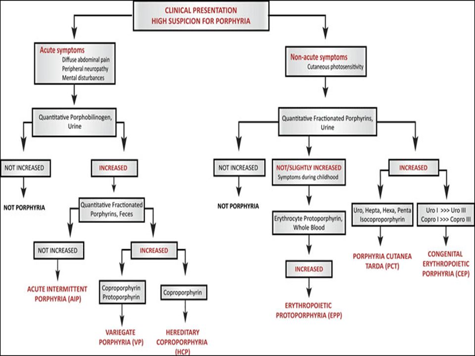
Porphyrias - Enzyme deficiencies and Clinical features
A group of rare disorders caused by deficiencies of enzymes of the heme biosynthetic pathway Caused by hereditary or acquired defects in heme synthesis Accumulation of porphyrins or porphyrin precursors in plasma and tissues increased excretion of of porphyrins or porphyrin precursors in the urine and feces Most porphyrias show a prevalent autosomal dominant pattern, except congenital eythropoietic porphyria, which is recessive Attacks of the disease are triggered by certain drugs, chemicals, and foods, and also by exposure to sun Treatment involves administration of hemin, which provides negative feedback for the heme biosynthetic pathway, and therefore, prevents accumulation of heme precursors

27
Classified into 2 groups based on the organ or cells affected
hepatic, can be acute or chronic, or Acute intermittent porphyria Variegate porphyria Hereditary coproporphyria Porphyria cutanea tarda Erythropoietic Congenital or hereditary erythropoietic porphyria Erythropoietic protoporphyria Those with tetrapyrrole intermediates show photosensitivity due to extended conjugated double bonds - Formation of superoxide radicals - Skin blisters, itches (pruritis) - Skin may darken, grow hair (hypertrichosis)
 - Skin may darken, grow hair (hypertrichosis)")
28
Types of Porphyrias

29
Acute intermittent porphyria (AIP)
Hepatic, autosomal dominant. Disease usually occurs after puberty Caused by a deficiency in Uroporphyrinogen synthase, which is involved in the conversion of porphobilinogen (PBG) to uroporphyrinogen III PBG, uroprophyrin, and 5-ALA accumulate in the plasma and the urine Hence urine turns dark on standing Clinical symptoms Abdominal pain Neuropsychiatric symptoms Smooth muscle spasms Hypertension Constipation Hypercholesterolemia No photosensitivity Due to accumulation of PBG and ALA on nerves in CNS
 to uroporphyrinogen III. PBG, uroprophyrin, and 5-ALA accumulate in the plasma and the urine. Hence urine turns dark on standing. Clinical symptoms. Abdominal pain. Neuropsychiatric symptoms. Smooth muscle spasms. Hypertension. Constipation. Hypercholesterolemia. No photosensitivity. Due to accumulation of PBG and ALA on nerves in CNS.")
30
Variegate Porphyria Caused by a deficiency of protoporphyrinogen oxidase ALA synthase is more active due to lack of sufficient heme, PBG, 5-ALA, uro and coproporphyrins are excreted in urine Urine is colored due to excretion of uro and coproporphyrins. Fecal excretion of uro and coproporphyrins is more. Clinical symptoms Photosensitivity is a constant symptom Other symptoms vary, hence the name. Alcohol and other drugs can aggravate the condition

31
Hereditary Coproporphyria (HCP)
Caused by partial deficiency of coproporphyrinogen III oxidase PBG is excreted in urine and Coproporphyrinogen III is excreted in urine and feces. Urine contains red colored pigment coproporphyrin, which is formed on exposure to light Fecal excretion of uro and coproporphyrins is more. Clinical symptoms Photosensitivity is a constant symptom Other symptoms similar to acute intermittant porphyria

32
Porphyria cutanea tarda (PCT)
Hepatic, autosomal dominant, most common porphyria Caused by partial deficiency of uroporphyrinogen decarboxylase, which is involved in the conversion of uroporphyrinogen III to coproporphyrinogen III Uroporphyrin I and III and PBG are excreted in urine Hence urine appears pinkish to brown Clinical symptoms Photosensitivity is a major symptom (cutaneous photosensitivity) Appears during 4-6 decades of life Hepatocellular damage from alcoholism is characteristic of this condition
 Appears during 4-6 decades of life. Hepatocellular damage from alcoholism is characteristic of this condition.")
33
Congenital or hereditary erythropoietic porphyria
Caused by partial deficiency of uroporphyrinogen III cosynthase Uroporphyrinogen I and coproporphyrinogen I are excreted in urine Urine turns red on standing due to formation of red uro and coproporphyrins from uroporphyrinogen I and coproporphyrinogen I , by the action of atmospheric O2. Clinical symptoms Photosensitivity is a major symptom Pink bones and teeth Hemolytic anemia and cutaneous lesions

34
Erythropoietic protoporphyria
Ferrochelatase enzyme is partially active Excess protoporphyrin IX in plasma and erythrocytes and so increased excretion of protoporphyrin IX in feces. Excretion of porphyrin or its precursors in urine is normal Clinical symptoms Photosensitivity is a major symptom (solar uriticaria) Liver cirrhosis Anemia
 Liver cirrhosis. Anemia.")
35
Acquired Porphyrias Lead poisoning
inhibition of ferrochelatase and ALA dehydratase displaces Zn+2 at enzyme active site Excretion of porphyrins in urine is up to 10mg/ day Automobile exhaust is a common source for lead Children Adults - developmental defects - severe abdominal pain - drop in IQ - mental confusion - hyperactivity - many other symptoms - insomnia - many other health problems Drugs which cause porphyria are Steroids Oral contraceptives Barbiturates Pesticides

36
Laboratory tests in analysis of porphyrias
Used to diagnose and monitor porphyrias. Done in urine, whole blood, erythrocytes, plasma, urine and feces For acute attacks, PBG and urine porphyrins on random samples If these are abnormal, then ALA, PBG, or porphyrins on 24 hours urine sample To distinguish VP and HCP, porphyrins are tested in feces For cutaneous porphyrias, porphyrins are tested in whole blood and urine. Enzyme testing such as PBG deaminase may be done to test detect latent porphyrias.

Similar presentations


. This.>")


 –Central to solar energy utilization Heme (Fe 2+ )>")


