Download presentation
Presentation is loading. Please wait.

1
Chapter 14 Translation

2
MESSENGER RNA Polypeptide chains are specified by open reading frames An open reading frame (ORF) is a contiguous, non-overlapping string of codons. Start codon: the first codon of an ORF. It specifies the first amino acid to be incorporated. It defines the reading frame for all subsequent codons. Stop codon: the last codon of an ORF. Polycistronic mRNAs: mRNAs that contain multiple ORFs. Monocistronic mRNAs: mRNAs that contain a single ORF.
 is a contiguous, non-overlapping string of codons. Start codon: the first codon of an ORF. It specifies the first amino acid to be incorporated. It defines the reading frame for all subsequent codons. Stop codon: the last codon of an ORF. Polycistronic mRNAs: mRNAs that contain multiple ORFs. Monocistronic mRNAs: mRNAs that contain a single ORF.")
3
Prokaryotic mRNAs have a ribosome binding site that recruits the translational machinery
RBS (ribosome binding site): a short sequence upstream of the start codon that facilitates binding of a ribosome. AGGAGG. Also referred to as a Shine-Dalgarno sequence. Interacts with 16S rRNA In case that the start codon of downstream ORF overlaps the stop codon of upstream ORF, for example, with AUGA, translation of two ORFs is linked. This is known as translational coupling.
: a short sequence upstream of the start codon that facilitates binding of a ribosome. AGGAGG. Also referred to as a Shine-Dalgarno sequence. Interacts with 16S rRNA. In case that the start codon of downstream ORF overlaps the stop codon of upstream ORF, for example, with AUGA, translation of two ORFs is linked. This is known as translational coupling.")
4
Eukaryotic mRNAs are modified at their 5’ and 3’ ends to facilitate translation
The 5’ cap is a methylated guanine nucleotide at 5’ end of mRNA. Recruits the ribosome. The ribosome moves in a 5’ 3’ direction until it encounters an AUG in a process called scanning. The Kozak sequence (PuNNAUGG) interacts with initiator tRNA. Poly-A tail promotes efficient recycling of ribosomes
 interacts with initiator tRNA. Poly-A tail promotes efficient recycling of ribosomes.")
5
TRANSFER RNA tRNAs are adaptors between codons and amino acids tRNAs are between 75 and 95 nt in length. 1. All tRNAs end at the 3’ terminus with the sequence 5’-CCA-3’ Several unusual bases are present in the primary structure of tRNAs ΨU (Pseudouridine), D (dihydrouridine), hypoxanthine, thymine, methylguanine -----probably improves tRNA function
, D (dihydrouridine), hypoxanthine, thymine, methylguanine -----probably improves tRNA function.")
6
tRNAs share a common secondary structure that resembles a cloverleaf
The acceptor stem is formed by pairing between the 5’ and 3’ ends of the mRNA molecules. It is the site of amino acid attachment. The ΨU loop. The unusual base ΨU is often found within the sequence 5’-TΨUCG-3’. The D loop The anticodon loop. The anticodon is always bracketed on the 3’ end by a purine and on its 5’ end by uracil. The variable loop. It varies in size from 3 to 21 bases.

7
tRNAs have an L-shaped three-dimensional structure
X-ray crystallography reveals an L-shaped tertiary structure. The terminus of the acceptor stem is about 70 Å away from the anticodon loop at the other end. Three kinds of interactions stabilize this structure: 1. The formation of the two extended regions of base pairing results in additional base stacking interactions. 2. unconventional hydrogen bonds between bases in different helical regions. 3. Interactions between the bases and the sugar-phosphate backbone.

8
ATTACHMENT OF AMINO ACIDS TO tRNA
tRNAs are charged by the attachment of an amino acid to the 3’ terminal adenosine nucleotide via a high-energy acyl linkage The carboxyl group of an amino acid is linked to 2’ or 3’-OH of the terminal adenosine nucleotide through the high energy acyl linkage. The energy released when the bond is broken drives the peptide bond formation. Aminoacyl-tRNA synthetases charge tRNAs in two steps 1. adenylylation – transfer of AMP 2. tRNA charging Class I tRNA synthetases attach a.a. to 2’-OH and are generally monomeric Class II enzymes attach a.a. to 3’-OH and are typically dimeric or tetrameric The amino acid rapidly equilibrates between attachment at 3’ and 2’-OH

11
An exception in some bacteria: amination of Glu-tRNAGln to Gln-tRNAGln
Each aminoacyl-tRNA synthetase attaches a single amino acid to one or more tRNAs Most organisms have 20 different tRNA synthetases. An exception in some bacteria: amination of Glu-tRNAGln to Gln-tRNAGln

12
tRNA synthetases recognize unique structural features of cognate tRNAs
Specificity determinants: the acceptor stem and the anticodon loop Discriminator in the acceptor stem: Changing a particular base pair in the acceptor stem converts the recognition specificity of a tRNA from one synthetase to another. The anticodon loop, including recognition of the anticodon itself, contributes to discrimination. However, in case of serine, AGC and UCA are completely different from each other. The specificity determinants should be outside of the anticodon. The set of tRNA determinants that enable synthetases to discriminate among tRNAs is referred to as the second genetic code.

14
Aminoacyl-tRNA formation is very accurate
Seletion of the correct amino acid is more difficult. Nevertheless, less than 1 in 1,000 tRNAs is charged with the incorrect amino acid. Tyrosine vs. phenylalanine -OH allows the synthetase to discriminate between the two amino acids. Valine vs. isoleucine -CH3 leads to 100 fold difference, which is unacceptable.

16
Some aminoacyl tRNA synthetases use an editing pocket to charge tRNAs with high accuracy
In addition to the catalytic pocket for adenylyation, isoleucyl tRNA synthetase has a nearby editing pocket for proofreading the product of adenylylation reaction. The pocket functions as a molecular sieve. AMP-isoleucine cannot enter the pocket whereas AMP-valine can fit into the pocket and is hydrolyzed.

17
The ribosome is unable to discriminate between correctly and incorrectly charged tRNAs
A mutant tRNA with a nucleotide substitution in the anticodon delivers its usual cognate amino acid to the wrong codon. Cysteinyl-tRNACys can be converted to alanine-tRNACys by chemical reduction. This reduction results in introduction of alanines at the cysteine codons. High fidelity of tRNA synthetases are required for accurate decoding of mRNAs.

18
THE RIBOSOME The ribosome is the macromolecular machine that directs the synthesis of proteins. More complex than the minimal replication and transcription machinery. The ribosome is composed of at least 3 RNAs and more than 50 proteins, with a molecular mass of greater than 2.5 megadaltons. The speed of translation is 20 amino acids (60 nt) per second in prokaryotes. Similar to the rate of RNA transcription 2-4 amino acids per second in eukaryotes
 per second in prokaryotes. Similar to the rate of RNA transcription. 2-4 amino acids per second in eukaryotes.")
19
Transcription and translation are coupled in prokaryotes.

20
The ribosome is composed of a large and a small subunit
Peptidyl transferase center: a part of the large subunit that is responsible for the formation of peptide bonds. Decoding center: a part of the small subunit where charged tRNA decode the codon units of the mRNA. 30S+50S=70S S+60S=80S 2/3 RNA + 1/3 protein

22
The large and Small subunits undergo association and dissociation during each cycle of Translation
The ribosome cycle: the sequence of association and dissociation of the ribosome.

23
Polyribosome (or polysome): an mRNA bearing multiple ribosomes
A single ribosome contacts with 30 nt, but mRNA can bind one ribosome for every 80 nucleotides. New Amino acids are attached to the C-terminus of the growing polypeptide chain Polypeptides are synthesized in the N- to C-terminal direction.

24
Peptide bonds are formed by transfer of the growing polypeptide chain from one tRNA to another
The ribosome catalyzes the formation of a peptide bond between the amino acids attached to tRNAs. Two consequences of the peptidyl transferase reaction: 1. The N-terminus of the protein is synthesized before the C-terminus. 2.The growing polypeptide chain is transferred from the peptidyl-tRNA to the aminoacyl-tRNA. No ATP is required, but ATP is spent during tRNA charging reaction.

26
Ribosomal RNAs are both structural and catalytic determinants of the ribosome
Ribosomal RNAs are not simply structural components but directly responsible for catalytic activity. The peptidyl transferase center and the decoding center are composed almost entirely of RNA. Most ribosomal proteins are on the periphery of the ribosome.

29
The peptidyl transferase center

30
The decoding center

31
The ribosome has three binding sites for tRNA
The A site is for the aminoacylated-tRNA. The P site is for the peptidyl-tRNA. The E site is for the exiting tRNA. Each tRNA binding site is at the interface between the large and the small subunits of the ribosome.

32
Channels through the ribosome allow the mRNA and growing polypeptide to enter and/or exit the ribosome There are two narrow channels in the small subunit, one for entry and the other one for exit of mRNA. only wide enough for unpaired RNA to pass through. There is a kink in the mRNA between the two codons. The incoming aminoacyl tRNA cannot bind to bases immediately adjacent to the vacant A site codon.

34
A channel in the large subunit provides an exit path for the newly synthesized polypeptide chain. The size of the channel limits the folding of the growing polypeptide chain. The polypeptide can form an alpha helix in the channel.

35
INITIATION OF TRANSLATION
Successful translation initiation needs three events The ribosome must be recruited to the mRNA A charged initiator tRNA must be placed into the P site of the ribosome The ribosome must be precisely positioned over the start codon.

36
Prokaryotic mRNAs are initially recruited to the small subunit by base-pairing to rRNA
The small subunit associates with the mRNA first by base-pairing between the RBS and the 16S rRNA. The small subunit is positioned such that the start codon will be in the P site when the large subunit joins the complex. The large subunit joins its partner only at the very end of the initiation process.

37
A specialized tRNA charged with a modified methionine binds directly to the prokaryotic small subunit Initiator tRNA: a special tRNA that base-pairs with the start codon (usually AUG or GUG). The initiator tRNA gets charged with N-formyl methionine. The charged initiator tRNA is referred to as fMet-tRNAifMet. Deformylase: removes the formyl group from the amino terminus during or after the synthesis of the polypeptide chain. Aminopeptidase often removes N-terminal methionine as well as one or two additional amino acids.
. The initiator tRNA gets charged with N-formyl methionine. The charged initiator tRNA is referred to as fMet-tRNAifMet. Deformylase: removes the formyl group from the amino terminus during or after the synthesis of the polypeptide chain. Aminopeptidase often removes N-terminal methionine as well as one or two additional amino acids.")
38
Three initiation factors direct the assembly of an initiation complex that contains mRNA and the initiator tRNA IF1 prevents tRNAs from binding to the portion of the small subunit that will become part of the A site. IF2 is a GTPase that interacts with three key components of the initiation machinery: the 30S, IF1, and charged initiator tRNA. It facilitates the association of the charged initiator tRNA with the small subunit IF3 binds to the small subunit and blocks it from reassociating with a large subunit. It occupies the E site. With all three IFs bound, the small subunit binds to mRNA and the initiator tRNA in either order. Base-pairing between start codon and the initiator leads to conformational change of the small subunit, resulting in the release of IF3. Binding of the large subunit Hydrolysis of GTP bound to IF2 Release of IF2/GDP and IF1 Formation of 70S initiation complex

44
Eukaryotic ribosomes are recruited to the mRNA by the 5’ cap
In eukaryotes, the small subunit associated with the initiator tRNA is recruited to the 5’ cap and scans along the mRNA until it reaches the first AUG. Four initiation fators (eIF1, eIF3, eIF5, and eIF1A) bind to the small subunit. The initiator tRNA is escorted by eIF2 which forms the ternary complex (eIF2-GTP-charged initiator tRNA). eIF2 positions the Met-tRNAiMet in the future P site, forming the 43S preinitiation complex. The mRNA is separately prepared for recognition by the small subunit. The cap is recognized by eIF4E. eIF4G and eIF4A are then recruited. 4E-BPs compete with eIF4G for the binding to eIF4E. eIF4B activates the RNA helicase activity of eIF4A, which unwinds any secondary structures. Interactions between initiation factors bring the mRNA to the 43S complex to form the 48S preinitiation complex.
 bind to the small subunit. The initiator tRNA is escorted by eIF2 which forms the ternary complex (eIF2-GTP-charged initiator tRNA). eIF2 positions the Met-tRNAiMet in the future P site, forming the 43S preinitiation complex. The mRNA is separately prepared for recognition by the small subunit. The cap is recognized by eIF4E. eIF4G and eIF4A are then recruited. 4E-BPs compete with eIF4G for the binding to eIF4E. eIF4B activates the RNA helicase activity of eIF4A, which unwinds any secondary structures. Interactions between initiation factors bring the mRNA to the 43S complex to form the 48S preinitiation complex.")
46
The start codon is found by scanning downstream from the 5’ end of the mRNA
Correct base-pairing between the start codon and the initiator tRNA changes conformation of the 43S complex and that of eIF5, which stimulates eIF2 to hydrolyze the bound GTP. eIF2-GDP, eIF1, eIF3, eIF5 are then released. eIF5B closely related to IF2 binds the initiator tRNA and stimulates association of 60S subunit. Binding of the large subunit leads to the release of remaining initiation factors by stimulating GTP hydrolysis by eIF5B, forming 80S initiation complex

48
Translation initiation factors hold eukaryotic mRNAs in circles
The poly-A tail contributes to efficient translation. Poly-A binding protein coats the poly-A tail and interacts with eIF4G, circularizing mRNA. The newly released ribosome is positioned to reinitiate translation on the same mRNA.

49
TRANSLATION ELONGATION
The correct addition of amino acids needs three key events to occur. 1. The correct aminoacyl-tRNA is loaded into the A site of the ribosome. 2. A peptide bond is formed between the aminoacyl-tRNA in the A site and the peptide chain that is attached to the peptidyl-tRNA in the P site. 3. The resulting peptidyl-tRNA in the A site and its associated codon must be translocated to the P site. Elongation factors control these events.

51
Aminoacyl-tRNAs are delivered to the A site by elongation factor EF-Tu
EF-Tu bound to GTP escorts aminoacyl-tRNAs to the ribosome. EF-Tu bound to GDP has little affinity for aminoacyl tRNAs. After tRNA enters the A site and a correct codon-anticodon match is made, the EF-Tu GTPase is activated by the factor binding center, which activates the IF2 GTPase when the large subunit joins the initiation complex.

53
The ribosome uses multiple mechanisms to select against incorrect aminoacyl-tRNAs
The energy difference a correct codon-anticodon pair and a near match cannot account for the high level of accuracy with the error rate of Three mechanisms select against incorrect pairings. 1. Two adjacent adenine residues in the 16S rRNA within the A site form additional hydrogen bonds within the minor groove.

54
2. The GTPase activity of EF-Tu is activated only by the proper interaction with the factor binding center, which is dependent on the correct codon-anticodon base pairing.

55
3. The third mechanism is a form of proofreading that occurs after EF-Tu is released. To participate in the peptidyl transferase reaction, the tRNA must rotate into the peptidyl transferase center in a process called accommodation. The 3’ end of tRNA moves almost 70A. This rotation is likely to place strain on the codon-anticodon interaction.

56
The Ribosome is a ribozyme.
The peptidyl transferase center is composed by the 23S rRNA of the large subunit. The N-terminus of L27 reaches into the active site. Experimental data suggest that although L27 facilitates peptide bond formation, it is not essential for peptidyl transferase activity. Entropic catalysis (entropy reduction): Base-pairing between 23S rRNA and 3’ CCA ends of the tRNAs in the A and P sites brings substrates in close proximity. The transferase reaction is catalyzed by the substrate-assisted catalysis, in which the peptidyl-tRNAs themselves carry critical catalytic elements. The 2’-OH of the P site tRNA may act as a part of a proton shuttle. Removal of this OH group results in 106-fold reduction in catalysis rates. The 2’-OH donates a proton to the 3’-OH of the peptidyl tRNA and accepts a proton from the attacking amino group.
: Base-pairing between 23S rRNA and 3’ CCA ends of the tRNAs in the A and P sites brings substrates in close proximity. The transferase reaction is catalyzed by the substrate-assisted catalysis, in which the peptidyl-tRNAs themselves carry critical catalytic elements. The 2’-OH of the P site tRNA may act as a part of a proton shuttle. Removal of this OH group results in 106-fold reduction in catalysis rates. The 2’-OH donates a proton to the 3’-OH of the peptidyl tRNA and accepts a proton from the attacking amino group.")
59
Peptide bond formation and the elongation factor EF-G drive translocation of the tRNAs and the mRNA
In the translocation step, the P-site tRNA must move to the E site, the A-site tRNA must move to the P site, and the mRNA must move by three nucleotides to expose the next codon. The initial translocation steps are coupled to the peptidyl transferase reaction. Translocation in the large subunit precedes translocation in the small subunit and tRNAs said to be in hybrid states. The completion of translocation requires the action of a second elongation factor EF-G. EF-G-GTP binds to the A site and the factor binding center stimulates GTP hydrolysis. GTP hydrolysis changes conformation of EF-G, allowing it to reach into the small subunit and trigger translocation of the A site tRNA. EF-G-GDP releases from the ribosome.

61
EF-G drives translocation by displacing the tRNA bound to the A site
EF-G-GDP occupies the A site of the decoding center Frame-shifting tRNAs having 4 nt anticodons move mRNA by four nt --- base-pairing is maintained during translocation. Movement of the P site tRNA into the E site disrupts base-pairing. A counterclockwise rotation of the small subunit occurs during translocation. Gates that separate A, P, and E sites must open during the translocation and close after EF-G is released. Molecular mimicry: EF-G-GDP and EF-Tu-GTP-tRNA have a very similar structure.

63
EF-Tu-GDP and EF-G-GDP must exchange GDP for GTP prior to participating in a new round of elongation
GDP has a lower affinity for EF-G than does GTP. The elongation factor EF-Ts acts as a GTP exchange factor for EF-Tu.

64
A cycle of peptide bond formation consumes two molecules of GTP and one molecule of ATP
Making a peptide bond costs two molecules of GTP and one of ATP. 1. one ATP is consumed for the formation of the aminoacyl-tRNA. 2. one GTP is consumed in the delivery of a charged tRNA and in ensuring the correct codon-anticodon recognition. 3. One more GTP is consumed in the EF-G-mediated process of translocation.

65
TERMINATION OF TRANSLATION
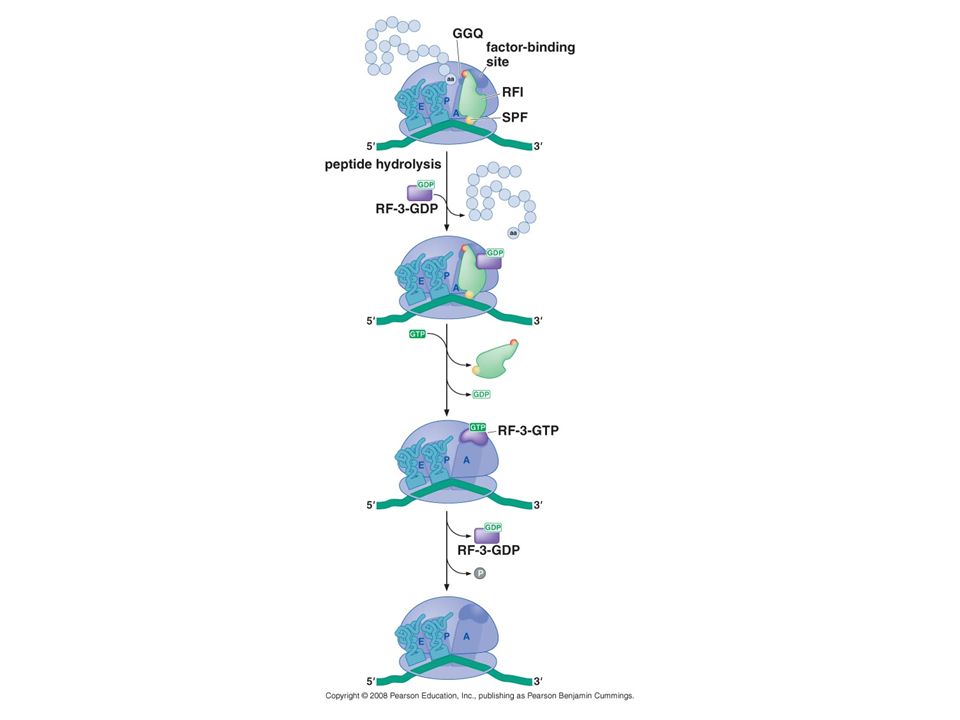
Release factors terminate translation in response to stop codons RFs (release factors) recognize stop codons and activate the hydrolysis of the polypeptide from the peptidyl-tRNA. 1. Class I release factors recognize the stop codons and trigger hydrolysis of the peptide chain. RF1 (for UAG and UAA) and RF2 (for UGA and UAA). eRF1 2. Class II release factors stimulate the dissociation of the class I factors from the ribosome after release of the polypeptide chain. RF3 and eRF3. regulated by GTP hydrolysis
 recognize stop codons and activate the hydrolysis of the polypeptide from the peptidyl-tRNA. 1. Class I release factors recognize the stop codons and trigger hydrolysis of the peptide chain. RF1 (for UAG and UAA) and RF2 (for UGA and UAA). eRF1. 2. Class II release factors stimulate the dissociation of the class I factors from the ribosome after release of the polypeptide chain. RF3 and eRF3. regulated by GTP hydrolysis.")
66
Short regions of class I release factors recognize stop codons and trigger release of the peptidyl chain Recognition of stop codons is mediated by a protein-RNA interaction. Swapping of short regions of RF1 and RF2 leads to changes in stop-codon specificity. Three amino acids (a peptide anticodon) are responsible for the specificity of stop codon recognition.
 are responsible for the specificity of stop codon recognition.")
67
RF1 binds to the A site of the ribosome.

68
The peptide anticodon is located very near the stop codon.

69
All class I release factors share a conserved GGQ motif that is essential for polypeptide release. The GGQ motif is located near the peptidyl transferase center.

70
Class I release factors functionally mimic a tRNA
Class I release factors functionally mimic a tRNA. The GGQ and the peptide anticodon occupy extreme ends of RF1.

71
GDP/GTP exchange and GTP Hydrolysis control the function of the class II Release factor
RF3: a class II release factor that accomplishes removal of the class I release factor from the ribosome. Has a higher affinity for GDP than GTP. RF3-GDP binds to the ribosome only in the presence of class I RF Conformational changes in the ribosome and class I factor induced by polypeptide release stimulate RF3 to exchange its bound GDP for GTP. The binding of GTP to RF3 leads to the formation of a high-affinity interaction with the ribosome, leading to the release of class I RF

73
The ribosome recycling factor mimics a tRNA
To participate in a new round of polypeptide synthesis, the ribosome gets separated from the tRNA and the mRNA and dissociate into its large and small subunits in a process called ribosome recycling. RRF (ribosome recycling factor) cooperates with EF-G and IF3 to recycle ribosomes after polypeptide release. RRF is a mimic of tRNA. However, it interacts with the ribosome in a manner very different from that of a tRNA EF-G and RRF lead to the release of the P-site tRNA directly from the P site, not moving it to the E site.
 cooperates with EF-G and IF3 to recycle ribosomes after polypeptide release. RRF is a mimic of tRNA. However, it interacts with the ribosome in a manner very different from that of a tRNA. EF-G and RRF lead to the release of the P-site tRNA directly from the P site, not moving it to the E site.")
77
REGULATION OF TRANSLATION
One advantage of translational control over transcriptional control is the ability to rapidly respond to stimuli. Translation is typically regulated at the level of initiation.

78
Protein or RNA binding near the ribosome-binding site negatively regulates bacterial translation initiation Protein or RNA can inhibit 30S subunit binding to the RBS. Some RNA-binding proteins recognize RNA structures adjacent to the RBS. An mRNA base-pairs with itself to mask the RBS. Translation of another gene disrupts the base-pairing and releases the translational inhibition.

79
Regulation of prokaryotic translation: Ribosomal proteins are translational repressors of their own synthesis Expression of ribosomal proteins and rRNA is regulated coordinately. Synthesis of ribosomal proteins (and ribosomes) is linked to cell’s growth rate.
 is linked to cell’s growth rate.")
80
Ribosomal protein genes are organized into several operons
Ribosomal protein genes are organized into several operons. When the copy number of a ribosomal protein operon increases, the amount of its mRNA increases but that of the ribosomal proteins stays nearly the same. Ribosomal proteins are repressors of their own translation. One (or a complex of two) of the encoded ribosomal proteins binds near the RBS of one of the most 5’-proximal genes.
 of the encoded ribosomal proteins binds near the RBS of one of the most 5’-proximal genes.")
82
Downstream genes are also repressed when the stop codon of an upstream gene is located very close to the start codon of a downstream gene or when mRNA folds into a structure which blocks internal RBSs from the ribosome binding.

83
Binding sites in rRNA and mRNA are similar in sequence and in secondary structure. The ribosomal protein biding is stronger to rRNA than to mRNA.

84
Global regulators of eukaryotic translation target key factors required for mRNA recognition and initiator tRNA ribosome binding Under conditions of reduced nutrients or other cellular stresses, translation in eukaryotic cells can be globally reduced. Phosphorylation of the alpha subunit of eIF2 inhibits the action of eIF2B, the GTP exchange factor for eIF2, leading to reduced levels of eIF2-GTP (which escorts the initiator tRNA to the 40S subunit). The eIF2a kinases are activated by a number of different conditions including amino acid starvation. 4E-BPs compete with eIF4G for binding to eIF4E, the cap-binding protein. Phosphorylation of 4E-BPs inhibits their binding to eIF4E. Phosphorylation of 4E-BPs is mediated by mTor, which is activated by growth factors that stimulate cell division. Rapamycin, an inhibitor of mTor, is a chemotherapy agent.
. The eIF2a kinases are activated by a number of different conditions including amino acid starvation. 4E-BPs compete with eIF4G for binding to eIF4E, the cap-binding protein. Phosphorylation of 4E-BPs inhibits their binding to eIF4E. Phosphorylation of 4E-BPs is mediated by mTor, which is activated by growth factors that stimulate cell division. Rapamycin, an inhibitor of mTor, is a chemotherapy agent.")
86
Spatial control of translation by mRNA-specific 4E-BPs
Binding to eIF4E is used to regulate translation of specific mRNAs. The Oscar mRNA is synthesized by nurse cells of the fly ovary and deposited into the anterior of the oocyte prior to fertilization. Oscar mRNA is then transported to the posterior of the oocyte. During the transport, translation of Oscar mRNA is repressed. The Oscar mRNA contains Bruno response elements in the 3’-UTR. Bruno recruits Cup, a 4E-BP. Cup is not abundant enough to inhibit global translation. Nanos is also regulated by Cup.

88
An iron-regulated, RNA-binding protein controls translation of ferritin
Iron is used as a cofactor in many proteins. Too little or too much iron can cause problems to the human body. The iron-binding protein ferritin stores and releases iron in a regulated manner. Ferritin translation is regulated by iron-binding protein called iron regulatory proteins (IRPs). In iron-deficient cells, IRPs bind to IRE (iron regulatory element) at the 5’ end of ferritin mRNA. IRP binding inhibits the ability of eIF4A/B to unwind the hairpin and blocks binding of 43S complex to the mRNA. When iron concentration is elevated in the cell, IRPs cannot bind to the IRE.
. In iron-deficient cells, IRPs bind to IRE (iron regulatory element) at the 5’ end of ferritin mRNA. IRP binding inhibits the ability of eIF4A/B to unwind the hairpin and blocks binding of 43S complex to the mRNA. When iron concentration is elevated in the cell, IRPs cannot bind to the IRE.")
89
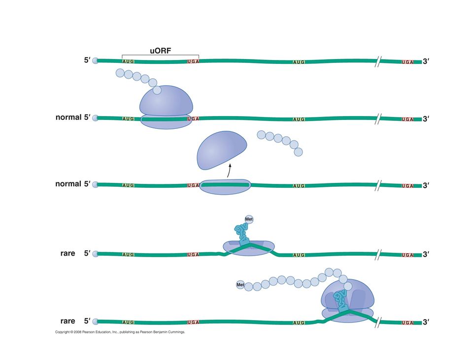
Translation of the yeast transcriptional activator Gcn4 is controlled by short upstream ORFs and ternary complex abundance Gcn4 is a yeast transcriptional activator that regulates genes involved in amino acid biosynthesis. Under conditions of low levels of amino acids, Gcn4 mRNA is translated, whereas under nonstarvation conditions it is not translated. Gcn4 mRNA has four upstream ORFs. The first uORF has a unique property that allows 50% of 40S subunits to remain bound to the mRNA after its translation and resume scanning.

90
For recognition of AUG by the small subunit of ribosome, the ternary complex eIF2-GTP-loaded initiator should bind to the 40S subunit. When amino acids are abundant, the ternary complex rebinds the scanning ribosome soon after translation of uORF1. Following reinitiation at one of the other uORFs, the ribosome fully dissociates from the mRNA and fails to translate the main Gcn4 ORF.

91
Under conditions of starvation, Gcn2 is activated and phosphorylates eIF2, leading to a reduced rate of the ternary complex formation and binding to the 40S subunit. The distance between uORF1 and the main ORF provides enough time for TC to bind to the 40S subunit, increasing the chance to translate Gcn4.

94
TRANSLATION-DEPENDENT REGULATION OF mRNA AND PROTEIN STABILITY
In some cases, the process of translation is used to detect defective mRNAs and elimenate ether defective mRNA or their protein products. The SsrA RNA rescues ribosomes that translate broken mRNAs Ribosomes stalled at the 3’end of a broken mRNA can be rescued by a chimeric tmRNA that is part tRNA and part mRNA. SsrA, a 457-nucleotide tmRNA, includes a region that resembles tRNAAla. The SsrA RNA can be charged with alanine. Translocation of peptidyl-SsrA RNA results in the release of the broken mRNA. The incomplete protein product is fused to a ten-amino-acid tag at its C-terminus, which is recognized by cellular proteases for its rapid degradation. This tmRNA can bind only to stalled ribosomes because additional room is created in the A site due to the missing 3’ end of the mRNA.

96
Eukaryotic cells degrade mRNAs that are incomplete or have premature stop codons
Translation is tightly linked to the process of mRNA decay in eukaryotic cells. Nonsense-mediated mRNA decay is the process by which mRNA containing a premature stop codon (a nonsense codon) is rapidly degraded. Normally, the exon junction complexes are displaced by the ribosome. If a premature stop codon is present, undisplaced complexes interact with the prematurely terminating ribosome, which activates an enzyme that removes the cap at the 5’ end of the mRNA. Removal of the cap causes rapid degradation of the mRNA by a 5’ 3’ exonuclease. Nonstop-mediated decay rescues ribosomes that translate mRNAs that lack a stop codon. The ribosome translates through the poly-A tail, adding multiple lysines to the end of protein. The stalled ribosome is bound by a protein (eRF3-like Ski7). This protein stimulates ribosome dissociation and recruit a 3’5 exonuclease. Proteins that contain poly-lysine at their carboxy-terminus are unstable and rapidly degraded. In theses processes, mRNA degradation requires translation of the damaged mRNA.
 is rapidly degraded. Normally, the exon junction complexes are displaced by the ribosome. If a premature stop codon is present, undisplaced complexes interact with the prematurely terminating ribosome, which activates an enzyme that removes the cap at the 5’ end of the mRNA. Removal of the cap causes rapid degradation of the mRNA by a 5’ 3’ exonuclease. Nonstop-mediated decay rescues ribosomes that translate mRNAs that lack a stop codon. The ribosome translates through the poly-A tail, adding multiple lysines to the end of protein. The stalled ribosome is bound by a protein (eRF3-like Ski7). This protein stimulates ribosome dissociation and recruit a 3’5 exonuclease. Proteins that contain poly-lysine at their carboxy-terminus are unstable and rapidly degraded. In theses processes, mRNA degradation requires translation of the damaged mRNA.")
101
14_UnFigure01.jpg

102
14_UnFigure04.jpg

103
14_UnFigure04a.jpg

104
14_UnFigure04b.jpg

106
Eric Klann & Thomas E. Dever

Similar presentations


>")
 Translation Sept 25, 2008 BIO 184 Dr. Tom Peavy.>")


 Protein Synthesis. Translation Slow rate of synthesis (18 amino acids per second) In bacteria translation and transcription are coupled.>")
 Lecture 8 Macromolecular Synthesis and Processing: Proteins (Text Chapter: 10)>")


