Download presentation
Presentation is loading. Please wait.

1
Chapter 3 Cells Physiology
Membrane Transport -Passive -Active Cell Cycle Protein Synthesis

2
Cellular Physiology: Membrane Transport
Membrane Transport – movement of substance into and out of the cell Transport is by two basic methods Passive transport No energy is required Active transport The cell must provide metabolic energy

3
Solutions and Transport
Solution – homogeneous mixture of two or more components Solvent – dissolving medium Solutes – components in smaller quantities within a solution

4
Intracellular fluid – nucleoplasm and cytosol
Extracellular fluid – fluid on the exterior of the cell Interstitial fluid – only the fluid that surrounds the cells

5
Selective Permeability
The plasma membrane allows some materials to pass while excluding others This permeability includes movement into and out of the cell

6
Passive Processes What determines whether or not a substance can passively permeate a membrane? Lipid solubility of substance Channels of appropriate size Carrier proteins PLAY Animation: Membrane Permeability

7
Passive Transport Processes
Diffusion Particles tend to distribute themselves evenly within a solution (reach equilibrium) Movement is from high concentration to low concentration, or down a concentration gradient Figure 3.8
 Movement is from high concentration to low concentration, or down a concentration gradient. Figure 3.8.")
8
Passive Transport Processes
Types of diffusion Simple diffusion (Dialysis) Unassisted process Solutes are lipid-soluble materials or small enough to pass through membrane pores
 Unassisted process. Solutes are lipid-soluble materials or small enough to pass through membrane pores.")
9
Passive Transport Processes - Diffusion
Nonpolar lipid- soluble (hydrophobic) substances diffuse directly through the phospholipid bilayer PLAY Animation: Diffusion (a) Simple diffusion of fat-soluble molecules directly through the phospholipid bilayer
 substances diffuse directly through the phospholipid bilayer. PLAY. Animation: Diffusion. (a) Simple diffusion of fat-soluble molecules. directly through the phospholipid bilayer.")
10
(a) Simple diffusion of fat-soluble molecules
Extracellular fluid Lipid- soluble solutes Cytoplasm (a) Simple diffusion of fat-soluble molecules directly through the phospholipid bilayer Figure 3.7a
 Simple diffusion of fat-soluble molecules. directly through the phospholipid bilayer. Figure 3.7a.")
11
Passive Transport Processes
Facilitated diffusion Substances require a protein carrier for passive transport. Still moves with the concetration gradient.

12
Diffusion through the Plasma Membrane
Figure 3.9

13
PhysioEx Lab Simple Diffusion Facilitated Diffusion

14
Passive Transport Processes
Types of diffusion Osmosis – simple diffusion of water across a semipermeable membrane

15
Passive Processes: Osmosis
Water diffuses through plasma membranes: Through the lipid bilayer Through protein channels

16
(d) Osmosis, diffusion of a solvent such as
Water molecules Lipid billayer Aquaporin (d) Osmosis, diffusion of a solvent such as water through a specific channel protein (aquaporin) or through the lipid bilayer Figure 3.7d
 Osmosis, diffusion of a solvent such as. water through a specific channel protein. (aquaporin) or through the lipid bilayer. Figure 3.7d.")
17
Passive Processes: Osmosis
Water concentration is determined indirectly by solute concentration because solute particles displace water molecules Osmolarity: The measure of total concentration of solute particles When solutions of different osmolarity are separated by a membrane, osmosis occurs until equilibrium is reached

18
Solute and water molecules move down their concentration gradients
(a) Membrane permeable to both solutes and water Solute and water molecules move down their concentration gradients in opposite directions. Fluid volume remains the same in both compartments. Left compartment: Solution with lower osmolarity Right compartment: Solution with greater osmolarity Both solutions have the same osmolarity: volume unchanged H2O Solute Solute molecules (sugar) Membrane Figure 3.8a
 Membrane permeable to both solutes and water. Solute and water molecules move down their concentration gradients. in opposite directions. Fluid volume remains the same in both compartments. Left. compartment: Solution with. lower osmolarity. Right. compartment: Solution with. greater osmolarity. Both solutions have the. same osmolarity: volume. unchanged. H2O. Solute. Solute. molecules. (sugar) Membrane. Figure 3.8a.")
19
(b) Membrane permeable to water, impermeable to solutes
Solute molecules are prevented from moving but water moves by osmosis. Volume increases in the compartment with the higher osmolarity. Both solutions have identical osmolarity, but volume of the solution on the right is greater because only water is free to move Left compartment Right compartment H2O Solute molecules (sugar) Membrane Figure 3.8b
 Membrane. Figure 3.8b.")
20
Importance of Osmosis When osmosis occurs, water enters or leaves a cell Change in cell volume disrupts cell function PLAY Animation: Osmosis

21
PhysioEx Lab Osmosis

22
Get Laptops ready to go.

23
Tonicity Tonicity: The ability of a solution to cause a cell to shrink or swell Isotonic: A solution with the same solute concentration as that of the cytosol Hypertonic: A solution having greater solute concentration than that of the cytosol Hypotonic: A solution having lesser solute concentration than that of the cytosol

25
Figure 3.9 (a) Isotonic solutions (b) Hypertonic solutions
(c) Hypotonic solutions Cells retain their normal size and shape in isotonic solutions (same solute/water concentration as inside cells; water moves in and out). Cells lose water by osmosis and shrink in a hypertonic solution (contains a higher concentration of solutes than are present inside the cells). Cells take on water by osmosis until they become bloated and burst (lyse) in a hypotonic solution (contains a lower concentration of solutes than are present in cells). Figure 3.9
 Hypotonic solutions. Cells retain their normal size and. shape in isotonic solutions (same. solute/water concentration as inside. cells; water moves in and out). Cells lose water by osmosis and. shrink in a hypertonic solution. (contains a higher concentration. of solutes than are present inside. the cells). Cells take on water by osmosis until. they become bloated and burst (lyse) in a hypotonic solution (contains a. lower concentration of solutes than. are present in cells). Figure 3.9.")
26
Passive Transport Processes
Filtration Water and solutes are forced through a membrane by fluid, or hydrostatic pressure A pressure gradient must exist Solute-containing fluid is pushed from a high pressure area to a lower pressure area

27
Active Transport Processes
Transport substances that are unable to pass by diffusion They may be too large They may not be able to dissolve in the fat core of the membrane They may have to move against a concentration gradient Two common forms of active transport Solute pumping Bulk transport

28
Active Transport Processes
Solute pumping Amino acids, some sugars and ions are transported by solute pumps ATP energizes protein carriers, and in most cases, moves substances against concentration gradients

29
Active Transport Processes
Figure 3.10

30
Active Transport Processes
Bulk transport Exocytosis Moves materials out of the cell Material is carried in a membranous vesicle Vesicle migrates to plasma membrane Vesicle combines with plasma membrane Material is emptied to the outside

31
Active Transport Processes
Figure 3.11

32
Active Transport Processes
Bulk transport Endocytosis Extracellular substances are engulfed by being enclosed in a membranous vesicle Types of endocytosis Phagocytosis – cell eating Pinocytosis – cell drinking

33
Active Transport Processes
Figure 3.12

34
Passive Membrane Transport – Review
Process Energy Source Example Simple diffusion Kinetic energy Movement of O2 through membrane Facilitated diffusion Movement of glucose into cells Osmosis Movement of H2O in & out of cells Filtration Hydrostatic pressure Formation of kidney filtrate

35
Active Membrane Transport – Review
Process Energy Source Example Active transport of solutes ATP Movement of ions across membranes Exocytosis Neurotransmitter secretion Endocytosis White blood cell phagocytosis Pinocytosis Absorption by intestinal cells

36
Osmosis Lab

37
Osmosis Lab Complete the lab calculations, graph and questions at home. Formal report due Wednesday, 9/26

38
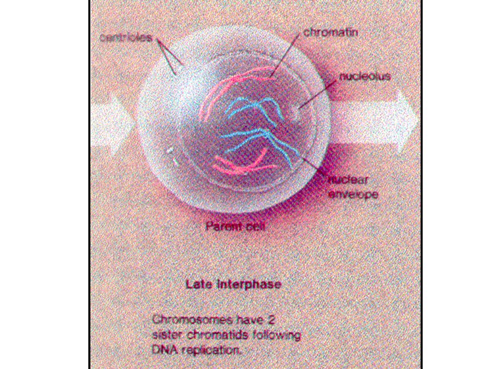
Cell Life Cycle Cells have two major periods Interphase Cell grows
Cell carries on metabolic processes Cell division Cell replicates itself Function is to produce more cells for growth and repair processes

39
Events of Cell Division
Mitosis Division of the nucleus Results in the formation of two daughter nuclei Cytokinesis Division of the cytoplasm Begins when mitosis is near completion Results in the formation of two daughter cells

40
Preparations for DNA Replication
Interphase No cell division occurs The cell carries out normal metabolic activity and growth

41
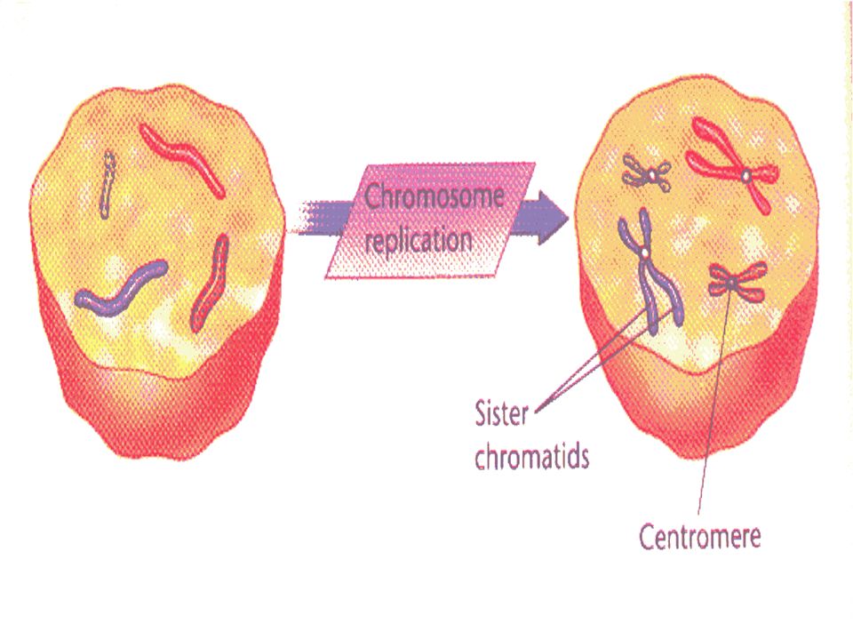
DNA Replication Genetic material duplicated and readies a cell for division into two cells Occurs toward the end of interphase DNA uncoils and each side serves as a template Figure 3.13

44
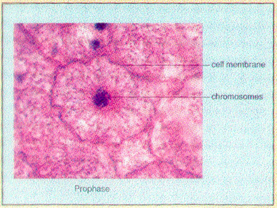
Stages of Mitosis Prophase Mitosis begins here
First part of cell division Centrioles migrate to the poles

47
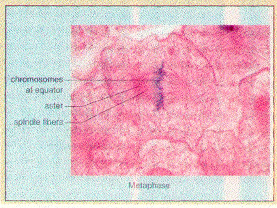
Stages of Mitosis Metaphase
Spindle from centrioles are attached to chromosomes at the centromeres that are aligned in the center of the cell

48
The chromatids appear to repell each other, taking the shape of an X.

51
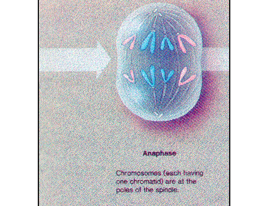
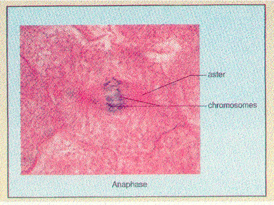
Stages of Mitosis Anaphase
Daughter chromosomes are pulled toward the poles The cell begins to elongate

52
Anaphase The separated sister chromatids (daughter chromosomes) appear to be pulled apart by the centromeres.
 appear to be pulled apart by the centromeres.")
55
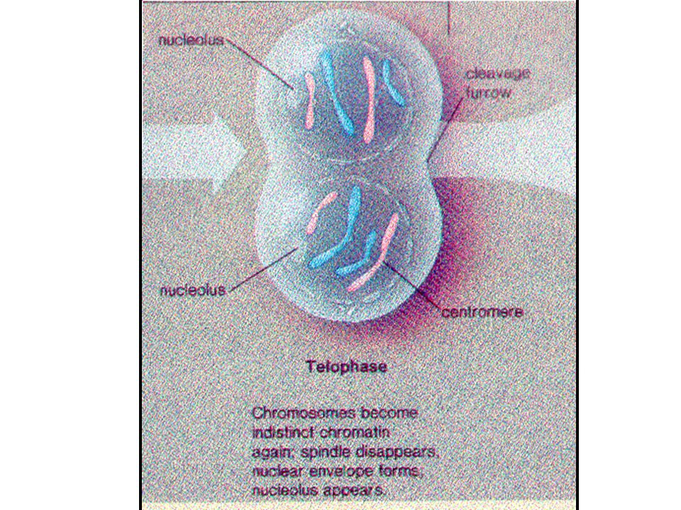
Telophase Daughter nuclei begin forming
A cleavage furrow (for cell division) begins to form
 begins to form.")
56
Telophase When the daughter chromosomes reach the end of the spindle. new nucleus forms.

59
Cytokinesis plasma membrane constricts (cleavage furrow) and the cytoplasm divides, called cytokinesis. The two resulting cells are called daughter cells.

61
Stages of Mitosis Figure 3.14; 1

62
Stages of Mitosis Figure 3.14; 2

63
Online Mitosis Lab

64
Protein Synthesis Gene – DNA segment that carries a blueprint for building one protein Proteins have many functions Building materials for cells Act as enzymes (biological catalysts) RNA is essential for protein synthesis
 RNA is essential for protein synthesis.")
65
Role of RNA Messenger RNA (mRNA) Transfer RNA (tRNA)
Carries the instructions for building a protein from the nucleus to the ribosome Has 3 base codons Transfer RNA (tRNA) Transfers appropriate amino acids to the ribosome for building the protein Has 3 base anticodons Ribosomal RNA (rRNA) Helps form the ribosomes where proteins are built
 Transfers appropriate amino acids to the ribosome for building the protein. Has 3 base anticodons. Ribosomal RNA (rRNA) Helps form the ribosomes where proteins are built.")
66
Transcription and Translation
Transfer of information from DNA’s base sequence to the complimentary base sequence of mRNA. In the nucleus Translation Base sequence of nucleic acid is translated to an amino acid sequence outside the nucleus Amino acids are the building blocks of proteins More Translation

67
Protein Synthesis Figure 3.15

Similar presentations


 Plasma membranes are selectively permeable some molecules pass through membrane; some don’t Types of Membrane Transport.>")


 Digest foods Dispose of wastes.>")


