Download presentation
Presentation is loading. Please wait.

1
The Eye

2
Anatomy of the Eye Sclera Choroid Retina Aqueous humor Iris
Blood vessels Pupil Cornea Optic nerve Lens Blind spot Ciliary muscle Vitreous humor Label the diagram of the eye!

3
Sclera and Cornea (outer layer)
Tough, white outer layer; Protects and supports the eye Tough, clear outer layer; Refracts (bends) light toward the pupil Sclera
 light toward. the pupil. Sclera.")
4
Choroid layer Did you know that many nocturnal animals also have an iridescent layer of tissue called the tapetum lucidum which helps reflect light back into the eye? Choroid - A black layer within the eye that prevents light from scattering and provides the retina with nutrients.

5
Anatomy of the Eye - Iris
The coloured part of the eye A muscle that contracts to control the size of the pupil; controls the amount of light entering the eye Iris scans can be used as a method of identification because each person has unique banding of colour in their eye

6
Anatomy of the Eye - Pupil
The hole in the middle of the iris Where light enters the eye Size changes depending on the amount of light present in the environment

7
Anatomy of the Eye - Lens
Transparent structure behind the iris Helps focus light rays onto the retina Changes shape based on the distance of the object

8
Aqueous Humour and Vitreous Humour
- Jelly-like material at the front of the eye behind the cornea; supplies cornea with nutrients, bends light and maintains shape Vitreous Humour - Bends light and gives the eye structure Iris scans – identification because each person’s iris has a different color pattern

9
Anatomy of the Eye - Retina
Nerve layer at the back of the eye Senses light and creates nerve impulses Location of the: Rods: detect black and white Cones: detect red, blue, & green

10
Rods and Cones Humans have 125 million rods and 6 million cones
Rods allow us to see black and white Peripheral vision appears to be black and white because the cones are concentrated at the fovea

11
Color blindness Color blindness occurs when
one or more cones are defective Red-green color blindness is most common Caused by a genetic defect on the X chromosome What numbers do you see?

12
Color blindness How do we see colour?

13
Normal Vision Reduced sensitivity to blue

14
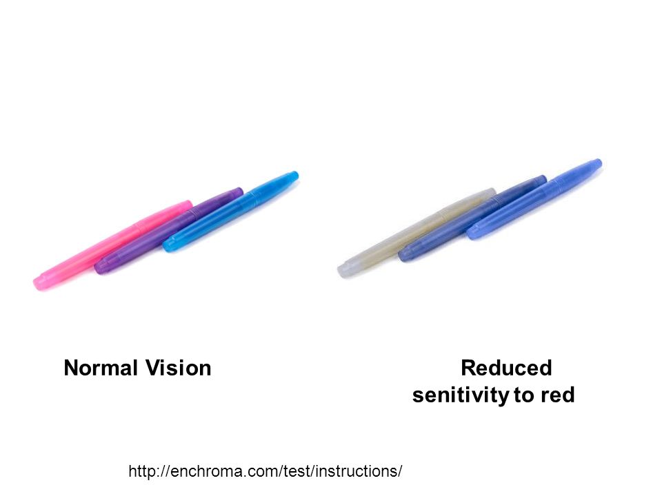
Normal Vision Reduced senitivity to red
15
Fovea centralis (macula)
Most sensitive area of the retina, highest concentration of CONES = greatest visual acuity To find the fovea, draw a straight line from the center of the lens

16
Anatomy of the Eye – Optic Nerve
Carries nerve signals from the retina to the brain, where the image is interpreted

17
Where the retina attaches to the optic nerve. There are
Blind Spot Where the retina attaches to the optic nerve. There are NO rods or cones in the blind spot Hold text book 30 cm away. Close your right eye and look at the black cross with the left eye. Slowly move your left eye towards the figure and when the dot disappears, the image is falling on the blind spot. You don’t notice the blind spot because one eye will compensate for the other. Blind Spot test! Open to page 472 Optic nerve carries info to the occipital lobe

18
Ciliary Muscle Ciliary Body
Alters the shape of the lens; important in accommodation

19
Pink eye (conjunctivitis) =
Conjunctiva Conjunctiva Thin layer on top of the cornea for protecting the eye; blocks entry of pathogens into the eye Pink eye (conjunctivitis) = infection of the conjunctiva
 = infection of the conjunctiva.")
20
Anatomy of the Eye – Eye Muscle
Helps to move the eye Eye muscles operate in antagonistic pairs (ex: one muscle must relax and the other muscle contracts to move the eye)
")
21
Who do you see?

22
Who do you see?

23
Who do you see?

24
Optical Illusions!

25
Vision Problems and Corrective Lenses
Hyperopia – “Far sightedness” Description: Person CAN see far objects; has difficulty seeing nearby objects Solution: Glasses with converging lenses

26
Vision Problems and Corrective Lenses
Myopia – “Near sightedness” Description: Person CAN see close objects; has difficulty seeing far objects Solution: Glasses with diverging lenses

27
Presbyopia

28
Astigmatism Irregularly shaped eye causes different focal lengths in the horizontal and vertical directions

29
Cataracts Clouding of the lens leading to decreased vision and eventual blindness.

Similar presentations


 What is the difference between a mirror an a lens? 2) Why do you think we have a lens in our eye instead of a mirror?>")
>")
>")


 than.>")


