Download presentation
Presentation is loading. Please wait.

1
OPHTHALMOLOGY EYE AND ITS DISEASES

2
OBJECTIVES References Functions of the eye; three steps of vision Why two eyes ? External features Eye ball (Cross section, coats, segments) Lacrimal apparatus and tear film Areas of special interest ( extraocular muscles, angle of ant. Chamber, retina, visual pathway) Bony orbit
 Lacrimal apparatus and tear film Areas of special interest ( extraocular muscles, angle of ant. Chamber, retina, visual pathway) Bony orbit.")
3
RECOMMENDED TEXTBOOKS PARSON KANSKI (CLINICAL OPHTHALMOLOGY) OPHTHALMOLOGY F0R UNDERGRADUATES BASIC AND CLINICAL SCIENCE COURSE (AAO) BASIC OPHTHALMOLOGY (AAO)
 OPHTHALMOLOGY F0R UNDERGRADUATES BASIC AND CLINICAL SCIENCE COURSE (AAO) BASIC OPHTHALMOLOGY (AAO)")
5
EYE BALL Sphere…24mm Two poles…Ant. (center of the cornea) …post. (between the optic disc & the fovea). …post. (between the optic disc & the fovea). Three concentric coats :..Outer fibrous..Middle vascular..Middle vascular..Inner nervous..Inner nervous
. …post. (between the optic disc & the fovea). Three concentric coats :..Outer fibrous..Middle vascular..Middle vascular..Inner nervous..Inner nervous.")
8
1-OUTER FIBROUS LAYER A- Cornea: ant. Transparent 1/6 th. A- Cornea: ant. Transparent 1/6 th. *Light Entry (transmission). *Light Entry (transmission). *Light Refraction (42 diopters out of total 60 diopters) *Light Refraction (42 diopters out of total 60 diopters) *Protective *Protective B- Sclera : post opaque white 5/6 th. B- Sclera : post opaque white 5/6 th. * Provides dark environment to improve contrast. * Provides dark environment to improve contrast. * Insertion of extraocular muscles. * Insertion of extraocular muscles. * Protective. * Protective. * Exit of optic nerve fibers thru a posterior opening (lamina cribrosa). * Exit of optic nerve fibers thru a posterior opening (lamina cribrosa). C- Limbus: corneoscleral junction C- Limbus: corneoscleral junction
. *Light Entry (transmission). *Light Refraction (42 diopters out of total 60 diopters) *Light Refraction (42 diopters out of total 60 diopters) *Protective *Protective B- Sclera : post opaque white 5/6 th. B- Sclera : post opaque white 5/6 th. * Provides dark environment to improve contrast. * Provides dark environment to improve contrast. * Insertion of extraocular muscles. * Insertion of extraocular muscles. * Protective. * Protective. * Exit of optic nerve fibers thru a posterior opening (lamina cribrosa). * Exit of optic nerve fibers thru a posterior opening (lamina cribrosa). C- Limbus: corneoscleral junction C- Limbus: corneoscleral junction.")
9
2- MIDDLE VASCULAR LAYER (UVEA) A. Iris : vertical muscular diaphragm behind the cornea, perforated at its center (pupil). It has two muscles(….). *Regulates amount of light entering the eye depending on ambient light condition. *Regulates amount of light entering the eye depending on ambient light condition. *Prevents peripheral rays entering (stop effect) *Prevents peripheral rays entering (stop effect) B. Ciliary body : posterior continuation of the iris. Triangular in section. * Gives attachment to the zonules which suspend the lens behind the pupil. * Gives attachment to the zonules which suspend the lens behind the pupil. * Contraction of ciliary muscles is responsible for the lens accommodation. * Contraction of ciliary muscles is responsible for the lens accommodation. * Secretion of aqueous humor from the ciliary epithelium. * Secretion of aqueous humor from the ciliary epithelium. C. Choroid : the posterior most part of the uvea.It lines the sclera & nourishes the outer sublayers of retina and the macula including the fovea.
. It has two muscles(….). *Regulates amount of light entering the eye depending on ambient light condition. *Regulates amount of light entering the eye depending on ambient light condition. *Prevents peripheral rays entering (stop effect) *Prevents peripheral rays entering (stop effect) B. Ciliary body : posterior continuation of the iris. Triangular in section. * Gives attachment to the zonules which suspend the lens behind the pupil. * Gives attachment to the zonules which suspend the lens behind the pupil. * Contraction of ciliary muscles is responsible for the lens accommodation. * Contraction of ciliary muscles is responsible for the lens accommodation. * Secretion of aqueous humor from the ciliary epithelium. * Secretion of aqueous humor from the ciliary epithelium. C. Choroid : the posterior most part of the uvea.It lines the sclera & nourishes the outer sublayers of retina and the macula including the fovea..")
10
Uveal Tract Parts

11
3-INNER NERVOUS LAYER (RETINA) Light sensitive layer changes light energy into an electrical impulse (Phototransduction). changes light energy into an electrical impulse (Phototransduction). Very thin transparent layer that lines the choroid. Anteriorly it reaches the Ora Serrata. Posteriorly it ends around the optic disc. Two main sublayers..Outer Retinal Pigment epithelium(RPE) &..Inner Sensory Retina (Neuroretina) which is subdivided into nine layers Macula?Fovea?
. Very thin transparent layer that lines the choroid. Anteriorly it reaches the Ora Serrata. Posteriorly it ends around the optic disc. Two main sublayers..Outer Retinal Pigment epithelium(RPE) &..Inner Sensory Retina (Neuroretina) which is subdivided into nine layers Macula Fovea .")
14
EYE BALL SEGMENTS & CHAMBERS The Crystalline Lens is suspended by zonules to the ciliary body. The lens & its zonules divide the eye ball into two segments: 1) Anterior segment: *Boundaries… *Boundaries… *Contains Aqueous Humor *Contains Aqueous Humor 2) Posterior segment: *Boundaries… *Boundaries… *contains Vitreous Humor *contains Vitreous Humor The anterior segment is further subdivided by the iris into A. Anterior chamber B. Posterior Chamber
 Anterior segment: *Boundaries… *Boundaries… *Contains Aqueous Humor *Contains Aqueous Humor 2) Posterior segment: *Boundaries… *Boundaries… *contains Vitreous Humor *contains Vitreous Humor The anterior segment is further subdivided by the iris into A. Anterior chamber B. Posterior Chamber.")
19
AQUEOUS HUMOR CIRCULATTION Dynamic equilibrium Secretion :from the Non Pigmented ciliary Epithelium...post. chamber…pupil…ant. Chamber (AC)… Drainage: Angle of the ant. Chamber…Trabecular meshwork…Canal of Schlemm…Collector channels…Episcleral venous plexus…Ciliary veins …Systemic circulation. Functions of the Aqueous humor: 1- Acts as blood or lymph to provide nutrients & take waste products from the avascular cornea and lens. 2- Light transmission and refraction. 3- Responsible for the IOP.
… Drainage: Angle of the ant. Chamber…Trabecular meshwork…Canal of Schlemm…Collector channels…Episcleral venous plexus…Ciliary veins …Systemic circulation. Functions of the Aqueous humor: 1- Acts as blood or lymph to provide nutrients & take waste products from the avascular cornea and lens. 2- Light transmission and refraction. 3- Responsible for the IOP..")
21
BONY ORBITS Two quadrilateral pyramidal cavities in the skull. Each has …Apex (posterior) Each has …Apex (posterior) …Base (anterior) …Base (anterior) …Four walls …Four walls Each contains...Eye ball Each contains...Eye ball …Extraocular muscles …Extraocular muscles …Vessels & nerves …Vessels & nerves …Lacrimal gland …Lacrimal gland …Lacrimal sac …Lacrimal sac …Orbital fat …Orbital fat Each communicates….with the cranial cavity thru: 1- Optic foramen 1- Optic foramen 2- Superior orbital fissure 2- Superior orbital fissure ….with the pterygoid fossa thru: ….with the pterygoid fossa thru: Inferior orbital fissure Inferior orbital fissure
 Each has …Apex (posterior) …Base (anterior) …Base (anterior) …Four walls …Four walls Each contains...Eye ball Each contains...Eye ball …Extraocular muscles …Extraocular muscles …Vessels & nerves …Vessels & nerves …Lacrimal gland …Lacrimal gland …Lacrimal sac …Lacrimal sac …Orbital fat …Orbital fat Each communicates….with the cranial cavity thru: 1- Optic foramen 1- Optic foramen 2- Superior orbital fissure 2- Superior orbital fissure ….with the pterygoid fossa thru: ….with the pterygoid fossa thru: Inferior orbital fissure Inferior orbital fissure.")
22
The bony orbit Walls

23
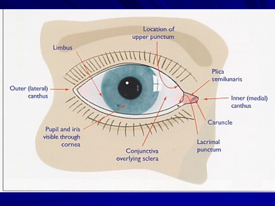
OCULAR ADNEXAE 1. Eye lids : two movable mucocutaneous folds, 2. Conjunctiva : mucous membrane lining the eye lids and covering the anterior part of sclera. 3. Lacrimal apparatus: A- Secretory system B- Drainage system. B- Drainage system.

24
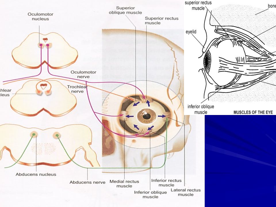
EXTRAOCULAR MUSCLES 6 = 4+2 6 = 4+2 From the Orbit to the Sclera From the Orbit to the Sclera L6. SO4 / 3

Similar presentations


 1. Cornea 2. Sclera Middle Tunic (pg. 470-474) 3. Choroid Coat 4. Ciliary Body 5. Lens & Accommodation 6. Aqueous.>")


>")

 separated by the palpebral fissue Eyelashes Tarsal glands Lacrimal apparatus Vision Accessory structures.>")


. 2. *The sclera is the.>")
