Download presentation
Presentation is loading. Please wait.

1
Copyright © 2008 Pearson Education, Inc., publishing as Pearson Benjamin Cummings PowerPoint ® Lecture Presentations for Biology Eighth Edition Neil Campbell and Jane Reece Lectures by Chris Romero, updated by Erin Barley with contributions from Joan Sharp Chapter 40 Basic Principles of Animal Form and Function

2
Overview: Diverse Forms, Common Challenges Anatomy is the study of the biological form of an organism Physiology is the study of the biological functions an organism performs Copyright © 2008 Pearson Education, Inc., publishing as Pearson Benjamin Cummings

3
Exchange with the Environment An animal’s size and shape directly affect how it exchanges energy and materials with its surroundings Exchange occurs as substances dissolved in the aqueous medium diffuse and are transported across the cells’ plasma membranes In vertebrates, the space between cells is filled with interstitial fluid, which allows for the movement of material into and out of cells Copyright © 2008 Pearson Education, Inc., publishing as Pearson Benjamin Cummings

4
Fig. 40-3 Exchange 0.15 mm (a) Single cell 1.5 mm (b) Two layers of cells Exchange Mouth Gastrovascular cavity
 Single cell 1.5 mm (b) Two layers of cells Exchange Mouth Gastrovascular cavity.")
5
Fig. 40-4 0.5 cm Nutrients Digestive system Lining of small intestine Mouth Food External environment Animal body CO 2 O2O2 Circulatory system Heart Respiratory system Cells Interstitial fluid Excretory system Anus Unabsorbed matter (feces) Metabolic waste products (nitrogenous waste) Kidney tubules 10 µm 50 µm Lung tissue
 Metabolic waste products (nitrogenous waste) Kidney tubules 10 µm 50 µm Lung tissue.")
6
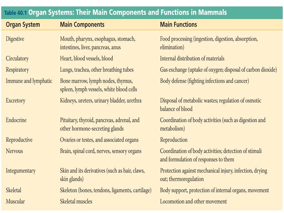
Most animals are composed of specialized cells organized into tissues that have different functions Tissues make up organs, which together make up organ systems Hierarchical Organization of Body Plans Copyright © 2008 Pearson Education, Inc., publishing as Pearson Benjamin Cummings

8
Tissue Structure and Function Epithelial Tissue – covers surfaces because cells are in contact – lines hollow organs, cavities and ducts – forms glands when cells sink under the surface Connective Tissue – material found between cells – supports and binds structures together – stores energy as fat – provides immunity to disease Muscle Tissue – cells shorten in length producing movement Nerve Tissue – cells that conduct electrical signals – detects changes inside and outside the body – responds with nerve impulses

9
Epithelial Tissue General Features: Closely packed cells forming continuous sheets Cells sit on basement membrane Apical (upper) free surface Avascular - nutrients diffuse in from underlying connective tissue Good nerve supply Rapid cell division Covering / lining versus glandular types Basic Functions: - protection - secretion - absorption - filtration
 free surface Avascular - nutrients diffuse in from underlying connective tissue Good nerve supply Rapid cell division Covering / lining versus glandular types Basic Functions: - protection - secretion - absorption - filtration")
10
Apical surface Basal surface Basal lamina 40 µm Basement Membrane Basal lamina –from epithelial cells –collagen fibers Holds cells to connective tissue Guide for cell migration during development

11
Classification of Epithelium Classified by arrangement of cells into layers –simple –stratified –pseudostratified Classified by shape of surface cells –squamous –cuboidal –columnar –transitional

12
Epithelial Tissue Classification

13
Epithelial Tissue Cuboidal epithelium Simple columnar epithelium Pseudostratified ciliated columnar epithelium Stratified squamous epithelium Simple squamous epithelium

14
Connective Tissue General Features: - Cells rarely touch due to extracellular matrix - Matrix - fibers & ground substance secreted by cells - Consistency varies from liquid, gel to solid - Does not occur on free surface - Good nerve & blood supply except cartilage & tendons Basic Functions: - support to body organs- storage - protection- binds body parts together

15
Connective Tissue 1. Ground substance: interstitial fluid, cell adhesion proteins, proteoglycans 2. Fibers: Collagen, Elastin, Reticular 3. Cells: - blastmast cells - cytemacrophages - clastblood cells

16
Connective Tissue 1. Loose connective tissue a. Adipose tissue 2. Dense (Fibrous) connective tissue a. Tendons b. Ligaments 3.Cartilage 4.Bone (Osseous ) Tissue 5. Blood Tissue
 connective tissue a. Tendons b. Ligaments 3.Cartilage 4.Bone (Osseous ) Tissue 5. Blood Tissue.")
17
Fig. 40-5c Connective Tissue Collagenous fiber Loose connective tissue Elastic fiber 120 µm Cartilage Chondrocytes 100 µm Chondroitin sulfate Adipose tissue Fat droplets 150 µm White blood cells 55 µm Plasma Red blood cells Blood Nuclei Fibrous connective tissue 30 µm Osteon Bone Central canal 700 µm

18
Collagenous fiber 120 µm Elastic fiber Loose connective tissue Nuclei Fibrous connective tissue 30 µm

19
Osteon Central canal Bone 700 µm Chondrocytes Chondroitin sulfate Cartilage 100 µm

20
Fat droplets Adipose tissue 150 µm White blood cells Plasma Red blood cells 55 µm Blood

21
Muscle Tissue Muscle tissue consists of long cells called muscle fibers, which contract in response to nerve signals It is divided in the vertebrate body into three types: – Skeletal muscle, or striated muscle, is responsible for voluntary movement – Smooth muscle is responsible for involuntary body activities – Cardiac muscle is responsible for contraction of the heart Copyright © 2008 Pearson Education, Inc., publishing as Pearson Benjamin Cummings

22
Fig. 40-5j Muscle Tissue 50 µm Skeletal muscle Multiple nuclei Muscle fiber Sarcomere 100 µm Smooth muscle Cardiac muscle Nucleus Muscle fibers 25 µm Nucleus Intercalated disk

23
Skeletal muscle Multiple nuclei Muscle fiber Sarcomere Smooth muscle Nucleus Muscle fibers NucleusIntercalated disk Cardiac muscle

24
Nervous Tissue Nervous tissue senses stimuli and transmits signals throughout the animal Nervous tissue contains: – Neurons, or nerve cells, that transmit nerve impulses – Glial cells, or glia, that help nourish, insulate, and replenish neurons Copyright © 2008 Pearson Education, Inc., publishing as Pearson Benjamin Cummings

25
Fig. 40-5n Glial cells Nervous Tissue 15 µm Dendrites Cell body Axon Neuron Axons Blood vessel 40 µm

26
Coordination and Control Control and coordination within a body depend on the endocrine system and the nervous system The endocrine system transmits chemical signals called hormones to receptive cells throughout the body via blood A hormone may affect one or more regions throughout the body Hormones are relatively slow acting, but can have long-lasting effects Copyright © 2008 Pearson Education, Inc., publishing as Pearson Benjamin Cummings

27
The nervous system transmits information between specific locations The information conveyed depends on a signal’s pathway, not the type of signal Nerve signal transmission is very fast Nerve impulses can be received by neurons, muscle cells, and endocrine cells Copyright © 2008 Pearson Education, Inc., publishing as Pearson Benjamin Cummings

28
Fig. 40-6 Stimulus Hormone Endocrine cell Signal travels everywhere via the bloodstream. Blood vessel Response (a) Signaling by hormones Stimulus Neuron Axon Signal Signal travels along axon to a specific location. Signal Axons Response (b) Signaling by neurons
 Signaling by hormones Stimulus Neuron Axon Signal Signal travels along axon to a specific location. Signal Axons Response (b) Signaling by neurons.")
29
Homeostasis Organisms use homeostasis to maintain a “steady state” or internal balance regardless of external environment In humans, body temperature, blood pH, and glucose concentration are each maintained at a constant level Copyright © 2008 Pearson Education, Inc., publishing as Pearson Benjamin Cummings

30
Mechanisms of homeostasis moderate changes in the internal environment For a given variable, fluctuations above or below a set point serve as a stimulus; these are detected by a sensor and trigger a response The response returns the variable to the set point Mechanisms of Homeostasis Copyright © 2008 Pearson Education, Inc., publishing as Pearson Benjamin Cummings Animation: Positive Feedback Animation: Positive Feedback Animation: Negative Feedback Animation: Negative Feedback

31
Fig. 40-8 Response: Heater turned off Stimulus: Control center (thermostat) reads too hot Room temperature decreases Set point: 20ºC Room temperature increases Stimulus: Control center (thermostat) reads too cold Response: Heater turned on
 reads too hot Room temperature decreases Set point: 20ºC Room temperature increases Stimulus: Control center (thermostat) reads too cold Response: Heater turned on.")
32
Feedback Loops in Homeostasis The dynamic equilibrium of homeostasis is maintained by negative feedback, which helps to return a variable to either a normal range or a set point Most homeostatic control systems function by negative feedback, where buildup of the end product shuts the system off Positive feedback loops occur in animals, but do not usually contribute to homeostasis Copyright © 2008 Pearson Education, Inc., publishing as Pearson Benjamin Cummings

33
Fig. 40-16 Sweat glands secrete sweat, which evaporates, cooling the body. Thermostat in hypothalamus activates cooling mechanisms. Blood vessels in skin dilate: capillaries fill; heat radiates from skin. Increased body temperature Decreased body temperature Thermostat in hypothalamus activates warming mechanisms. Blood vessels in skin constrict, reducing heat loss. Skeletal muscles contract; shivering generates heat. Body temperature increases; thermostat shuts off warming mechanisms. Homeostasis: Internal temperature of 36–38°C Body temperature decreases; thermostat shuts off cooling mechanisms.

34
Fig. 42-27 Breathing control centers Cerebrospinal fluid Pons Medulla oblongata Carotid arteries Aorta Diaphragm Rib muscles

35
Fig. 45-12 Homeostasis: Blood glucose level (about 90 mg/100 mL) Glucagon STIMULUS: Blood glucose level falls. Alpha cells of pancreas release glucagon. Liver breaks down glycogen and releases glucose. Blood glucose level rises. STIMULUS: Blood glucose level rises. Beta cells of pancreas release insulin into the blood. Liver takes up glucose and stores it as glycogen. Blood glucose level declines. Body cells take up more glucose. Insulin
 Glucagon STIMULUS: Blood glucose level falls. Alpha cells of pancreas release glucagon. Liver breaks down glycogen and releases glucose. Blood glucose level rises. STIMULUS: Blood glucose level rises. Beta cells of pancreas release insulin into the blood. Liver takes up glucose and stores it as glycogen. Blood glucose level declines. Body cells take up more glucose. Insulin.")
36
Calcitonin Thyroid gland releases calcitonin. Stimulates Ca 2+ deposition in bones Reduces Ca 2+ uptake in kidneys STIMULUS: Rising blood Ca 2+ level Blood Ca 2+ level declines to set point Homeostasis: Blood Ca 2+ level (about 10 mg/100 mL) Blood Ca 2+ level rises to set point STIMULUS: Falling blood Ca 2+ level Stimulates Ca 2+ release from bones Parathyroid gland Increases Ca 2+ uptake in intestines Active vitamin D Stimulates Ca 2+ uptake in kidneys PTH Figure 45.11
 Blood Ca 2+ level rises to set point STIMULUS: Falling blood Ca 2+ level Stimulates Ca 2+ release from bones Parathyroid gland Increases Ca 2+ uptake in intestines Active vitamin D Stimulates Ca 2+ uptake in kidneys PTH Figure")
Similar presentations