Download presentation
Presentation is loading. Please wait.

1
Practical Blood Bank Lab 3 Rh Grouping

2
Practical Aspects of Rh Grouping
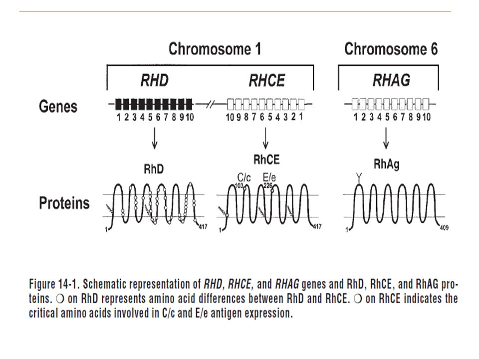
The Rh blood group system is the most polymorphic of the human blood groups, consisting of at least 45 independent antigens and, next to ABO, is the most clinically significant in transfusion medicine Rh grouping -in routine- use for donors and patients involves testing for Rh (D) antigen only. However tests for other important Rh antigens e.g. C, c, E and e may be done for Rh genotyping. The method of Rh grouping mainly depends on the type of reagents available, for which the manufacturers’ instructions have to be strictly followed.
 antigen only. However tests for other important Rh antigens e.g. C, c, E and e may be done for Rh genotyping. The method of Rh grouping mainly depends on the type of reagents available, for which the manufacturers’ instructions have to be strictly followed.")
4
Reagents for Rh (D) Grouping
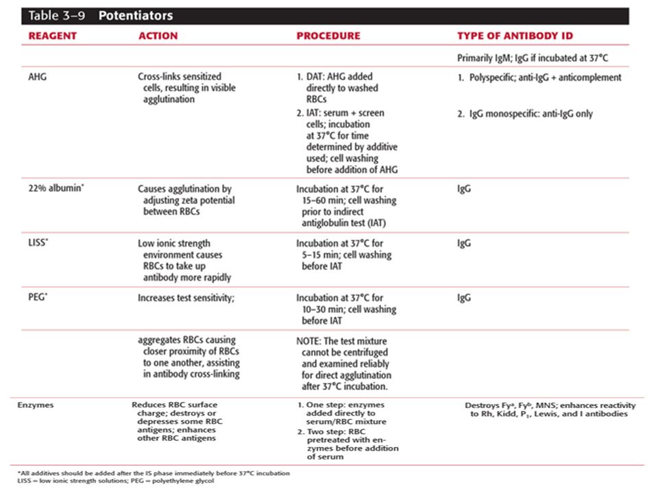
Polyclonal human anti-D serum (IgG) Potentiating or enhancing substances such as albumin, enzymes and AHG reagent are used to bring about agglutination with human IgG anti-D. Anti-D serum (IgG) for saline or rapid tube test (high protein medium) This contains macromolecular additives and give reliable results. Anti-D for saline tube test 2 types Anti-D IgM Anti-D IgG, Chemically modified a potentiator is a reagent that enhances sensitization of an antigen. Potentiators are used in the clinical laboratory for performing blood banking procedures that require enhancement of agglutination in order to detect the presence of antibodies or antigens in a patient's blood sample. Examples of potentiators include albumin, LISS (low ionic-strength saline) and PEG (polyethylene glycol) [1]. Potentiators are also known as enhancement reagents.
 Potentiating or enhancing substances such as albumin, enzymes and AHG reagent are used to bring about agglutination with human IgG anti-D. Anti-D serum (IgG) for saline or rapid tube test (high protein medium) This contains macromolecular additives and give reliable results. Anti-D for saline tube test 2 types. Anti-D IgM Anti-D IgG, Chemically modified. a potentiator is a reagent that enhances sensitization of an antigen. Potentiators are used in the clinical laboratory for performing blood banking procedures that require enhancement of agglutination in order to detect the presence of antibodies or antigens in a patient s blood sample. Examples of potentiators include albumin, LISS (low ionic-strength saline) and PEG (polyethylene glycol) [1]. Potentiators are also known as enhancement reagents.")
5
Monoclonal antibodies
IgM anti-D monoclonal reagent IgM and IgG anti-D monoclonal reagent Blend of IgM monoclonal + IgG polyclonal reagent These antibodies are highly specific, react equally well at 20°C as well as 37°C and are reliable for slide and rapid test tube technique. IgM anti-D monoclonal reagent cannot be used for Du testing by indirect antiglobulin test (IAT) while IgM + IgG monoclonal reagent and blend of IgM monolconal and IgG polyclonal can be used for Du testing.
 while IgM + IgG monoclonal reagent and blend of IgM monolconal and IgG polyclonal can be used for Du testing.")
7
Controls for Rh (D) grouping
Known Group 0 Rh (D) positive and group 0 Rh (D) negative cells may be used as controls with monoclonal anti-D reagent. Alternatively, AB serum or diluents control provided with the anti-D reagent or 22% bovine serum albumin may be used as negative control with the test cells
 positive and group 0 Rh (D) negative cells may be used as controls with monoclonal anti-D reagent. Alternatively, AB serum or diluents control provided with the anti-D reagent or 22% bovine serum albumin may be used as negative control with the test cells.")
8
Rh (D) Grouping In most of the blood transfusion laboratories, Rh (D) grouping is performed along with the ABO grouping and same techniques as used for ABO grouping may also be employed for Rh typing
 grouping is performed along with the ABO grouping and same techniques as used for ABO grouping may also be employed for Rh typing")
9
Slide Technique This technique may be used in emergency Rh (D) typing if a centrifuge is not available. The slide test is not recommended for routine test as it may not pick up weak reactions, thus giving negative results.

10
Tube Technique Saline Agglutination test for Rh (D) Typing Procedure
Prepare 2-5% washed red cell suspension of test sample. Place 1 drop of anti-D in cleaned tube labelled D. Place 1 drop of 22% bovine albumin /control reagent in another tube labeled C. Add 1 drop of 2-5% test cell suspension to each tube. Mix well and centrifuge at 1000 rpm for 1 minute (in case of using IgG anti-D, incubate at 37°C for 10min. and centrifuge (spin tube method) or incubate at 37°C for 60 minutes (sedimentation method). Resuspend the cell button and look for agglutination. All negative results must be confirmed under microscope
 or incubate at 37°C for 60 minutes (sedimentation method). Resuspend the cell button and look for agglutination. All negative results must be confirmed under microscope.")
11
Interpretation Positive test : Agglutination in anti-D and smooth suspension in control tube. Negative test : Smooth suspension in all the tubes (test and control) Test is considerable invalid if both test and control tubes show a positive reaction. In such case, the test should be repeated using saline IgM anti-D. For all microscopically negative reactions in donor grouping, Du testing should be performed, hereas some workers suggest that if the two anti D reagents used are potent and specific, it is not necessary to perform Du testing.
 Test is considerable invalid if both test and control tubes show a positive reaction. In such case, the test should be repeated using saline IgM anti-D. For all microscopically negative reactions in donor grouping, Du testing should be performed, hereas some workers suggest that if the two anti D reagents used are potent and specific, it is not necessary to perform Du testing.")
12
Albumin technique for Rh (D) typing
Principle Albumin increases the dielectric constant of the medium and thus reduces the zeta potential. Due to this effect, the electrical repulsion between the red blood cells is less and the cells agglutinate. Mostly 22% bovine albumin is used, as higher concentrations can cause rouleaux formation. Zeta potential is electric potential in the interfacial double layer (DL) at the location of the slipping plane versus a point in the bulk fluid away from the interface. In other words a potential is the potential difference between the dispersion medium and the stationary layer of fluid attached to the dispersed particle.
 at the location of the slipping plane versus a point in the bulk fluid away from the interface. In other words a potential is the potential difference between the dispersion medium and the stationary layer of fluid attached to the dispersed particle.")
13
The zeta (ζ) potential is the electrostatic potential at the boundary dividing the compact layer and the diffuse layer. The zeta potential serves as an important parameter in characterizing the electrostatic interaction between particles in dispersed systems and the properties of dispersion as affected by this electrical phenomenon.(9-12)
.")
14
Schematic representaion of the ionic cloud concept and it relevance to hemagglutinaiton induced by IgM and IgG antibodies. Compare the size of the IgG antibody with that of IgM molecule. The size of the IgG molecule is not large enough to span the distance between two adacent RBCs.

15
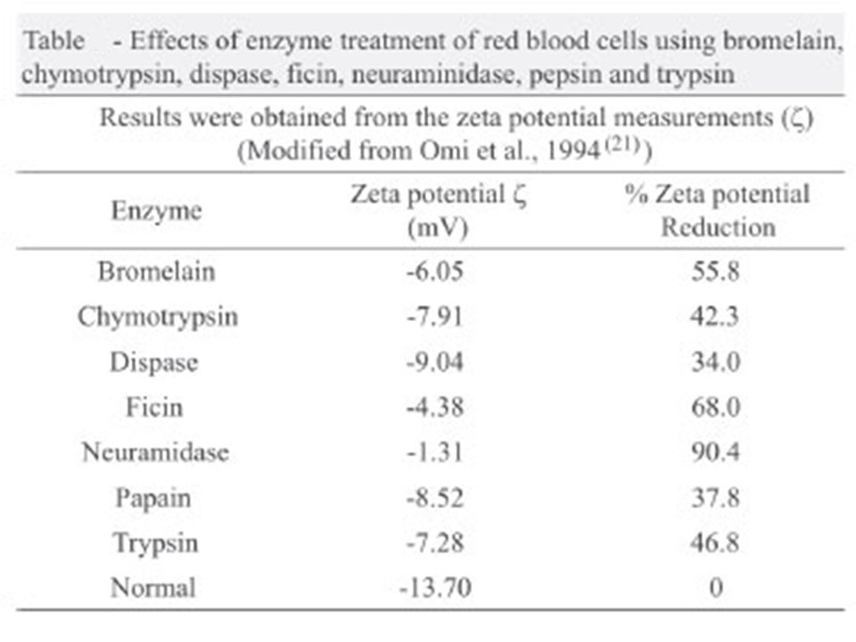
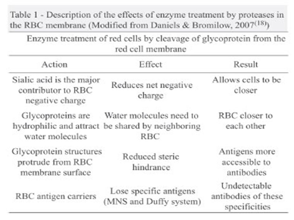
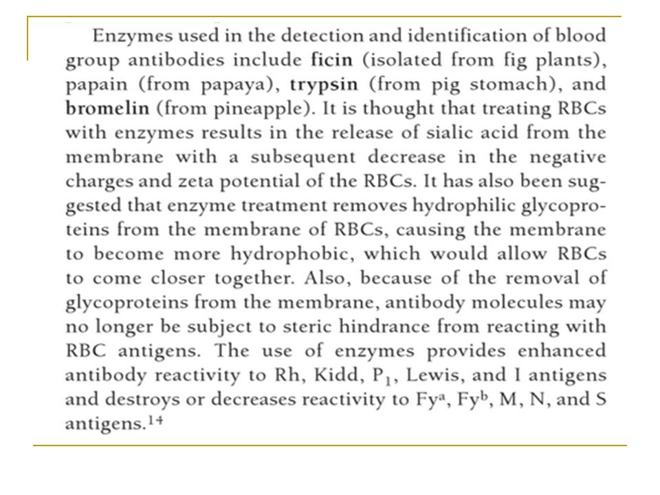
Enzyme agglutination technique for Rh (D) typing
Proteolytic enzymes such as papain, trypsin, bromelain and ficin digest the cell membrane partially and expose Rh antigens to react with IgG antibodies. When the membrane is partially removed, it brings about a loss of negative electric charge on the red cell which is responsible for keeping the cells a set distance apart, hence small IgG molecules are able to span the gap between cells and bring about agglutination.

19
Rh(D) Grouping In Haemolytlc Disease of the Newborn
In haemolytic disease of the newborn, the baby’s red cells may be coated with immunoglobulin and a saline reactive Rh antiserum is usually necessary for testing. When the cells are heavily coated with antibody, no free antigenic sites remain for reaction, resulting in a negative test. This is suspected when the infant’s cells show a positive direct antiglobulin test (DAT) and a negative test with anti-D reagent. In such instances it is recommended that the antibody should be eluted by gentle elution (heating at 45°C for 30 minutes) to expose the antigenic sites before testing.
 and a negative test with anti-D reagent. In such instances it is recommended that the antibody should be eluted by gentle elution (heating at 45°C for 30 minutes) to expose the antigenic sites before testing.")
20
Rh Discrepancies Rh –ve persons mistyped, & transfused with Rh +ve blood have 70% chance of becoming immunized False +ve reactions can be identified by testing an Rh control with the patient’s red cells each time an Rh typing is performed Rh Control: The Rh control is an autocontrol that reveals whether or not the patient's red cells are agglutinating in the absence of the D antigen, i.e., are clumping whether they are D-positive or D-negative. The control consists of testing the patient's cells with an Rh control medium (supplied by the manufacturer) that contains everything that is present in the anti-D typing sera except the anti-D.
 that contains everything that is present in the anti-D typing sera except the anti-D.")
21
Causes of false positive reactions
Positive direct antiglobulin test Polagglutinable red cells Cold agglutinins or Rouleaux formation

22
1- Positive direct antiglobulin test
The presence of Ab on patient’s red cells can cause a false +ve reaction with slide and tube anti-D High protein medium reduces zeta potential allowing red cells to move closer The cell bound Ab can cross link cells and cause agglutination

23
2- Polyagglutinable red cells
Rh–ve red cells that are polyagglutinable due to T or Tn activation Agglutination occurs if anti-T or anti-Tn present in the anti-D reagent Most anti-D reagents do not contain these antibodies T activation occurs most often in association with infections with Streptococcus pneumoniae, Clostridium perfringens, or Vibrio cholerae, though the same findings can be present in influenza virus infections. Enzymes from the bacteria cleave sialic acid residues from glycophorins A and B, exposing the T antigen to full view (while also decreasing expression of MNS blood group antigens carried on the same glycophorin chains). - See more at:
. - See more at:")
24
3- Cold agglutinins or Rouleaux formation
Rh typing is performed using serum suspended red cells If individual’s serum contains cold agglutinin or abnormal protein, false positive reactions can occur

25
False Negatives False negatives are not readily identifiable, but can occur in the following instances: The most common is the result of too heavy cell suspension due to too many cells for the amount of antibody in the antisera. They may also rarely be caused by extremely strong positive DAT. In this case a the patient's D antigen sites are coated in vivo and there are no sites left for commercial anti-D to attach to. This can be fixed by heating cells gently to elute off antibody without damaging cells, then re-test.

26
Resolving Rh Problems Erroneous Results in Rh Grouping
Perform clerical checks for validity of labels and requisition forms. Obtain a fresh blood sample of patient. Check patient records for history, diagnosis, pregnancy, medication and previous transfusion. Check equipment and reagents for proper quality control. Check the antisera and controls. Perform alternative procedures such as washing of red cells with warm saline, enzyme treatment of red cells, absorption / elution studies. Carry out family studies.

27
Lectin Reactions Clinical and laboratory data are use to predict with polyagglutination also Lectins are used in to work up with suspected cases of polyagglutination Arachis hypogea : peanut الفول السوداني Glycine Max: Soyabean فول الصويا Salvia Sclarea: دنو ، دقن الفار Salvia horminum: Dolichos biflorus:

28
Thank You

Similar presentations

 and Elution/Eluate Testing>")


SBB Blood Centers of the Pacific>")


 Course code: MLHE-201 Supervisor: Prof. Dr Magda Sultan Date : 26/12/2013 Outcome : The student will know : -The types of.>")

>")


