Download presentation
Presentation is loading. Please wait.

3
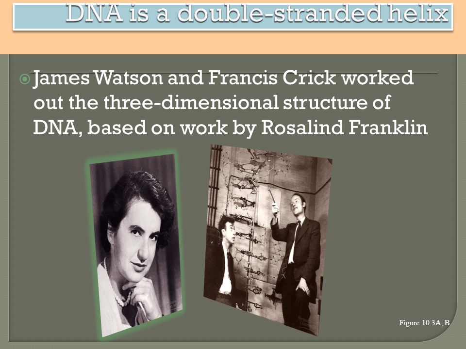
James Watson and Francis Crick worked out the three-dimensional structure of DNA, based on work by Rosalind Franklin Figure 10.3A, B

4
Deoxyribonucleic Acid Double helix Carries genetic information Located in the nucleus The monomer is a nucleotide A phosphate A ribose sugar A nitrogenous base

5
What are the bases in DNA A – adenine T – thymine C – cytosine G – guanine Base pair rules

7
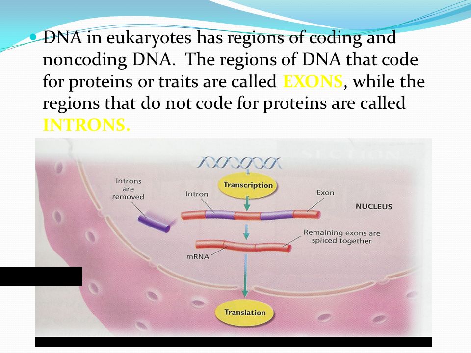
DNA in eukaryotes has regions of coding and noncoding DNA. The regions of DNA that code for proteins or traits are called EXONS, while the regions that do not code for proteins are called INTRONS. cytoplasm

8
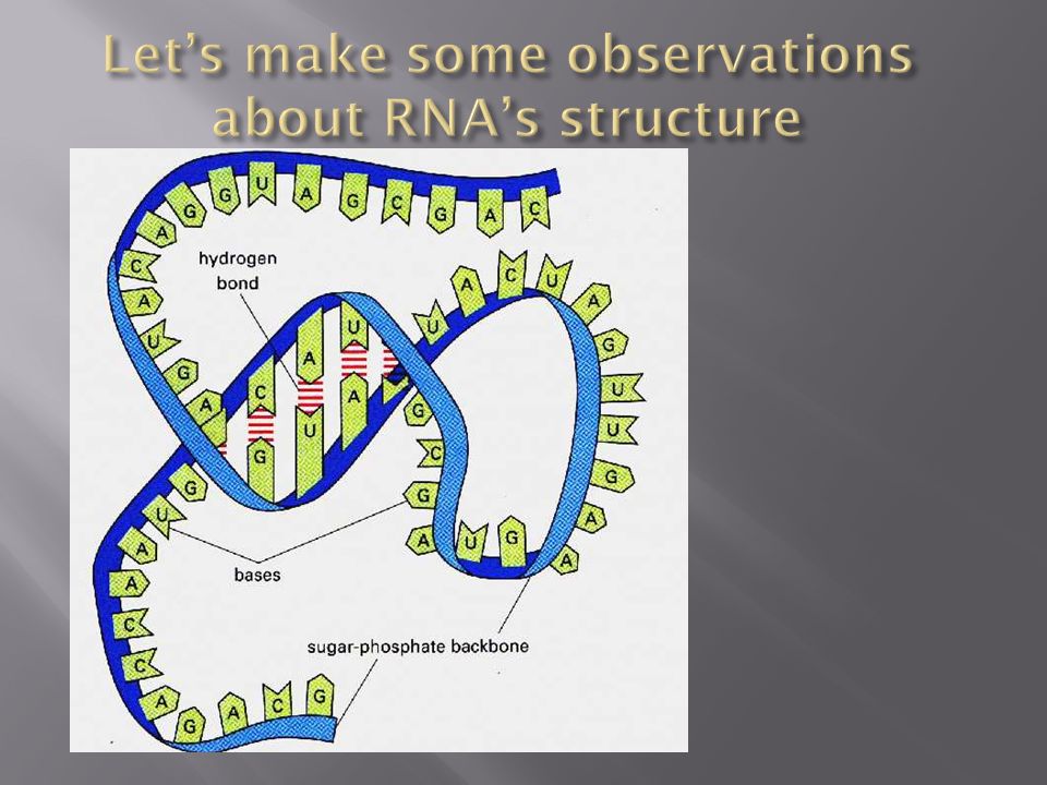
Translation RNA Single stranded Does not contain thymine but has uracil instead. tRNA carries 3 base pair code for specific amino acid. Amino acids compose polypeptid chains. One or more polypeptide chains compose a protein proteins provide the “blueprints” for our characteristics and functions.

11
RNA stands for: Ribonucleic acid RNA is found: Nucleus and Cytoplasm

12
Adenine (A) Cytosine (C) Guanine (G) Uracil (U)
 Cytosine (C) Guanine (G) Uracil (U)")
13
Messenger RNA (mRNA) Ribosomal RNA (rRNA) Transfer RNA (tRNA)
 Ribosomal RNA (rRNA) Transfer RNA (tRNA)")
14
Single stranded Many different shapes “Cheap copy” of DNA

16
Pre-mRNA – the original sequence of RNA created during transcription mRNA reaches the ribosomes

18
In Eukaryotes only Introns- non-coded sections Exons- codes for a protein Before RNA leaves the nucleus, introns are removed and exons are spliced together A cap and poly A tail are added to ends of the sequence mRNA leaves the nucleus through the nuclear pores

20
ii

21
Two subunits to the ribosome 3 grooves on the ribosome (A, P, E) A: tRNA binding site P: polypeptite bonding site E: exit site
 A: tRNA binding site P: polypeptite bonding site E: exit site")
22
Production of proteins from mRNA mRNA goes to the ribosomes in the cytoplasm or the RER and produces proteins

23
1. mRNA leaves the nucleus and binds to a ribosome 2. the 5’ end of mRNA binds to ribosome

24
4. Amino acids attached to a tRNA molecule and are brought over to the mRNA. 5. This tRNA has an anticodon that matches the codon on the mRNA strand Anticodon: Group of 3 unpaired nucleotides on a tRNA strand. (binds to mRNA codon)
.")
25
6. tRNA binds to the mRNA sequence and adds an amino acid 7. Each amino acid matches up with 1-6 tRNA molecules 8. tRNA leaves and amino acids bond together through a polypeptide bond

26
The two ribosomal subunits come together with the mRNA and the first tRNA molecule which attaches to the start codon (AUG). This is the only tRNA that will attach to the P site. The first amino acid is always methionine.

27
The tRNA anticodon will hydrogen bind to the mRNA codon in the A site.

28
The amino acid in the P site will form a peptide bond with the amino acid in the A site.

29
The tRNA's and the mRNA move down one site. The empty tRNA is released from the exit site.

30
This process will repeat hundreds of times.

31
Translation is terminated with the stop codon is reached. There are three different stop codons UGA, UAA, UAG. The release factor recognizes the stop codon and releases the polypeptide strand. All the factors break apart and are reused.

32
Untwisting and replication of DNA each strand is a template for a new strand Figure 10.4B helicase DNA polymerase

33
DNA replication begins at many specific sites How can entire chromosomes be replicated during S phase? Figure 10.5A Parental strand Origin of replication Bubble Two daughter DNA molecules Daughter strand

34
Each strand of the double helix is oriented in the opposite direction Figure 10.5B 5 end3 end 5 end P P P P P P P P

35
–The DNA is transcribed into RNA, which is translated into the polypeptide Figure 10.6A DNA RNA Protein TRANSCRIPTION TRANSLATION The information constituting an organism’s genotype is carried in its sequence of bases

Similar presentations


. Learning Objectives Describe how DNA is used to make protein Explain process of transcription Explain process.>")


 Nucleic acid that composes chromosomes and carries genetic information.>")


