Download presentation
Presentation is loading. Please wait.

1
Skeletal Tissues

2
Bone Function

3
Bone Structure Compact Bone – dense, hard bone made up of cylinder- shaped units called osteons or Haversian systems which permit delivery of nutrients and removal of wastes Spongy or Cancellous Bone – made up of needle-like bony spicules called trabeculae that act as braces to provide support and osteocytes (bone cells); their arrangement varies depending on the individual bones and the nature, magnitude, and direction of stresses; site of red blood cell formation (hematopoiesis) Periosteum – dense, fibrous blood vessel rich membrane that covers bone; continuous with the ligaments and tendons; supplies the bone with nutrients and oxygen as well as removes wastes and transport newly created blood cells
; their arrangement varies depending on the individual bones and the nature, magnitude, and direction of stresses; site of red blood cell formation (hematopoiesis) Periosteum – dense, fibrous blood vessel rich membrane that covers bone; continuous with the ligaments and tendons; supplies the bone with nutrients and oxygen as well as removes wastes and transport newly created blood cells")
4
Bone Structure

5
Microscopic Structure of Bone

6
Microscope Structure of Bone Compact Bone- contains many cylinder-shaped structural units called osteons or Haversian system Osteons (Haversian Systems)consist of: a. Lamellae – cylinder-shaped layers of calcified matrix b. Lacunae - “little lakes”; small spaces containing tissue fluid in which bone cells (osteocytes) reside c. Canaliculi – canals radiating in all directions from the lacunae and connecting them to each other and into a larger canal (Haversian canal) d. Haversian Canal – extends lengthwise through the center of each Haversian system; contains blood vessels, lymphatic vessels and nerves; serves as a conduit for the blood vessels to pass through the bone tissue; aka osteonic canals * Volkmann’s canals run almost perpendicularly to the Haversian canals and allow blood vessels to pass through them; aka perforating canals ** The Haversian systems ensure that even dense, hard bone has access to the blood vessels of the circulatory system.
 reside c. Canaliculi – canals radiating in all directions from the lacunae and connecting them to each other and into a larger canal (Haversian canal) d. Haversian Canal – extends lengthwise through the center of each Haversian system; contains blood vessels, lymphatic vessels and nerves; serves as a conduit for the blood vessels to pass through the bone tissue; aka osteonic canals * Volkmann’s canals run almost perpendicularly to the Haversian canals and allow blood vessels to pass through them; aka perforating canals ** The Haversian systems ensure that even dense, hard bone has access to the blood vessels of the circulatory system..")
9
Microscope Structure of Bone (cont’d) Cancellous bone (spongy bone) contains no osteons!!!! Spongy bone contains needle-like bony spicules called trabeculae. Osteocytes here receive nutrients and remove wastes through diffusion

10
Bone Tissue Bone (osseous) tissue is a distinctive type of connective tissue due to its extracellular matrix which is hard and calcified. Great strength and minimal weight make it ideal for support and protection. The extracellular matrix consists of inorganic salts and an organic matrix. The majority of the inorganic salts are calcium and phosphate. (hydroxyapatite crystals) Magnesium, sodium, sulfate and fluoride are also in bone. The organic matrix consists of collagenous fibers and a mixture of polysaccharides and protein called ground substance. Chondroitin sulfate is an important part of the ground substance and is required for repair and maintenance of bone and cartilage. The organic matrix gives bone a degree of plastic-like resilience to reasonable limits of stress to prevent injuries.
 Magnesium, sodium, sulfate and fluoride are also in bone. The organic matrix consists of collagenous fibers and a mixture of polysaccharides and protein called ground substance. Chondroitin sulfate is an important part of the ground substance and is required for repair and maintenance of bone and cartilage. The organic matrix gives bone a degree of plastic-like resilience to reasonable limits of stress to prevent injuries..")
11
Four Types of Bones (These have varying amounts of cancellous and compact bone in their structure.) Long Bones – extended longitudinal axes and uniquely shaped articular ends; provide structural support; weight-bearing; femur and humerus Short Bones - cube or box-shaped; length and width are about the same; carpals and tarsals; allow a wider range of movement than long bones Flat Bones – broad and thin; usually have a curved surface; scapula, ribs and sternum, skull, pelvis Irregular Bones – usually clustered in groups; shapes and sizes vary; vertebral bones, facial bones; skull, kneecaps *Irregular bones not found in groups are called sesamoid bones and embedded within a tendon; include the patella (kneecap), first metatarsal of the foot and pisiform (small, pea-shaped bone of wrist)
 Long Bones – extended longitudinal axes and uniquely shaped articular ends; provide structural support; weight-bearing; femur and humerus Short Bones - cube or box-shaped; length and width are about the same; carpals and tarsals; allow a wider range of movement than long bones Flat Bones – broad and thin; usually have a curved surface; scapula, ribs and sternum, skull, pelvis Irregular Bones – usually clustered in groups; shapes and sizes vary; vertebral bones, facial bones; skull, kneecaps *Irregular bones not found in groups are called sesamoid bones and embedded within a tendon; include the patella (kneecap), first metatarsal of the foot and pisiform (small, pea-shaped bone of wrist)")
12
Sesamoid Bones of the Human Body

13
Parts of Long Bone

14
Diaphysis – main shaftlike portion of the long bone Epiphyses – both ends of the long bone; provide space for muscle attachment and stability; consists of spongy bone and filled with red marrow Articular Cartilage – thin layer of hyaline cartilage that covers articular (joint) surfaces of epiphyses Medullary Cavity – tubelike hollow space in the diaphysis ; filled with yellow marrow rich in fat in adults; and red marrow (myeloid tissue) in infants and children Endosteum – thin epithelial membrane lining the medullary cavity Nutrient Artery – principal artery and major supplier of oxygen and nutrients to the shaft of the bone and the marrow Periosteum – dense, white fibrous membrane that covers bone except at joint surfaces; its fibers penetrate the bone providing anchoring for muscles; provides the bone with blood vessels; essential for bone survival and formation Parts of Long Bone
 surfaces of epiphyses Medullary Cavity – tubelike hollow space in the diaphysis ; filled with yellow marrow rich in fat in adults; and red marrow (myeloid tissue) in infants and children Endosteum – thin epithelial membrane lining the medullary cavity Nutrient Artery – principal artery and major supplier of oxygen and nutrients to the shaft of the bone and the marrow Periosteum – dense, white fibrous membrane that covers bone except at joint surfaces; its fibers penetrate the bone providing anchoring for muscles; provides the bone with blood vessels; essential for bone survival and formation Parts of Long Bone")
15
Periostium (cont’d) Periosteum – tough outer membrane that covers bone; continuous with the ligaments and tendons; supplies the bone with nutrients and oxygen as well as removes wastes and transports newly created blood cells * It is the “Life Support Sheath” for bone with a network of blood vessels – provides nutrient blood for living bone cells (osteocytes) and carries away carbon dioxide * The periosteum is essential for bone cell survival and bone formation, a process that continues throughout life.
 Periosteum – tough outer membrane that covers bone; continuous with the ligaments and tendons; supplies the bone with nutrients and oxygen as well as removes wastes and transports newly created blood cells * It is the Life Support Sheath for bone with a network of blood vessels – provides nutrient blood for living bone cells (osteocytes) and carries away carbon dioxide * The periosteum is essential for bone cell survival and bone formation, a process that continues throughout life.")
16
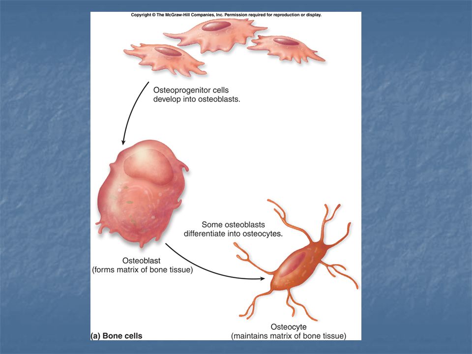
Types of Bone Cells Osteoblasts – synthesize and secrete the organic matrix (osteoid); provide framework for the deposition of calcium and phosphate that results in mineralized bone (calcification) Osteoclasts – giant multinucleate cells responsible for erosion of bone minerals; every 24 hour period has an alteration of primarily osteoblast, then osteoclast activity that results in continuous change and remodeling of bone tissue Osteocytes – mature, nondividing osteoblasts that have been surrounded by matrix and now lie within the lacunae
; provide framework for the deposition of calcium and phosphate that results in mineralized bone (calcification) Osteoclasts – giant multinucleate cells responsible for erosion of bone minerals; every 24 hour period has an alteration of primarily osteoblast, then osteoclast activity that results in continuous change and remodeling of bone tissue Osteocytes – mature, nondividing osteoblasts that have been surrounded by matrix and now lie within the lacunae")
17
Types of Bone Cells

18
Osteoblast – Osteoclast Activity The alteration of activity between osteoblasts and osteoclasts maintains homeostasis of calcium ion concentrations in the blood – “blood-calcium levels” in the blood. The alteration of activity between osteoblasts and osteoclasts maintains homeostasis of calcium ion concentrations in the blood – “blood-calcium levels” in the blood. Homeostasis of blood-calcium levels is essential for: Homeostasis of blood-calcium levels is essential for: Bone formation Bone formation Normal blood clottiing Normal blood clottiing Transmission of nerve impulses Transmission of nerve impulses Maintenance of skeletal and cardiac muscle contraction Maintenance of skeletal and cardiac muscle contraction

20
Bone Marrow The site for production of blood cells (haematopoiesis) Found in certain long bones and in the spaces of some spongy bone; forms ‘stem cells’ which develop into any of the three types of blood cells - red blood cells, white blood cells and platelets Two types of marrow: a) red – production of red blood cells; myeloid tissue; all bones in an infant or child contain this; is found in adults in the spongy bone of the ribs, vertebrae, pelvis, sternum, femur and humerus b) yellow – replaces red marrow in adults; saturated with fat and does not produce blood cells; found in hollow interior of the middle portion of long bones Yellow marrow can convert to red during times of decreased blood supply such as exposure to radiation, toxins and disease. Yellow marrow can convert to red during times of decreased blood supply such as exposure to radiation, toxins and disease.

21
Bone Marrow

22
Bone Marrow Transplant

23
Bone Development

24
Cartilage Bone As a fetus the bones are cartilage and fibrous structures shaped like bone. Water, protein and insoluble fibers are the main building blocks. It has no blood vessels to transport energy for its living cells; however due to high osmotic pressure, nutritional particles are diffused to joint cartilage cells from the surrounding fluid. The shaft (diaphysis) area of the bone is where the actual bone starts to form and blood vessels grow. The blood vessels continue their growth into the cartilage and the transformation into bone tissue commences. In the center of the shaft a "pre-marrow" forms.
 area of the bone is where the actual bone starts to form and blood vessels grow. The blood vessels continue their growth into the cartilage and the transformation into bone tissue commences. In the center of the shaft a pre-marrow forms..")
25
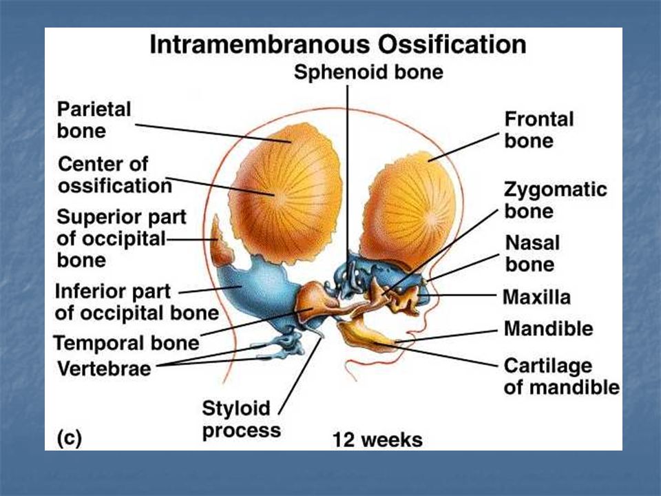
Specific Types of Bone Formation Intramembranous Ossification: a. Bone derived from sheets of connective tissue membrane b. Flat bones of the skull and clavicles are formed this way, as well as patella and the suture joints of the skull c. Appositional growth – addition of osseous tissue to outer surface; increase in width/thickness/diameter Endochondral Ossification: (Long Bones) a. Bone derived from cartilage b. Most bones of the body form this way c. Interstitial growth – interior expansion (bone grows lengthwise) d. Appositional growth – increase in width/diameter
 a. Bone derived from cartilage b. Most bones of the body form this way c. Interstitial growth – interior expansion (bone grows lengthwise) d. Appositional growth – increase in width/diameter.")
28
Endochondral Ossification Primary Ossification Center – blood vessel enters cartilage at midpoint of diaphysis and ossification progresses from the diaphysis toward the epiphyses and the bone gets longer Secondary Ossification Center – occurs in the epiphyses and growth occurs toward the diaphysis; no medullary cavity forms; occurs in epiphyseal plate and spongy bone forms; leads to growth of epiphyses

29
At the final stage of bone development the epiphyseal (growth) plates have moved to the ends of the bone, and a growth in length is no longer possible. Spongy bone has formed in the epiphysis.

31
Bone Growth and Resorption Bones grow in length and diameter due to remodeling of bone by the activity of osteoblasts and osteoclasts. The rate of bone growth (ossification) is faster during childhood and adolescence than bone destruction (resorption). In early adulthood, the two processes remain balanced so the rate of bone formation equals the rate of bone destruction. Between the ages of 35 and 40, the process reverses and bone loss begins to exceed bone gain. http://classes.midlandstech.edu/carter p/Courses/bio210/skel3a.htm http://classes.midlandstech.edu/carter p/Courses/bio210/skel3a.htm http://classes.midlandstech.edu/carter p/Courses/bio210/skel3a.htm http://classes.midlandstech.edu/carter p/Courses/bio210/skel3a.htm
 is faster during childhood and adolescence than bone destruction (resorption). In early adulthood, the two processes remain balanced so the rate of bone formation equals the rate of bone destruction. Between the ages of 35 and 40, the process reverses and bone loss begins to exceed bone gain. p/Courses/bio210/skel3a.htm p/Courses/bio210/skel3a.htm p/Courses/bio210/skel3a.htm p/Courses/bio210/skel3a.htm.")
32
Cartilage Classified as connective tissue Fibers embedded in a firm gel to allow for flexibility (Much more flexible than bone due to gel-like matrix which lacks calcium and other mineral salts) No canal system and no blood vessels penetrate it Three types: hyaline, elastic, and fibrocartilage Hyaline- (most abundant) - covers surfaces of bones (articular), tip of nose and rings of trachea, connects the ribs with the sternum Elastic – (elasticity as well as firmness) - external ear, epiglottis, eustachian tubes Fibrocartilage – (greatest tensile strength – strong & rigid) intervertebral disks and near points of attachment of some large tendons to bones
 No canal system and no blood vessels penetrate it Three types: hyaline, elastic, and fibrocartilage Hyaline- (most abundant) - covers surfaces of bones (articular), tip of nose and rings of trachea, connects the ribs with the sternum Elastic – (elasticity as well as firmness) - external ear, epiglottis, eustachian tubes Fibrocartilage – (greatest tensile strength – strong & rigid) intervertebral disks and near points of attachment of some large tendons to bones")
33
Types of Cartilage Types of Cartilage The type of protein fiber in the cartilage matrix determines the type of cartilage.

34
Bone Repair and Fractures Fracture – a break in the continuity of a bone; initiates the repair process due to bone death or damage to the periosteal and osteon blood vessels. Dead bone is either removed by osteoclasts or serves as a scaffolding for the deposition of repair tissue called callus.

35
Bone Repair (continued) Callus – forms after a fracture; serves as a protective collar for the bone and binds the broken ends of the fracture inside and outside; eventually replaced with normal bone Hematoma – (Bruise) Blood pooling at the site of injury helps trigger the bone repair process.
 Callus – forms after a fracture; serves as a protective collar for the bone and binds the broken ends of the fracture inside and outside; eventually replaced with normal bone Hematoma – (Bruise) Blood pooling at the site of injury helps trigger the bone repair process.")
36
Bone Diseases and Disorders Metabolic bone diseases: disorders of bone remodeling a. Osteoporosis – excessive loss of calcified matrix, bone mineral, and collagenous fibers; causes a reduction in total bone mass; decreasing estrogen and testosterone levels reduce osteoblastic activity and new bone growth; in women decreasing estrogen levels associated with menopause cause accelerated bone resorption; inadequate intake of calcium or vitamin D can result in decreased bone mass and osteoporosis b. Paget’s Disease – characterized by rapid and disorganized bone remodeling c. Osteomyelitis – bacterial infection of bone and marrow tissue; associated with other infections and with direct contamination due to open fracture or wound

37
Osteoporosis

38
Neoplasms – bone tumors affecting skeletal tissue Rickets – Vitamin D deficiency causes epiphyseal plate to thicken but not to calcify so the growing bone becomes deformed and bends under weight

39
Regulation of Blood Calcium Levels Bone stores about 98% of the body’s calcium therefore bones regulate blood-calcium levels. Homeostasis of calcium ions is vital for blood clotting, nerve impulse transmission, and muscle contraction. Primary homeostatic mechanisms involved in the regulation process are: parathyroid hormone(PTH) and calcitonin. The Parathyroid hormone, made by the parathyroid glands, maintains the level of calcium in the blood. When the levels are low, osteoclasts are stimulated to break bone down. Renal (kidney) absorption of calcium also occurs. Vitamin D synthesis increases. This aids in the absorption of calcium into the bloodstream. High levels of calcium in the blood stimulate the thyroid gland to produce calcitonin – a hormone that inhibits osteoclasts and stimulates the bone building osteoblasts to deposit calcium in the bones.
 and calcitonin. The Parathyroid hormone, made by the parathyroid glands, maintains the level of calcium in the blood. When the levels are low, osteoclasts are stimulated to break bone down. Renal (kidney) absorption of calcium also occurs. Vitamin D synthesis increases. This aids in the absorption of calcium into the bloodstream. High levels of calcium in the blood stimulate the thyroid gland to produce calcitonin – a hormone that inhibits osteoclasts and stimulates the bone building osteoblasts to deposit calcium in the bones..")
40
Thyroid Gland

41
Parathyroid glands

Similar presentations


 femur, humerous Short - (cube/box) carpal, tarsal Flat – (broad/thin) skull, scapulae Irregular – (varies/groups)>")


. BONE FUNCTION: Support and Protection bones shape and form body structures bones support and protect softer,>")


 Joints Cartilages Ligaments Divided into two divisions Axial skeleton –>")


