Download presentation
Presentation is loading. Please wait.

1
Hemostasis & blood coagulation

2
Learning objectives Hemostais Platelets and their role in hemostasis Clotting : nomenclature of clotting factors Clotting cascade: extrinsic, intrinsic, common pathways Fibrin stabilization Synthesis of clotting factors Role of vit K in clotting

3
Platelets and their role in hemostasis
MORPHOLOGY COUNT THROMBOPOIESIS LIFE SPAN, ROLE OF SPLEEN FUNCTIONS RELATED DISORDERS

4
MORPHOLOGY SMALL, GRANULATED, LACK NUCLEI, DIAMETER-2-4µ, GLYCOPROTEIN COAT ON MEMBRANE. EXTENSIVELY INVAGINATED MEMBRANE WITH INTRICATE CANALICULAR SYSTEM IN CONTACT WITH ECF RING OF MICROTUBULES AROUND THEIR PERIPHERY

5
MEMBRANE RECEPTORS FOR:
COLLAGEN, ADP, VESSEL WALL VON WILLEBRAND FACTOR, FIBRINOGEN. CYTOPLASMIC CONTENTS: ACTIN, MYOSIN, GLYCOGEN, LYSOSOMES, ENZYME SYSTEMS FOR SYNTEHESIS OF ATP, ADP, PROSTAGLANDINS, GRANULES.

6
Contents of GRANULES: Some clotting factors like thrombin Platelet derived growth factor (PDGF) Platelet activating factor (PAF) von Willebrand factor PROPERTIES: ADHESION, AGGREGATION, AGGLUTINATION

7
THROMBOPOIESIS COUNT: 150000-400000 cells/cubic mm bld Location
adults - red bone marrow Half life 4 days Differentiation hemocytoblasts (stem cells) develop into large megakaryocytes megakaryocytes have lots of ER & Golgi megakaryocyte pinches off tiny cytoplasmic packets (4000 plts /megak)
 develop into large megakaryocytes. megakaryocytes have lots of ER & Golgi. megakaryocyte pinches off tiny cytoplasmic packets (4000 plts /megak)")
8
Megakaryocyte in bone marrow (note it has a large nucleus)
8 8

9
In a stained blood smear platelets are often seen as eosinophilic clumps
9 9

10
REGULATION normally negative feedback homeostasis
primarily the liver and kidney releases thrombopoietin (TPO) to increase platelet production liver disease leads to abnormally low platelet production from insufficient TPO WBCs release an interleukin - also weakly stimulates platelet production
 to increase platelet production. liver disease leads to abnormally low platelet production from insufficient TPO. WBCs release an interleukin - also weakly stimulates platelet production.")
11
LIFE SPAN : 9-12 DAYS ROLE OF SPLEEN: STORAGE (25-40% sequestered ) DESTRUCTION SPLENOMEGALY- increased destruction -THROMBOCYTOPENIA SPLENECTOMY- THROMBOCYTOSIS

12
FUNCTIONS HAEMOSTASIS Definition:
SPONTANEOUS ARREST OF BLEEDING FROM SMALL INJURED VESSELS (arterioles, capillaries, venules)
")
13
FUNCTIONS A. HEMOSTASIS STEPS OF HEMOSTASIS: 1. VASOCONSTRICTION
2. PLATELET PLUG FORMATION 3. COAGULATION

14
Primary hemostasis : refers to the formation of a temporary hemostatic platelet plug that stops loss of blood Secondary hemostasis (definitive hemostasis, or clotting): formation of a strong fibrin clot sealing the site of injury
: formation of a strong fibrin clot sealing the site of injury.")
15
VASOCONSTRICTION DIRECT: LOCAL MYOGENIC SPASM (more the trauma more is the spasm eg: in crush injury) REFLEX SYMPATHETIC SEROTONIN, THROMBAXANE A2

16
16 16

17
Platelet Function: Platelet Activation (is classically described using the following terms)
Process Brief Description Platelet activation Platelets bound to vWF become ‘activated’ and release mediators including ADP, thrombin, serotonin, platelet activating factor, vWF. They synthesize and release thromboxane (TXA2) All these mediators promote platelet adhesion to vessel wall, and platelet aggregation (see next slide) TXA2, thrombin and serotonin also cause vasoconstriction locally Thrombin also activates the clotting cascade 17 17
 All these mediators promote platelet adhesion to vessel wall, and platelet aggregation (see next slide) TXA2, thrombin and serotonin also cause vasoconstriction locally. Thrombin also activates the clotting cascade")
18
Platelet Adhesion: Process Brief Description Platelet adhesion
Platelets adhere to subendothelial collagen; This process requires von Willebrand factor (vWF); vWF comes from injured endothelial cells and activates platelets; Both subendothelial collagen and platelets have receptors that bind vWF; 18 18
; vWF comes from injured endothelial cells and activates platelets; Both subendothelial collagen and platelets have receptors that bind vWF;")
19
Platelet Aggregation:
Process Brief Description Platelet aggregation Platelets link up with each other forming a loose mass of platelets; ADP, thrombin, TXA2, serotonin, platelet activating factor [PAF] from platelets all promote platelet aggregation Fibrinogen (a clotting factor) cross links platelets Platelet membranes have fibrinogen receptors Note – platelet activation, adhesion and aggregation interact and reinforce each other 19 19
 cross links platelets. Platelet membranes have fibrinogen receptors. Note – platelet activation, adhesion and aggregation interact and reinforce each other")
20
ADHESION OF PLTS TO COLLAGEN VIA vWF
DAMAGE TO VESSEL WALL COLLAGEN GETS EXPOSED ADHESION OF PLTS TO COLLAGEN VIA vWF ACTIVATION OF PLATELETS RELEASE OF ADP FROM SECRETORY VESICLES SYNTHESIS OF THROMBAXANE A2 FROM ARACHIDONIC ACID ON PLASMA MEMBRANE AGGREGATION OF PLATS LOOSE PLT PLUG

21
Vessel damage, collagen exposed
Platelet activation Discharge of chemical mediators Synthesis of Thromboxane A2 Serotonin, thromboxane A2, Thromboxane A2, ADP Contraction of Vascular smooth muscle Platelet aggregation Vasoconstriction Platelet plug Temporary arrest of bleeding

22
PLATELET PLUG attracted to & stick to damaged vessel walls
platelets enlarge & get sticky platelets release attractant chemicals that attract more platelets Positive Feedback Cycle produces a bigger & faster reaction platelets release chemicals that attract more platelets to site How limits to injured site ? ADP stimulates release of prostacyclin & nitric oxides, from intact endothelium prevents aggregation of platelets.

23
FUNCTIONS CONTD SEROTONIN- VASOCONSTRICTION
PLATELET PLUG- TEMPORARY SEALING OF VESSLES PLATLET FACTOR 3- FOR CLOTTING ACTIN, MYOSIN- CLOT RETRACTION PDGF- WOUND HEALING & REPAIR OF ENDOTHELIUM

24
OTHER LESS IMP FUNCTIONS
PHAGOCYTOSIS OF: DYES, CARBON, MICRO-ORGANISMS, OLD CELLS ETC. AS ANTIGEN PRESENTING CELL (APC) SECRETES VARIOUS CYTOKINES: HELP IN HAEMOPOIESIS, FUNCTIONING OF NK CELLS ETC
 SECRETES VARIOUS CYTOKINES: HELP IN HAEMOPOIESIS, FUNCTIONING OF NK CELLS ETC.")
26
THROMBOCYTOPENIA CAUSES – Splenomegaly, infections, toxins, radiation, rare genetic diseases OUTCOME - increased bruising (purpura) & increased risk of serious hemorrhaging Thrombaesthenic purpura: due to weak platelets.
 & increased risk of serious hemorrhaging. Thrombaesthenic purpura: due to weak platelets.")
27
Consequence of thrombocytopenia
Prolongation of ‘bleeding time’ – excessive bleeding from relatively minor injuries.. However, ‘clotting time’ is normal (details later in Practicals) Note extensive bruising (purpura) 27 27
 Note extensive bruising (purpura)")
28
BLOOD COAGULATION

29
CLOT TRANSFORMATION OF SOLUBLE BLOOD INTO SOLID GEL
CLOT OR THROMBUS CONSISTS OF INSOLUBLE PROTEIN POLYMER OF FIBRIN and TRAPPED BLOOD CELLS

30
WHAT PREVENTS INTRAVASCULAR CLOTTING ?
Physiologic anticlotting or antithrombic mechanisms prevent inappropriate or excess clotting are: CLOTTING FACTORS ARE INACTIVE DEGRDATION OF ACTIVATED CLOTTING FACTORS CONTINUOUS MOTION OF BLOOD AT A NORMAL FLOW RATE

31
SMOOTH INTACT ENDOTHELIUM PGI2 FROM ENDOTHELIAL CELLS.
GLYCOCALYX THROMOMODULIN RECEPTORS ON ENDOTHELIAL WALL CIRCULATING ANTICOAGULANTS-Antithrombin III – heparin complex (inhibits IXa, Xa, XIa and XIIa) FIBRINOLYTIC SYSTEM- Protein C – protein S complex inactivates Va, VIIIa and inactivates inhibitor of tPA
 FIBRINOLYTIC SYSTEM- Protein C – protein S complex inactivates Va, VIIIa and inactivates inhibitor of tPA.")
32
V Proaccelerin, labile factor VII Proconvertin
CLOTTING FACTORS I Fibrinogen II Prothrombin III Tissue Factor IV Calcium V Proaccelerin, labile factor VII Proconvertin

33
VIII Antihemophiliac factor A, antihemophilic globulin (AHG)
IX Christmas Factor, Antihemophilic factor B X Stuart-Prower Factor XI Plasma thromboplastin antecedent (PTA) XII Hageman Factor XIII fibrin stabilizing factor
 XII Hageman Factor. XIII fibrin stabilizing factor.")
34
OTHER FACTORS von Willebrand factor Protein C Protein S Thrombomodulin
Antithrombin III Prekallikrein High mol wt kininogens

35
SOME IMP FACTS ABOUT COAGULATION
inactive clotting factors present in blood at all times most factors produced by liver or platelets cascading effect 1 enzyme used to activate next enzyme in series --> greatly amplifies final step Ca++ - used as an inorganic catalyst for many of these coagulation steps 2 PATHWAYS INITATE CLOTTING: Initiator is different for extrinsic & intrinsic pathway Most clotting factors are produced in the liver Vitamin K is required for the synthesis of clotting factors II, VII, IX and X

36
Mechanism of clotting:
inactive prothrombin converted into thrombin (active enzyme) by E & I pathway Common pathway: thrombin converts soluble plasma protein fibrinogen into insoluble fibrin Positive Feedback thrombin stimulates part of its own production pathway
 by E & I pathway. Common pathway: thrombin converts soluble plasma protein fibrinogen into insoluble fibrin. Positive Feedback. thrombin stimulates part of its own production pathway.")
37
INTRINSIC: ACTIVATION BY CELLULAR ELEMENTS WITHIN BLOOD
INITIATOR: EXPOSED COLLAGEN, CONTACT FACTOR, ELECTRONEGATIVE SURFACE (test tube or capillary tube) EXTRINSIC: ACTIVATION BY CELLULAR ELEMENT OUTSIDE THE BLOOD (from tissue injury) INITIATOR: TISSUE FACTOR
 EXTRINSIC: ACTIVATION BY CELLULAR ELEMENT OUTSIDE THE BLOOD (from tissue injury) INITIATOR: TISSUE FACTOR.")
38
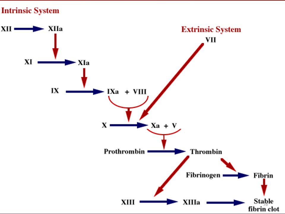
‘a’ denotes activated form of the clotting factor
38 38
39
The Clotting Cascade Note details not contained in the previous slide; TPL – tissue thromboplastin; TFI – tissue factor inhibitor; ‘a’ denotes activated form of the factor. 39 39

40
INTRINSIC PATHWAY EXTRINSIC PATHWAY
COMMON PATHWAY 40 40

41
Intrinsic and Extrinsic Pathways compared
Intrinsic Pathway Extrinsic Pathway Stimulus for activation Factor XIIa comes into contact with subendothelial collagen; Electronegative surface if collected in glass tube Blood vessel injury; tissue thromboplastin from tissues enters bloodstream and activates Factor VII Why this name? The entire cascade is intrinsic to the circulatory system and the vessel wall – thus this name Called “extrinsic” because it is activated by a factor extrinsic to plasma (i.e., tissue thromboplastin) Inhibitor Thrombomodulin-thrombin complex and activated protein C Tissue factor pathway inhibitor (TFPI) forms a complex with tissue thromboplastin and Factor VIIa and Factor Xa Time Longer of the two cascades; aPTT is seconds Shorter of the two cascades; PT is seconds 41 41
 Inhibitor. Thrombomodulin-thrombin complex and activated protein C. Tissue factor pathway inhibitor (TFPI) forms a complex with tissue thromboplastin and Factor VIIa and Factor Xa. Time. Longer of the two cascades; aPTT is seconds. Shorter of the two cascades; PT is seconds")
43
CLOT RETRACTION Def - consolidation or tightening of clot
Method - fibrin threads contract/shrink Benefits - pull damaged edges of vessel wall together, repair is faster; tighter, stronger clot Timing - retraction occurs within min.

46
Learning outcomes At the end of this class student should be able to Define hemostasis. Know the difference between primary hemostasis & definitive hemostasis. Describe the structure & function of platelets Normal count, factors regulating thrombopoiesis

47
Common causes of thrombocytoenia & thrombocytosis
Role of platelets in hemostasis Clotting factors with their roman numerals & common names, & where they are produced from ? Cascade of events in extrinsic & intrinsic mechanism of clotting, and initiating factors. Role of vitamin K in clotting.

Similar presentations


 Thrombocytes. PLATELETS (PLT) Thrombocytes.>")


>")


