Download presentation
Presentation is loading. Please wait.

1
Identification of Cancer Stem Cells using Flow Cytometry Analysis تعيين الخلايا الجذعية السرطانية باستخدام تحليل التدفق الخلوي Dr. Ayat Al-Ghafari Biochemistry Department Monday 25-5-1436

2
Outlines 1.Cancer 2.Heterogeneity of cancer 3.Cancer stem cells (CSCs) hypothesis 4.Flow cytometry Overview Instrument major parts How does flow cytometer work
 hypothesis 4.Flow cytometry Overview Instrument major parts How does flow cytometer work")
3
Cancer Cancer or malignant neoplasm disease is a group of diseases which is characterized by: 1.The uncontrolled growth of cells (proliferation) 2.Uncontrolled movement (migration and metastasis) 3.Ability to invade neighboring tissues (invasion) 4.Resistance to death (necrosis and apoptosis)
 2.Uncontrolled movement (migration and metastasis) 3.Ability to invade neighboring tissues (invasion) 4.Resistance to death (necrosis and apoptosis)")
4
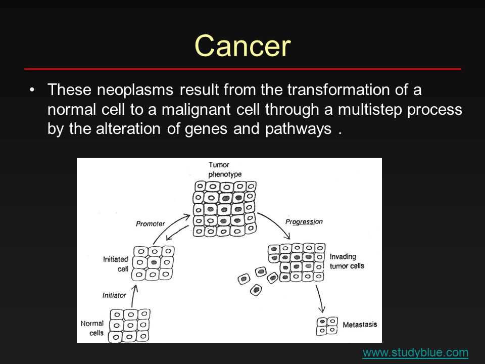
Cancer These neoplasms result from the transformation of a normal cell to a malignant cell through a multistep process by the alteration of genes and pathways. www.studyblue.com
5
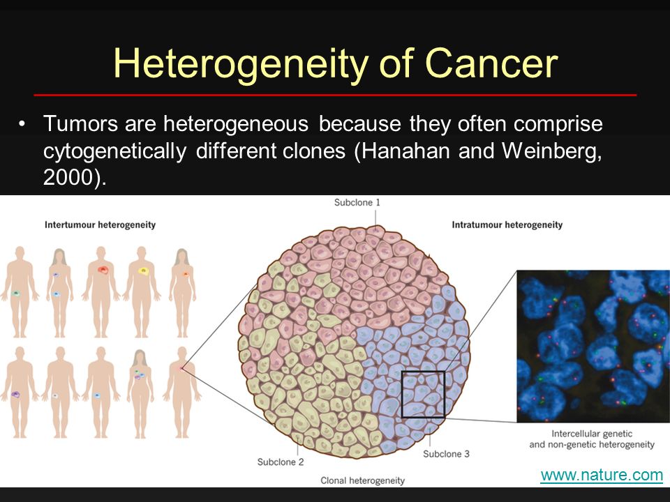
Heterogeneity of Cancer Tumors are heterogeneous because they often comprise cytogenetically different clones (Hanahan and Weinberg, 2000). www.nature.com
6
Heterogeneity of Cancer This heterogeneity results in differences in clinical tumor behavior and responses to treatment. The first evidence of the heterogeneity of tumors was demonstrated in 1961 by Southam and Brunschwig when they harvested cancer cells from recurrent tumors and auto- transplanted the cells at different sites (Southam and Brunschwig, 1961). This suggested hypothesis that a subset of these cells, not all of them, had the ability to initiate the tumor.
. This suggested hypothesis that a subset of these cells, not all of them, had the ability to initiate the tumor..")
7
Heterogeneity of Cancer By the early 1990s, it was clear from analysis of cell surface expressed proteins (CD proteins) that tumor cells were heterogeneous in terms of their protein expression. Lapidot et al., found that CD34 + CD38 - cells from patients with AML were able to initiate tumor when they injected to NOD-SCID mice while CD34 + CD38 + did not initiate the tumor (Lapidot et al., 1994).
..")
8
Heterogeneity of Cancer These findings confirmed that not all the cells in the tumor are responsible for its progression and lead to the establishment of a new theory called the cancer stem cells (CSCs) hypothesis. CSCs hypothesis proposes that growth and progression of many cancers are driven by small subpopulations of CSCs rather than all the cells in the tumor (Reya et al., 2001).
..")
9
Cancer stem cells Subset of cells within tumors with the ability to self-renew and differentiate to the phenotypically diverse drug resistant tumor cell population to drive tumorigenesis Normal stem cells Rare cells within organs with the ability to self- renew and differentiate to all types of cells within the organ to drive organogenesis

10
Cancer Stem Cells (CSCs) Hypothesis Several methods are generally used to characterize CSCs: 1.The isolation of a side population or sub-population (SP) based on cell surface marker expression by flow cytometry 2.Identification of a small population of cells by their ability to differentiate 3.Identification of a small population of cells by their ability to form spheres (colonies)
 Hypothesis Several methods are generally used to characterize CSCs: 1.The isolation of a side population or sub-population (SP) based on cell surface marker expression by flow cytometry 2.Identification of a small population of cells by their ability to differentiate 3.Identification of a small population of cells by their ability to form spheres (colonies)")
11
Flow Cytometry It is a powerful technique to identify multiple parameters of a single cell within a heterogeneous population

12
Flow cytometry Ploidy analysis Flow Cytometry Cell Counting Flow cytometry GFP expression analysis

13
Flow Cytometry Overview It performs its analysis by passing thousands of cells /seconds through a laser beam and capturing the light that emerges from each cell as they passing through. Then the data gathered can be analyzed statistically by flow cytometry software that report cellular characteristics such as size, complexity, phenotype, and health. The flow cytometer contains several parts as shown by the following image.

14
Flow Cytometry Instrument Overview

15
1.Fluidics system Which presents the sample to the interrogation point and takes away the waste. 2.Lasers Which are the light source for scatter and fluorescence.

16
Flow Cytometry Instrument Overview 3.Optical system Which gather and direct the light. 4.Detectors Which received the light.

17
Flow Cytometry Instrument Overview 5.The external computer system Which converts the signals from the detector into digital data and perform the necessary analyses.

18
Flow Cytometry Steps (1) Interrogation point
 Interrogation point")
19
Flow Cytometry Steps (2) Hydrodynamic focusing
 Hydrodynamic focusing")
20
Flow Cytometry Steps (3) Forward Scatter
 Forward Scatter")
21
Flow Cytometry Steps (4) Forward Scatter Detector
 Forward Scatter Detector")
22
Flow Cytometry Steps (5) Side Scatter Detector
 Side Scatter Detector")
23
Flow Cytometry Steps (5) Side Scatter Detector 2-dimensional dot scatter plot
 Side Scatter Detector 2-dimensional dot scatter plot")
24
Flow Cytometry Steps (5) Side Scatter Detector There is a correlation between the data obtained from both the forward and the side scatter
 Side Scatter Detector There is a correlation between the data obtained from both the forward and the side scatter")
25
Flow Cytometry Steps (5) Side Scatter Detector
 Side Scatter Detector")
26
Flow Cytometry Steps (6) Florescence Detector It gives more information about the structure and function
 Florescence Detector It gives more information about the structure and function")
28
Example

29
They wanted to evaluate the effect of two drugs MBO-SC and MBO-asGCS on the expression of CD44+ in breast CSCs

30
References 1.HANAHAN, D. & WEINBERG, R. 2000. The hallmarks of cancer. Cell, 100, 57-70. 2.SOUTHAM, C. & BRUNSCHWIG, A. 1961. Quantitative studies of autotransplantation of human cancer. Cancer, 14, 971–978. 3.LAPIDOT, T., SIRARD, C., VORMOOR, J., MURDOCH, B., HOANG, T., CACERES-CORTES, J., MINDEN, M., PATERSON, B., CALIGIURI, M. A. & DICK, J. E. 1994. A cell initiating human acute myeloid leukaemia after transplantation into SCID mice. Nature, 367, 645–648. 4.REYA, T., MORRISON, S. J., CLARKE, M. F. & WEISSMAN, I. L. 2001. Stem cells, cancer, and cancer stem cells. Nature, 414, 105-111. 5.https://www.lifetechnologies.com/sa/en/home/brands/invitrogen.html

Similar presentations


 of blood...>")
 is the fifth most frequent cancer in the world and the third most common cause of cancer mortality.>")
 Optical Light Scatter and Flow Cytometry.>")


 Excise cancer from Breast Isolate Cancer cells No Tumor Inject mice 10 3 cells 10 4 cells.>")


, Ph.D. ext 5632; References: 1.Chapter 23 Cancer in “Molecular Cell Biology”>")

