Download presentation
Presentation is loading. Please wait.

1
Light microscope

2
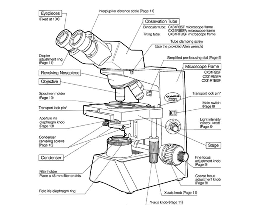
Light Microscope 1. The light microscope operates on the principle that light energy will pass through and around a thin object, such as a microorganism and, with the aid of lenses, form a magnified impression on the visual sensory layer of the eye.

3
2. The light microscope uses two sets of lenses ( ocular and objective) and is therefore referred to as a compound microscope.
 and is therefore referred to as a compound microscope..")
4
3. The microscope shown is binocular because it has paired ocular lenses. The two sets of lenses and their housing are supported by a base and an arm.

5
4. The stage is the platform on which the specimen or a slide supporting the specimen is placed. Clips hold the slide in place. The slide may be moved by fingers or by a mechanical apparatus associated with the stage.

6
5. A light microscope is usually equipped with a built in light source controlled by an on/off switch on the base. The light is projected upward through a condensor which houses a series of lenses under the stage.

7
These lenses concentrate the passing light into a strong beam which is projected onto the specimen above. The condenser lenses are controlled by a controlling knob.

9
6. Mounted on the condenser is the iris diaphragm, a series of thin brass sheets adjusting the size of the aperture through which the light passes . Aperture – hole or opening through which light travels

10
The diaphragm is operated by a lever which spreads the sheets apart or overlaps them.

11
This action controls the angular width of the light beam, increasing the contrast between the light and dark portions of the specimen.

12
7. The concentrated beam of light passes through and around the thin or thinly cut specimen placed on a glass slide and usually covered with a glass or plastic coverslip.

13
8. Light passing up through and around the specimen enters the objective placed in the light path. Four objectives, housed on a movable turret, are shown.

14
Each objective has a certain magnifying power; for example a scanning objective magnifies 4 times, a low power objective magnifies ten times, a high power objective magnifies 40 times, and an oil immersion objective magnifies 100 times.

15
It is wise to start out with the scanning objective for orientation, then switch to progressively higher powers. Upon passing through the objective, the light then enters the tube of the microscope where an image forms.

16
An ocular or eyepiece is located at the top of the microscope
An ocular or eyepiece is located at the top of the microscope. The oculars magnify the image projected ten times. Thus, the image visualized by the eye can be magnified, 40, 100, 400, and 1000 times, the product of the magnifying powers of the ocular and the objective in use.

17
9. Light microscopes have two focusing controls
9. Light microscopes have two focusing controls. The coarse adjustment control is generally the larger of two grooved knobs at the lower side of the microscope’s arm. It is used to bring the specimen roughly in focus

18
A more refined, precise focus can be achieved by rotation of the smaller fine adjustment knob.

Similar presentations
- focuses image under lowest power. Cannot use with other lenses. 2.Fine adjustment knob (E)- used to focus images under.>")


>")


 First lens First lens 2. Arm: connects base and body.>")

