Download presentation
Presentation is loading. Please wait.

2
Current Concepts in Pathology of Soft Tissue Sarcoma Nuzhat Husain & Nidhi Verma Indian J Surg Oncol Received: 16 December 2011 / Accepted: 2 February 2012 / Published online: 3 April 2012 Indian Association of Surgical Oncology 2012

3
Abstract Soft tissue sarcomas (STS) constitute a heterogeneous category of soft tissue neoplasia composed mostly of uncommon tumors of diverse histology, different biology and varied outcomes. Substantial developments in immunohistochemistry (IHC), cytogenetics and molecular genetics of STS have caused a significant change in the classification and diagnosis of these tumors with a direct implication for clinical management and prognosis. In this review we discuss newer developments impacting diagnosis and prediction.
, cytogenetics and molecular genetics of STS have caused a significant change in the classification and diagnosis of these tumors with a direct implication for clinical management and prognosis. In this review we discuss newer developments impacting diagnosis and prediction..")
4
WHO Classification of Soft Tissue Tumors WHO working group divides STS into four categories: Benign, Intermediate (locally aggressive), Intermediate (metastasizing) Malignant
, Intermediate (metastasizing) Malignant")
5
Grading the staging of STS is largely determined by grade. Unfortunately there is no generally agreed up on scheme for grading STS. The most widely used STS grading systems are French Federation of Cancer Centers Sarcoma Group (FNCLCC) and National Cancer Institute (NCI) Both systems have 3 grades and are based on mitotic activity, necrosis and differentiation. The FNCLCC system is easier to use and appears slightly better in predicting prognosis.
 and National Cancer Institute (NCI) Both systems have 3 grades and are based on mitotic activity, necrosis and differentiation. The FNCLCC system is easier to use and appears slightly better in predicting prognosis..")
6
Grading of malignant peripheral nerve sheath tumor, embryonal and alveolar rhabdomyosarcoma, angiosarcoma, extraskeletal myxoid chondrosarcoma, alveolar soft part sarcoma, clear cell sarcoma, and epithelioid sarcoma is not recommended. Morphological Categories… architectural pattern, appearance of the cells, and the characteristics of the stroma Immunohistochemical characteristics

8
Pitfalls in the Diagnosis of STS. Certain reactive processes may mimic sarcomas. Therefore it is important to determine whether the lesion under study is a reactive process or a neoplasm. For example in nodular fascitis and ischemic fascitis proliferating fibroblasts that surround a central hypocellular zone of fibrinoid change. Cells comprising reactive lesions often have the appearance of tissue culture fibroblasts with large vesicular nuclei, prominent nucleuli and cytoplasmic basophilia. No atypical mitosis or nuclear atypia.

9
It is not always possible to classify soft tissue tumors precisely based on biopsy material. especially FNA and core needle biopsy specimens. pathologists should make every attempt to classify lesions in small biopsy specimens, on occasion stratification into very basic diagnostic categories, such as lymphoma, carcinoma, melanoma, and sarcoma, In some cases, precise classification is only possible in open biopsies or resection specimens.

10
In many cases, it is unreasonable to expect a precise classification of these tumors based on an intraoperative consultation. Intraoperative consultation is useful in assessing if “lesional” tissue is present and in constructing a differential diagnosis that can direct proper resection of tissue. The role of frozen section in STS is limited only to assure the surgeon that he has obtained representative, adequate and viable tissue for diagnosis or to evaluate the margins.

11
Grading of sarcomas in small needle core biopsies is also frequently erroneous. Inappropriate sampling may lead to high grade lesion being diagnosed as low grade sarcoma. Core biopsy containing necrosis usually implies high grade sarcoma but necrosis should be coagulative and distinguished from hyaline change. Necrosis reflective of prior therapy or surgical intervention does not upgrade a lesion. Grading becomes unreliable if chemotherapy and radiotherapy is given prior to biopsy.

12
IHC Pathologists must proceed cautiously and consider IHC results in the context of all available data in a given case of soft-tissue tumors (aberrant antigen expression)... pitfalls that can be introduced by technical factors, such as tissue processing and fixation as well as the IHC procedures. IHC in fact adds confusion to the diagnostic process in 5 % to 10 % of cases.

13
IHC… With the implementation of immunohistochemistry, cytogenetics and molecular genetic analysis significant changes have been made regarding the classification and diagnosis of STS. direct implications for management and prognosis. Many new entities have been recognized (desmoplastic small round cell tumor and intimal sarcomas are examples). Some sarcoma entities lost in importance (e.g. the so-called malignant fibrous histiocytoma, Hemangiopericytoma categories)
. Some sarcoma entities lost in importance (e.g. the so-called malignant fibrous histiocytoma, Hemangiopericytoma categories).")
14
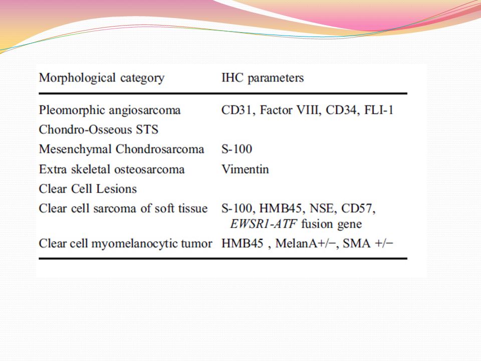
Table 2 STS: mophological categories and IHC parameters

18
The diagnostic approach consists in ruling out a nonmesenchymal tumor, followed by trying to define mesenchymal cell lineage… A panel of immunostains, rather than single marker, should always be performed. The extent of sub-categorization of tumors should be driven by clinical relevance. For instance in cases of pleomorphic sarcoma treatment protocols do not discriminate between histologic subtypes. How far should one go with ancillary studies in trying to determine specific lineage and what are the clinical/prognostic implications is a question best answered by need driven requirements of treating oncologists.

19
IHC … initial battery of IHC markers should always include a broad keratin (pancytokeratin), S100, and vimentin. CD30, LCA … Synovial sarcoma is a unique soft tissue sarcoma showing both mesenchymal and epithelial differentiation. Despite its name, it is neither related to nor does it arise from synovial cells. IHC is particularly useful in the diagnosis of the monophasic spindle cell variant, which is more difficult to distinguish from other spindle cell sarcomas.

20
Epithelioid sarcoma: Cytokeratins and EMA are strongly positive in almost all cases, mostly coexpressed with vimentin. CD34 is positive in about 50 % with a strong membranous staining pattern. When positive, this stain is helpful in excluding carcinomas which are CD34 negative. Round cell tumours including Ewing’s sarcoma/ Primitive Neuroectodermal Tumors, alveolar rhabdomyosarcoma, desmoplastic small round cell tumor, poorly differentiated synovial sarcoma, and Merkel cell carcinoma…

21
It is hoped that detection of tumor-specific alterations and validation through genetic analysis on larger samples will lead to development of new IHC antibodies. These new markers detect tumor-specific fusion proteins that are either over expressed or aberrantly expressed as a result of a translocation. Examples of such antibodies are ALK-1, WT-1, and FLI-1.

22
Molecular genetics of STS polymerase chain reaction-based techniques. fluorescence in situ hybridization (FISH). comparative genomic hybridization. expression arrays. direct genome sequencing. DNA methylation analysis. identified new histogenetic concepts and built up robust diagnostic methods…
. comparative genomic hybridization. expression arrays. direct genome sequencing. DNA methylation analysis. identified new histogenetic concepts and built up robust diagnostic methods….")
23
The majority of sarcomas carry nonspecific genetic changes in a background of a complex karyotype. Many of these alterations have been detected in a small number of cases and confirmatory tests are not yet commercially available. Their diagnostic utility is therefore limited with the exception of some fluorescence in situ hybridization probes, which are being used for the diagnosis of synovial sarcoma, ES/PNET, and alveolar rhabdomyosarcoma.

24
Soft tissue sarcomas can be divided in two categories: simple karyotypes complex karyotypes simple : 15–20% bear specific reciprocal translocations which can be used as diagnostic markers. Some others have specific somatic mutations (e.g., cKIT and PDGF receptor alpha in GIST. Specific amplifications ( MDM2 and CDK4 amplification in the liposarcoma category ) MYCN amplification in Neuroblastoma, Translocation FKHR (FOXO1A) in alveolar rhabdomyosarcoma…
 MYCN amplification in Neuroblastoma, Translocation FKHR (FOXO1A) in alveolar rhabdomyosarcoma….")
25
The Ewing sarcoma breakpoint region 1 (EWSR1; also known as EWS) : one of the most commonly involved genes in sarcoma translocations. In fact, it is involved in a broad variety of mesenchymal lesions complex karyotypes account for about 50% of sarcomas. This sarcoma category includes most of spindle cell/pleomorphic sarcomas.

26
Apart from the obvious relevance of specific genetic aberrations for establishing a correct diagnosis, which is a prominent prognostic parameter in itself, the importance of the genetic aberrations on clinical outcome of STS is elusive. The currently used prognostication systems rely on relatively crude clinical parameters, such as tumor size, and histopathologic parameters such as morphologic subtype, grade, necrosis, vascular invasion, and growth pattern, all of which suffer from inter-observer variability.

27
there is no reason to believe that the pattern of genetic aberrations should be of less importance for the management of soft tissue sarcomas than for that of leukemias and lymphomas, where genomic analyses today constitutes an integral part of the routine diagnostic and prognostic profiling. However, there is clearly a need for further analyses, using cytogenetic as well as high resolution genomic techniques, on larger series and to allow proper evaluation of the prognostic importance of characteristic genomic changes.

28
The role of predicting response of targeted therapy has been well established for KIT and PDGFRA mutations in gastrointestinal stromal tumors. The role of the vascular endothelial growth factor and its receptor in angiosarcoma, epithelioid hemangioendothelioma and hemangiopericytoma / solitary fibrous tumor has been evaluated. efficacy of bevacizumab, an anti-VEGF antibody, sunitinib, sorafenib, and pazopanib, VEGFR tyrosine kinase inhibitors…

29
Finally The application of molecular and immunohistochemical techniques needs to be under strict protocol and results should be interpreted in the morphological context in order to avoid disastrous mistakes in tumor classification.

Similar presentations


>")


 to Identify Genetic Changes in Fine Needle Biopsy of Lung Lesions Prepared by Jin Jen NCI.>")
University of Utah, School of Medicine, Department of Pathology, Salt lake City, UT and Cytogenetics/Molecular.>")