Download presentation
Presentation is loading. Please wait.

1
II Structure and Catalysis 5. Amino Acids, Peptides, and Proteins 6. The Three-Dimensional Structure of Proteins 7. Protein Function 8. Enzymes 9. Carbohydrates and Glycobiology 10. Lipids 11. Biological Membranes and Transpot 12. Biosignaling

2
Essential Question What is the structure, chemistry, and biological function of lipids?

3
Key Questions 1.What is the structure and chemistry of fatty acids? 2.What is the structure and chemistry of triacylglycerols? 3.What is the structure and chemistry of glycerophospholipids? 4.What are shingolipids, and how are they important for higher animals? 5.What are waxes? And how are they used? 6.What are steroids, and what are their cellular functions?

4
The biological functions of the lipids 1.Energy storage (fats and oils) 2.Major structural elements of biological membrane (phospholipids and sterols). 3.Others (enzyme cofactors, light-absorbing pigments, hormone, electron carrier)
.")
5
Numeral Prefixes 一 mono- 二 di-, bi- 三 tri- 四 tetra- 五 penta- 六 hexa- 七 hepta- 八 octa- 九 ennea-, nona- 十 deca- 十一 hendeca-, undeca- 十二 dodeca- 十三 trideca- 十四 tetradeca- 十五 pentadeca- 十六 hexadeca- 十七 heptadeca- 十八 octadeca- 十九 nonadeca- 二十 eicosa-

6
*** Storage Lipids *** Structural Lipids in Membranes *** Lipids as Signals, Cofactors, and Pigments *** Separation and Analysis of Lipids

7
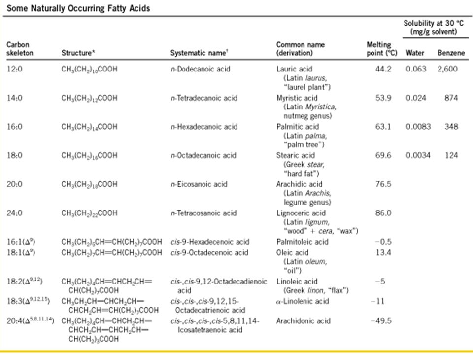
Fatty Acids; are carboxylic acids, hydrocarbon derivatives with unbranched hydrocarbon chains from 4 to 36 carbons long and fully saturated or nonsaturated. The fatty acids are at about the same low oxidation or high reduction state as the hydrocarbons in fossil fuels. Thus, the cellular oxidation of fatty acids (to CO 2 and H 2 O) is high exergonic.
 is high exergonic..")
9
Fatty Acids Are Hydrocarbon Derivatives

10
Two Types of Fatty Acids 1. Triacylglycerols 2. Waxes

11
The simplest lipids constructed from fatty acids are the triacylglycerols, are composed of three fatty acids each in ester linkage with a single glycerol. Simple triacylglycerols of 16:0, 18:0, and 18: l, for example, are tristearin, tripalmitin, and triolein, respectively. Triacylglycerols Are Fatty Acid Esters of Glycerol

12
Triacylglycerols Provide Stored Energy and Insulation As stored fuels, triacylglycerols have two significant advantages over polysaccharides such as glycogen and starch. 1.The carbon atoms of fatty acids are more reduced than those of sugars, and oxidation of triacylglycerols yields more than twice as much energy, gram for gram, as that of carbohydrates. 2. Furthermore, because triacylglycerols are hydrophobic and therefore unhydrated, the organism that carries fat as fuel does not have to carry the extra weight of water of hydration that is associated with stored polysaccharides.

13
Triacylglycerols Provide Stored Energy and Insulation

14
Triacylglycerols stored under skin serve not only as energy stores but as insulation against low temperatures

15
Two Types of Fatty Acids 1. Triacylglycerols 2. Waxes

16
Waxes Serve as Energy Stores and Water Repellents Biological waxes are esters of long-chain saturated and unsaturated fatty acids (having 14 to 36 carbon atoms) with long-chain alcohols (having 16 to 30 carbon atoms). Their melting points (60 to 100 °C) are generally higher than those of triacylglycerols.
 are generally higher than those of triacylglycerols..")
17
Waxes Serve as Energy Stores and Water Repellents Major Functions of Waxes; 1.Chief storage form of metabolic fuel. 2.Water-repellent (on feather) and their firm consistency (beeswax)
 and their firm consistency (beeswax).")
18
Waxes Serve as Energy Stores and Water Repellents

19
*** Storage Lipids *** Structural Lipids in Membranes *** Lipids as Signals, Cofactors, and Pigments *** Separation and Analysis of Lipids

21
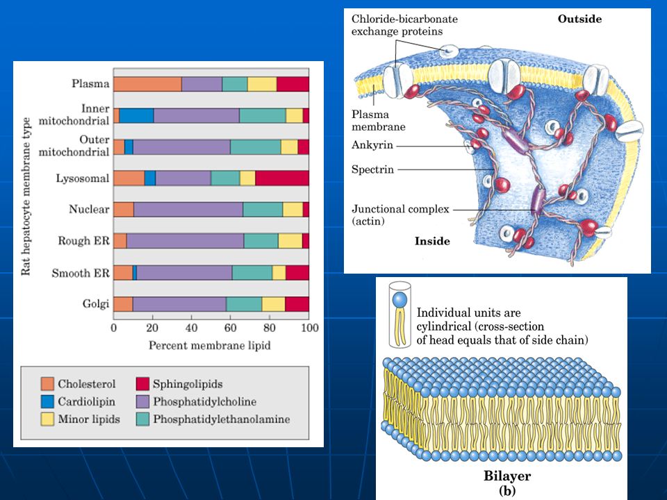
Three general types of membrane lipids: 1.Glycerophospholipids, in which the hydrophobic regions are composed of two fatty acids joined to glycerol; 2.Sphingolipids, in which a single fatty acid is joined to a fatty amine, sphingosine; 3.Sterols, compounds characterized by a rigid system of four fused hydrocarbon rings.

22
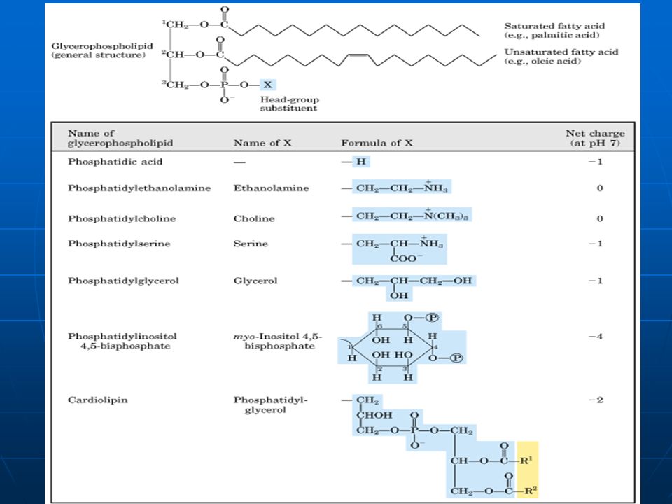
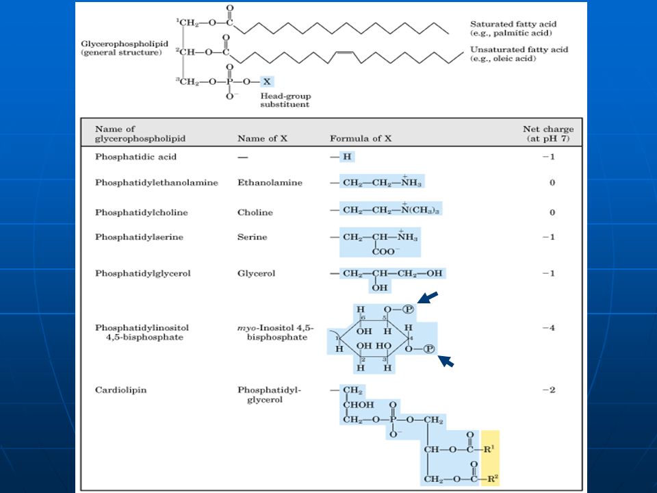
Glycerophospholipids, are membrane lipids in which two fatty acids are attached in ester linkage to the first and second carbons of glycerol, and a highly polar or charged group is attached through a phosphodiester linkage to the third carbon. Glycerophospholipids Are Derivatives of Phosphatidic Acid

24
Three general types of membrane lipids: 1.Glycerophospholipids, in which the hydrophobic regions are composed of two fatty acids joined to glycerol; 2.Sphingolipids, in which a single fatty acid is joined to a fatty amine, sphingosine; 3.Sterols, compounds characterized by a rigid system of four fused hydrocarbon rings.

25
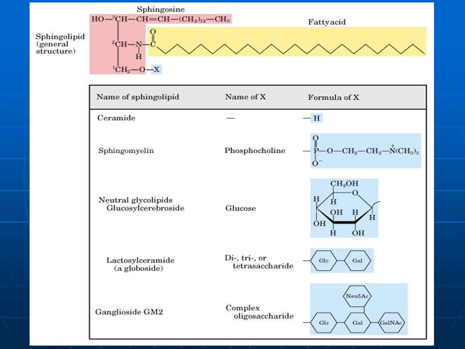
Sphingolipids Are Derivatives of Sphingosine Sphingolipids are composed of one molecule of the long-chain amino alcohol sphingosine, one molecule of a long-chain fatty acid, a polar head alcohol, and sometimes phosphoric acid in diester linkage at the polar head group.

27
Sphingolipids Are Derivatives of Sphingosine Three subclasses of sphingolipids; 1. Sphingomyelins 2. Glycosphingolipids 3. Gangliosides (all derivatives of ceramide, but differing in their head groups)
.")
28
Sphingomyelins are present in plasma membranes of animal cells; the myelin sheath which surrounds and insulates the axons of myelinated neurons is a good source of sphingomyelins.

29
Sphingomyelins contain phosphocholine or phosphoethanolamine as their polar head group, and are therefore classified as phospholipids, together with glycerophospholipids.

30
Sphingolipids Are Derivatives of Sphingosine Three subclasses of sphingolipids; 1. Sphingomyelins 2. Glycosphingolipids 3. Gangliosides (all derivatives of ceramide, but differing in their head groups)
.")
31
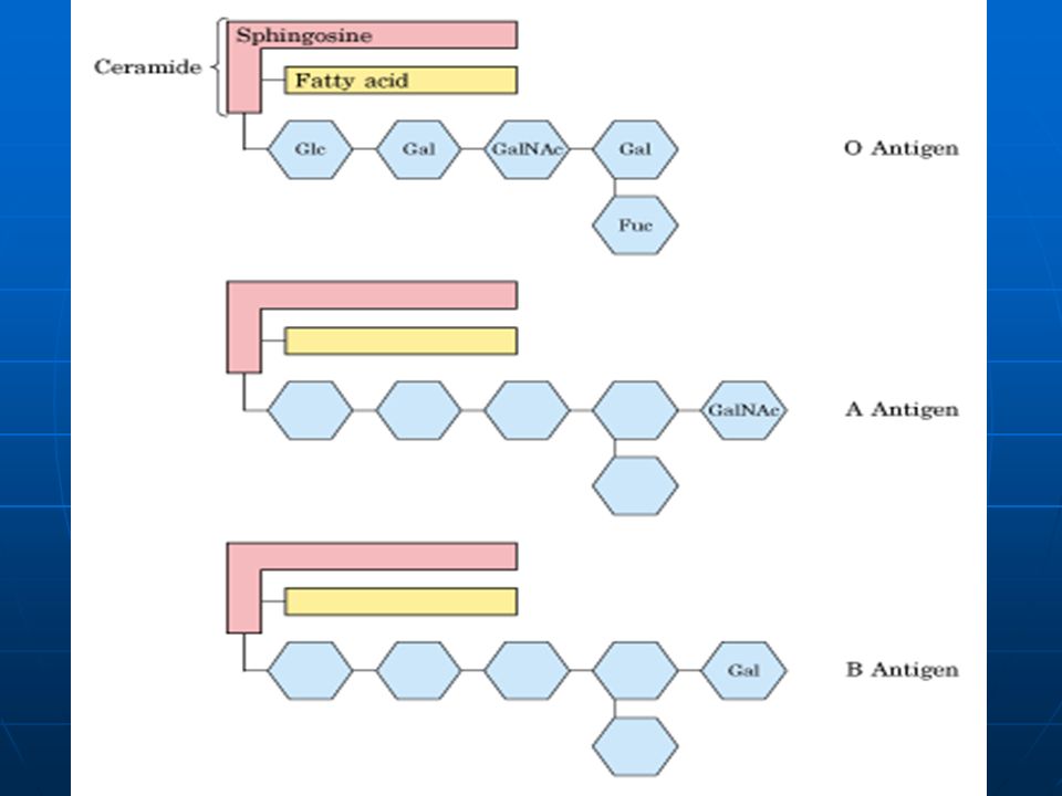
The sugar-containing sphingolipids are called glycosphingolipids. These glycosphingolipids occur largely in the outer face of the plasma membrane.

32
Sphingolipids Are Sites of Biological Recognition The human blood groups (O, A, B) are determined in part by the sugar head groups in these glycosphingolipids. The same three types of complex sugar groups are also found attached to certain blood proteins of individuals of blood types O, A, and B, respectively.

34
Sphingolipids Are Derivatives of Sphingosine Three subclasses of sphingolipids; 1. Sphingomyelins 2. Glycosphingolipids 3. Gangliosides (all derivatives of ceramide, but differing in their head groups)
.")
35
Gangliosides, the most complex sphingolipids, contain very large polar heads made up of several sugar units. Gangliosides make up about 6% of the membrane lipids in the gray matter of the human brain, and they are present in lesser amounts in the membranes of most nonneural animal tissues.

36
Three general types of membrane lipids: 1.Glycerophospholipids, in which the hydrophobic regions are composed of two fatty acids joined to glycerol; 2.Sphingolipids, in which a single fatty acid is joined to a fatty amine, sphingosine; 3.Sterols, compounds characterized by a rigid system of four fused hydrocarbon rings.

37
Sterols are structural lipids consisting of four fused rings, three with six carbons and one with five. Cholesterol, the major sterol in animal tissues, is amphipathic, with a polar head group and a nonpolar hydrocarbon body about as long as a 16-carbon fatty acid in its extended form.

38
Sterols Have Four Fused Hydrocarbon Rings The sterols serve as precursors for a variety of products with specific biological activities. Bile acids, and variety of steroid hormones are produced from cholesterol by oxidation of the side chain at C-17.

39
*** Storage Lipids *** Structural Lipids in Membranes *** Lipids as Signals, Cofactors, and Pigments *** Separation and Analysis of Lipids

40
Lipids present in much smaller amounts, have active roles in the metabolic traffic as metabolites and messengers. 1.Some serve as potent signals, as hormones carried in the blood from one tissue to another, or as intracellular messengers generated in response to an extracellular signal (hormone or growth factor). 2.Others function as enzyme cofactors in electron-transfer reactions in chloroplasts and mitochondria, or in the transfer of sugar moieties in a variety of glycosylation reactions. 3.lipids with a system of conjugated double bonds: pigment molecules that absorb visible light.
. 2.Others function as enzyme cofactors in electron-transfer reactions in chloroplasts and mitochondria, or in the transfer of sugar moieties in a variety of glycosylation reactions. 3.lipids with a system of conjugated double bonds: pigment molecules that absorb visible light..")
41
1.Phosphatidylinositols (Intracellular Signals) 2.Eicosanoids (Messages to nearby Cells) 3.Steroids (Messages between Tissues) 4.Vitamins A and D (Hormone Precursors) 5.Vitamins E and K (Cofactors Cofactors) 6.Dolichols (Activate Sugar Precursors)
 2.Eicosanoids (Messages to nearby Cells) 3.Steroids (Messages between Tissues) 4.Vitamins A and D (Hormone Precursors) 5.Vitamins E and K (Cofactors Cofactors) 6.Dolichols (Activate Sugar Precursors)")
42
Phoshatidylionsitols Acts as Intracellular Signals

44
1.Phosphatidylinositols (Intracellular Signals) 2.Eicosanoids (Messages to nearby Cells) 3.Steroids (Messages between Tissues) 4.Vitamins A and D (Hormone Precursors) 5.Vitamins E and K (Cofactors) 6.Dolichols (Activate Sugar Precursors)
 2.Eicosanoids (Messages to nearby Cells) 3.Steroids (Messages between Tissues) 4.Vitamins A and D (Hormone Precursors) 5.Vitamins E and K (Cofactors) 6.Dolichols (Activate Sugar Precursors)")
45
Eicosanoids Are Potent Biological Effectors Eicosanoids are fatty acid derivatives with a variety of extremely potent hormonelike actions on various tissues of vertebrate animals. Unlike hormones, they are not transported between tissues in the blood, but act on the tissue in which they are produced. This family of compounds is known to be involved in reproductive function; in the inflammation, fever, and pain associated with injury or disease; in the formation of blood clots and the regulation of blood pressure; in gastric acid secretion; and in a variety of other processes important in human health or disease.

46
Three classes of eicosanoids: 1. prostaglandins, 2. thromboxanes, 3. leukotrienes.

47
Eicosanoids Are Potent Biological Effectors

48
The prostaglandins (PG) contain a five-membered ring of carbon atoms originally part of the chain of arachidonic acid. They derive their name from the tissue in which they were first recognized (the prostate gland). Major function; Some prostaglandins stimulate contraction of the smooth muscle of the uterus during labor or menstruation. Others affect blood flow to specific organs, the wake-sleep cycle, and the responsiveness of certain tissues to hormones such as epinephrine and glucagon. Prostaglandins in a third group elevate body temperature (producing fever) and cause inflammation, resulting in pain.
. Major function; Some prostaglandins stimulate contraction of the smooth muscle of the uterus during labor or menstruation. Others affect blood flow to specific organs, the wake-sleep cycle, and the responsiveness of certain tissues to hormones such as epinephrine and glucagon. Prostaglandins in a third group elevate body temperature (producing fever) and cause inflammation, resulting in pain..")
49
The thromboxanes, first isolated from blood platelets (also known as thrombocytes), have a six-membered ring containing an ether. They are produced by platelets and act in formation of blood clots and the reduction of blood flow to the site of a clot.

50
Leukotrienes, found first in leukocytes, contain three conjugated double bonds. They are powerful biological signals; for example, they induce contraction of the muscle lining the airways to the lung. Overproduction of leukotrienes causes asthmatic attacks. The strong contraction of the smooth muscles of the lung that occurs during anaphylactic shock is part of the potentially fatal allergic reaction in individuals hypersensitive to bee stings, penicillin, or various other agents.

51
1.Phosphatidylinositols (Intracellular Signals) 2.Eicosanoids (Messages to nearby Cells) 3.Steroids (Messages between Tissues) 4.Vitamins A and D (Hormone Precursors) 5.Vitamins E and K (Cofactors) 6.Dolichols (Activate Sugar Precursors)
 2.Eicosanoids (Messages to nearby Cells) 3.Steroids (Messages between Tissues) 4.Vitamins A and D (Hormone Precursors) 5.Vitamins E and K (Cofactors) 6.Dolichols (Activate Sugar Precursors)")
52
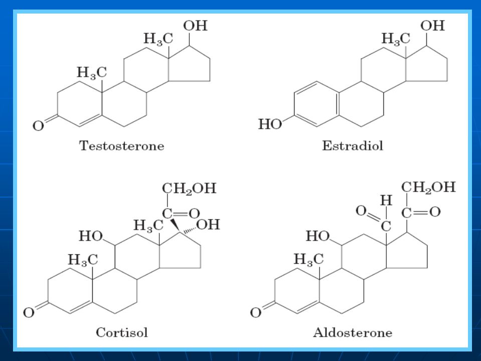
Steroid Hormones Carry Messages between Tissues Steroids derived from cholesterol: Testosterone, the male sex hormone, is produced in the testes. Estradiol, one of the female hormones, is produced in the ovaries and placenta. Cortisol and aldosterone are hormones produced in the cortex of the adrenal gland; they regulate glucose metabolism and salt excretion, respectively.

54
1.Phosphatidylinositols (Intracellular Signals) 2.Eicosanoids (Messages to nearby Cells) 3.Steroids (Messages between Tissues) 4.Vitamins A and D (Hormone Precursors) 5.Vitamins E and K (Cofactors) 6.Dolichols (Activate Sugar Precursors)
 2.Eicosanoids (Messages to nearby Cells) 3.Steroids (Messages between Tissues) 4.Vitamins A and D (Hormone Precursors) 5.Vitamins E and K (Cofactors) 6.Dolichols (Activate Sugar Precursors)")
55
Vitamins: The compounds that are essential to the health of humans and other vertebrates but cannot be synthesized by these animals and must be therefore be obtained in the diet.

56
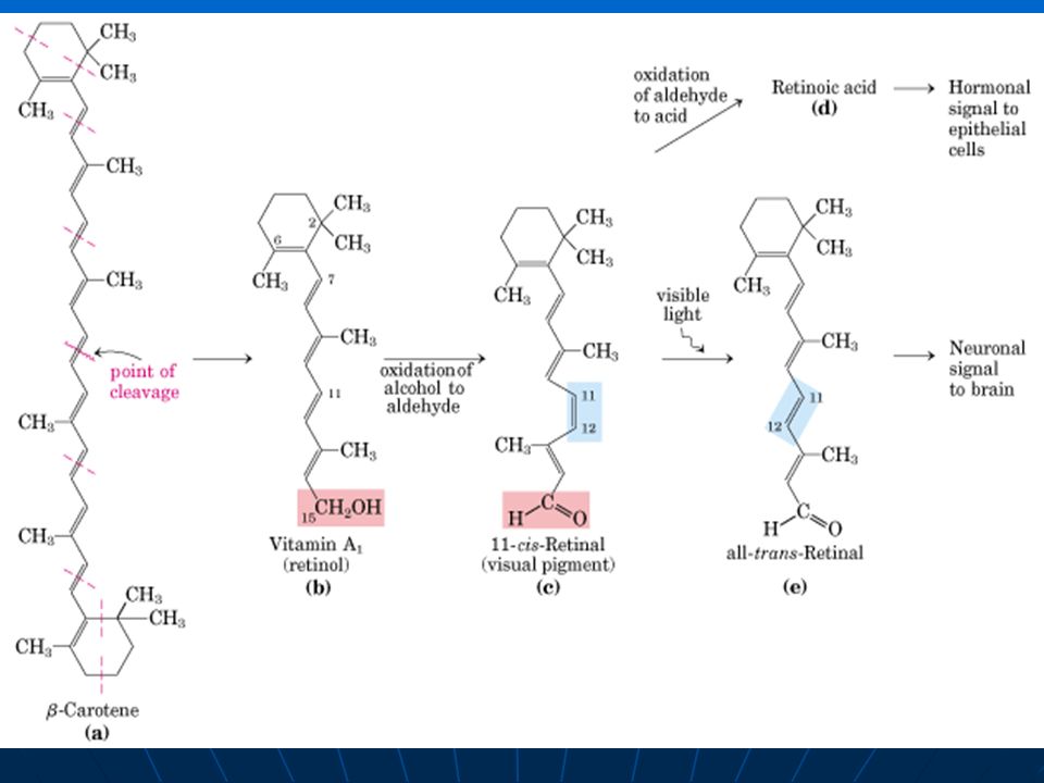
Vitamin A (retinol) is a pigment essential to vision. Deficiency of vitamin A leads to a variety of symptoms in humans, which include dry skin, dry eyes, dry mucous membranes, retarded development and growth, sterility in male, and night blindness. Fish liver oils, eggs, whole milk are good vitamin A dietary sources. Carrots, sweet potatoes and other yellow vegetables contain rich β–carotene, a precursor of vitamin A Vitamins A and D Are Hormone Precursors

58
Vitamin D (cholecalciferol) is a derivative of cholesterol and the precursor to a hormone essential in calcium and phosphate metabolism in vertebrate animals. Vitamin D3 itself is not biologically active, but it is the precursor of 1,25-dihydroxycholecalciferol, a potent hormone that regulates the uptake of calcium in the intestine and the balance of release and deposition of bone calcium and phosphate. Vitamin D3 is normally formed in the skin in a photochemical reaction driven by the ultraviolet component of sunlight. It is also abundant in fish liver oils, and is added to commercial milk as a nutritional supplement.

59
Vitamins A and D Are Hormone Precursors

60
1.Phosphatidylinositols (Intracellular Signals) 2.Eicosanoids (Messages to nearby Cells) 3.Steroids (Messages between Tissues) 4.Vitamins A and D (Hormone Precursors) 5.Vitamins E and K (Cofactors) 6.Dolichols (Activate Sugar Precursors)
 2.Eicosanoids (Messages to nearby Cells) 3.Steroids (Messages between Tissues) 4.Vitamins A and D (Hormone Precursors) 5.Vitamins E and K (Cofactors) 6.Dolichols (Activate Sugar Precursors)")
61
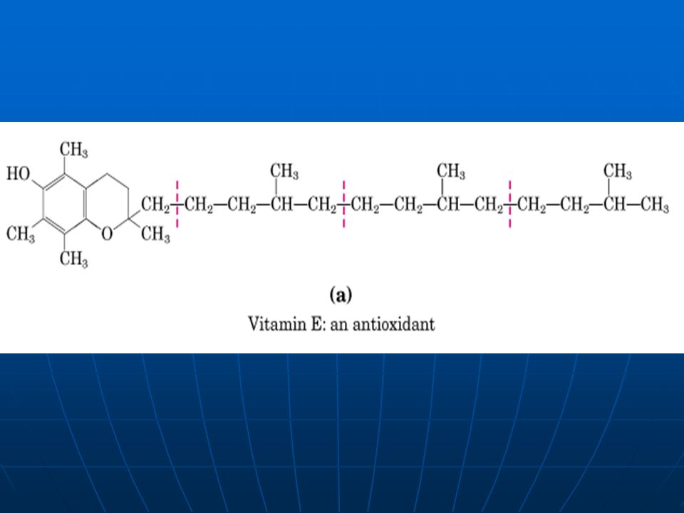
Vitamin E is the collective name for a group of closely related lipids called tocopherols, all of which contain a substituted aromatic ring and a long hydrocarbon side chain. Tocopherols are found in hens' eggs and vegetable oils, and are especially abundant in wheat germ. Deficiency of vitamin E is very rare in humans, but when laboratory animals are fed diets depleted of vitamin E, they develop scaly skin, muscular weakness and wasting, and sterility. Vitamins E and K Are Oxidation- Reaction Cofactors

63
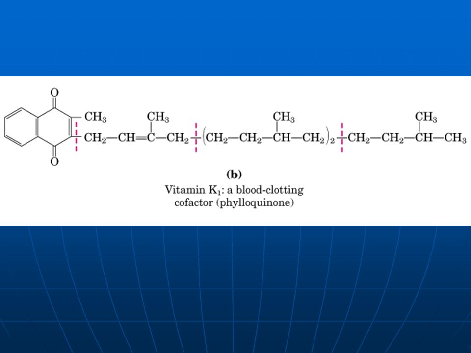
Vitamin K is a lipid cofactor required for normal blood clotting. Vitamin Kl is found in green plant leaves, and a related form, vitamin K2 is formed by bacteria residing in the animal intestine. The vitamin K acts in the formation of prothrombin, a blood plasma protein essential in blood-clot formation. Deficiency of vitamin K results in slowed blood clotting, which can be fatal to a wounded animals. Vitamins E and K Are Oxidation- Reaction Cofactors

65
1.Phosphatidylinositols (Intracellular Signals) 2.Eicosanoids (Messages to nearby Cells) 3.Steroids (Messages between Tissues) 4.Vitamins A and D (Hormone Precursors) 5.Vitamins E and K (Cofactors) 6.Dolichols (Activate Sugar Precursors)
 2.Eicosanoids (Messages to nearby Cells) 3.Steroids (Messages between Tissues) 4.Vitamins A and D (Hormone Precursors) 5.Vitamins E and K (Cofactors) 6.Dolichols (Activate Sugar Precursors)")
66
Dolichols; derived from four or more linked isoprene units. These compounds have strong hydrophobic interactions with membrane lipid, anchoring the attached sugars to the membrane where they participate in sugar-transfer reactions.

67
*** Storage Lipids *** Structural Lipids in Membranes *** Lipids as Signals, Cofactors, and Pigments *** Separation and Analysis of Lipids

68
Purification and Structural Analysis 1.Isolation 2.Purification 3.Structural Analysis

69
Lipid Extraction Requires Organic Solvents Neutral lipids are readily extracted with ethyl ether, chloroform, or benzene, solvents in which lipid clustering driven by hydrophobic interactions does not occur. Membrane lipids are more effectively extracted by more polar organic solvents, such as ethanol or methanol, which reduce the hydrophobic interactions among lipid molecules but also weaken the hydrogen bonds and electrostatic interactions that bind membrane lipids to membrane proteins.

70
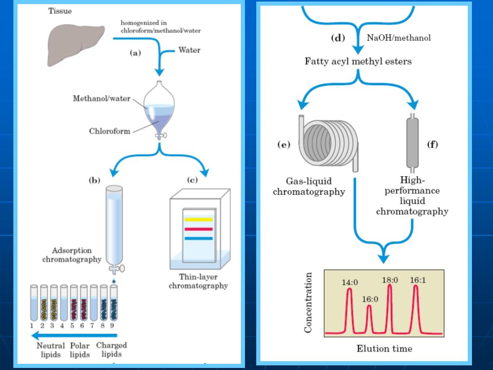
Separation, and identification of cellular lipids. (a)Tissue is homogenized in a chloroform/ methanol/water (1:2:0.8) mixture. (b)Absorption chromatography on a column of silica gel (Si(OH) 4 ). (c)Thin-layer chromatography (TLC), Stain by rhodamine and iodine fumes. (d)Transesterified in a warm aqueous solution of NaOH and methanol, producing a mixture of fatty acyl methyl esters. (e)Separated on the basis of chain length and degree of saturation by gas-liquid chromatography. (f)Separation by HPLC (g)Precise determination of molecular mass, by mass spectroscopy.
Tissue is homogenized in a chloroform/ methanol/water (1:2:0.8) mixture. (b)Absorption chromatography on a column of silica gel (Si(OH) 4 ). (c)Thin-layer chromatography (TLC), Stain by rhodamine and iodine fumes. (d)Transesterified in a warm aqueous solution of NaOH and methanol, producing a mixture of fatty acyl methyl esters. (e)Separated on the basis of chain length and degree of saturation by gas-liquid chromatography. (f)Separation by HPLC (g)Precise determination of molecular mass, by mass spectroscopy..")
72
1.Carbohydrates (three major size classes) 2.starch and glycogen 3.cellulose and chitin 4.Biological functions of Lipids 5.Structure and functions of fatty acids (triacylglycerols and waxes) 6.Structures and functions of three Types of Membrane Lipids (examples).
 2.starch and glycogen 3.cellulose and chitin 4.Biological functions of Lipids 5.Structure and functions of fatty acids (triacylglycerols and waxes) 6.Structures and functions of three Types of Membrane Lipids (examples).")
Similar presentations
 Membrane/Structural.>")


 Reference: Lehninger Biochemistry Advance Biochemistry Isfahan University of.>")


 are the major components of triacylglycerols, glycerophospholipids, and sphingolipids.>")


