Download presentation
Presentation is loading. Please wait.

1
Embryonic period

2
Embryonic period or period of Organogenesis
Is the period from the third to the eighth weeks of development and is the time when each of the three germ layers, ectoderm, mesoderm, and endoderm, gives rise to a number of specific tissues and organs.

3
By the end of the embryonic period, the main organ systems have been established, rendering the major features of the external body form recognizable by the end of the second month.

4
Derivatives of the ectoderm germ layer
(a) the central nervous system; (b) the peripheral nervous system; (c) the sensory epithelium of the ear, nose, and eye; and (d) the epidermis, including the hair and nails. ( e ) In addition, it gives rise to subcutaneous glands, the mammary glands, the pituitary gland, and enamel of the teeth.
 the central nervous system; (b) the peripheral nervous system; (c) the sensory epithelium of the ear, nose, and eye; and. (d) the epidermis, including the hair and nails. ( e ) In addition, it gives rise to subcutaneous glands, the mammary glands, the pituitary gland, and enamel of the teeth.")
5
CNS( Neural plate , Neural fold and Neural groove )
Appears at the beginning of the 3rd week as a slipper – shaped plate of thickened ectoderm ( Neural plate ) in the mid-dorsal region in front o the primitive pit . Its lateral edges soon elevate to form the ( Neural folds ) . the depressed mid-region forms the neural groove
 in the mid-dorsal region in front o the primitive pit . Its lateral edges soon elevate to form the ( Neural folds ) . the depressed mid-region forms the neural groove.")
6
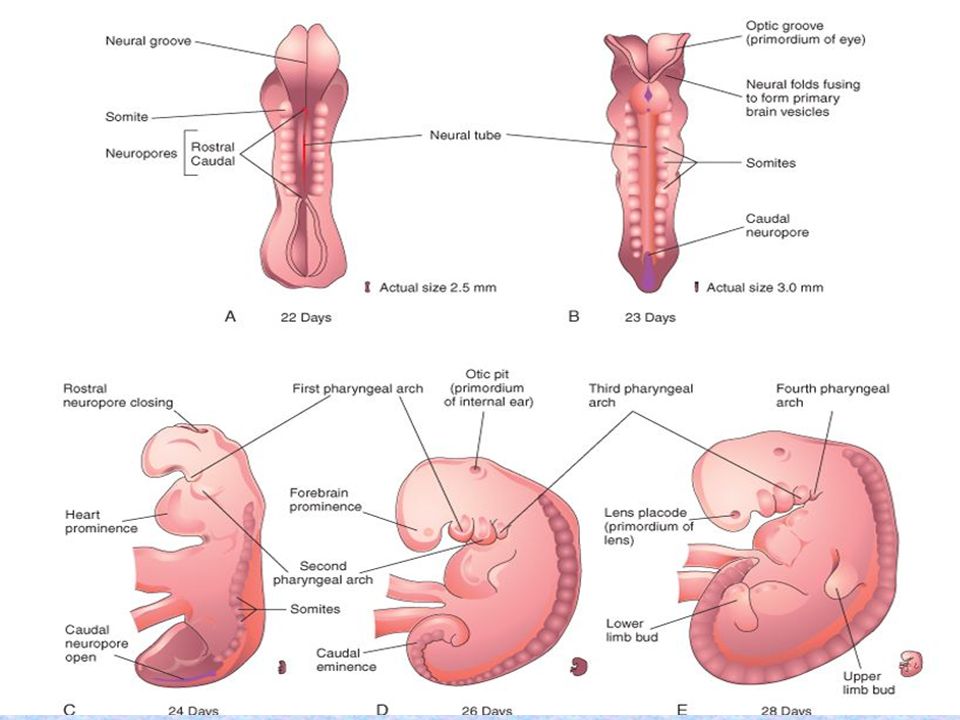
Neural tube Gradually, the neural folds approach each other in the midline, where they fuse Fusion begins in the cervical region (fifth somite) and proceeds cranially and caudally . As a result, the neural tube is formed. Until fusion is complete, the cephalic and caudal ends of the neural tube communicate with the amniotic cavity by way of the cranial and caudal neuropores, respectively . Closure of the cranial neuropore occurs at approximately day 25 (18- to 20-somite stage), whereas the posterior neuropore closes at day 27 (25-somite stage).
 and proceeds cranially and caudally . As a result, the neural tube is formed. Until fusion is complete, the cephalic and caudal ends of the neural tube communicate with the amniotic cavity by way of the cranial and caudal neuropores, respectively . Closure of the cranial neuropore occurs at approximately day 25 (18- to 20-somite stage), whereas the posterior neuropore closes at day 27 (25-somite stage).")
8
Closure o the neural folds results in:
Neurulation is complete, and the central nervous system is represented by a closed tubular structure with a narrow caudal portion, the spinal cord, and a much broader cephalic portion characterized by a number of dilations, the brain vesicles .

9
Closure of the neural folds results in:
As the neural folds elevate and fuse, cells at the lateral border or crest of the neuroectoderm begin to dissociate from their neighbors. The neural crest will undergo an epithelial-to-mesenchymal transition as it leaves the neuroectoderm by active migration and displacement to enter the underlying mesoderm.

10
Neural Crest Derivatives
1. Connective tissue and bones of the face and skull 2. Cranial nerve ganglia 3. C cells of the thyroid gland 4.Conotruncal septum in the heart 5.Odontoblasts 6. Dermis in face and neck 7.Spinal (dorsal root) ganglia 8.Sympathetic chain and preaortic ganglia 9. Parasympathetic ganglia of the gastrointestinal tract 10. Adrenal medulla 11. Schwann cells 12.Glial cells 13.Meninges (forebrain) 14. Melanocytes 15. Smooth muscle cells & blood vessels of the face and forebrain
 ganglia. 8.Sympathetic chain and preaortic ganglia. 9. Parasympathetic ganglia of the gastrointestinal tract. 10. Adrenal medulla. 11. Schwann cells. 12.Glial cells. 13.Meninges (forebrain) 14. Melanocytes. 15. Smooth muscle cells & blood vessels of the face and forebrain.")
11
Closure o the neural folds results in:
Two bilateral ectodermal thickenings, the otic placodes and the lens placodes, become visible in the cephalic region of the embryo. During further development, the otic placodes invaginate and form the otic vesicles, which will develop into structures needed for hearing and maintenance of equilibrium . At approximately the same time, the lens placodes appear. These placodes also invaginate and, during the fifth week, form the lenses of the eyes

12
Derivatives of the Mesodermal Germ Layer
Supporing tissues such as connective tissue , carilage & bone Striated and smooh musculature Blood and lymph cells and the walls of the heart , blood and lymph vessels Kidneys , gonads and their corresponding ducts Corical portion of the suprarenal gland Spleen

13
Mesodermal Germ Layer cells of the mesodermal germ layer form a thin sheet of loosely woven tissue on each side of the midline (day 17).
.")
14
Paraxial mesoderm cells close to the midline proliferate and form a thickened plate of tissue known as Paraxial mesoderm

15
Paraxial mesoderm By the beginning of the third week, paraxial mesoderm begins to be organized into segments. These segments, known as Somitomeres, first appear in the cephalic region of the embryo, and their formation proceeds cephalocaudally. In the head region, somitomeres form in association with segmentation of the neural plate into neuromeres and contribute to mesenchyme in the head

16
Paraxial mesoderm From the occipital region caudally, somitomeres further organize into somites. The first pair of somites arises in the occipital region of the embryo at approximately the 20th day of development . From here, new somites appear in craniocaudal sequence at a rate of approximately three pairs per day until, at the end of the fifth week, 42 to 44 pairs are present . There are 4 occipital, 8 cervical, 12 thoracic, 5 lumbar, 5 sacral, and 8 to 10 coccygeal pairs. The first occipital and the last five to seven coccygeal somites later disappear, while the remaining somites form the axial skeleton .

18
Because somites appear with a specified periodicity, the age of an embryo can be accurately determined during this early time period by counting somites

19
Stages in development of a somite
A. Mesoderm cells are arranged around a small cavity

20
Stages in development of a somite
B. Cells of the ventral and medial walls of the somite lose their epithelial arrangement and migrate in the direction of the notochord. These cells collectively constitute the sclerotome. Cells at the dorsolateral portion of the somite migrate as precursors to limb and body wall musculature. Dorsomedial cells migrate beneath the remaining dorsal epithelium of the somite to form the myotome.

21
Stages in development of a somite
C. Cells forming the myotome continue to extend beneath the dorsal epithelium . Each segmentally arranged myotome contributes to muscles of the back The remaining dorsal epithelium forms the dermatome . dermatomes disperse to form the dermis and subcutaneous tissue of the skin

22
Stages in development of a somite
D. After ventral extension of the myotome, dermatome cells lose their epithelial configuration and spread out under the overlying ectoderm to form dermis.

23
Each somite forms its own :
Sclerotome (the tendon cartilage and bone component), its own Myotome (providing the segmental muscle component), and its own Dermatome, which forms the dermis of the back. Each myotome and dermatome also has its own segmental nerve component.
, its own Myotome (providing the segmental muscle component), and. its own Dermatome, which forms the dermis of the back. Each myotome and dermatome also has its own segmental nerve component.")
24
Intermediate mesoderm
temporarily connects paraxial mesoderm with the lateral plate It differentiates into urogenital structures. In cervical and upper thoracic regions, it forms segmental cell clusters (future nephrotomes), whereas more caudally, it forms an unsegmented mass of tissue, the nephrogenic cord. Excretory units of the urinary system and the gonads develop from this partly segmented, partly unsegmented intermediate mesoderm
, whereas more caudally, it forms an unsegmented mass of tissue, the nephrogenic cord. Excretory units of the urinary system and the gonads develop from this partly segmented, partly unsegmented intermediate mesoderm.")
25
Lateral Plate Mesoderm
the mesoderm layer remains thin laterally and is known as the lateral plate. With the appearance and coalescence of intercellular cavities in the lateral plate, this tissue is divided into two layers : (a) a layer continuous with mesoderm covering the amnion, known as the somatic or parietal mesoderm layer, and (b) a layer continuous with mesoderm covering the yolk sac, known as the splanchnic or visceral mesoderm layer .Together, these layers line a newly formed cavity, the intraembryonic cavity, which is continuous with the extraembryonic cavity on each side of the embryo.
 a layer continuous with mesoderm covering the amnion, known as the somatic or parietal mesoderm layer, and. (b) a layer continuous with mesoderm covering the yolk sac, known as the splanchnic or visceral mesoderm layer .Together, these layers line a newly formed cavity, the intraembryonic cavity, which is continuous with the extraembryonic cavity on each side of the embryo.")
26
Lateral Plate Mesoderm
Mesoderm from the parietal layer, together with overlying ectoderm, will form the lateral and ventral body wall. The visceral layer and embryonic endoderm will form the wall of the gut. Mesoderm cells of the parietal layer surrounding the intraembryonic cavity will form thin membranes, the mesothelial membranes, or serous membranes, which will line the peritoneal, pleural, and pericardial cavities and secrete serous fluid Mesoderm cells of the visceral layer will form a thin serous membrane around each organ

27
Blood vessels form in two ways
vasculogenesis , in which vessels arise from blood islands,

28
Blood vessels form in two ways
B. angiogenesis , in which new vessels sprout from existing ones

29
Extraembryonic blood vessel formation in the villi, chorion, connecting stalk, and wall of the yolk sac in a presomite embryo of approximately 19 days. The first blood islands appear in mesoderm surrounding the wall of the yolk sac at 3 weeks of development and slightly later in lateral plate mesoderm and other regions These islands arise from mesoderm cells that are induced to form hemangioblasts. Hemangioblasts are induced by vascular endothelial growth factor (VEGF), which is secreted by surrounding mesoderm cells. Hemangioblasts in the center of blood islands form hematopoietic stem cells, the precursors of all blood cells, whereas peripheral hemangioblasts differentiate into angioblasts, the precursors to blood vessels.
, which is secreted by surrounding mesoderm cells. Hemangioblasts in the center of blood islands form hematopoietic stem cells, the precursors of all blood cells, whereas peripheral hemangioblasts differentiate into angioblasts, the precursors to blood vessels.")
30
the first blood cells arise in the blood islands of the yolk sac, but this population is transitory.
The definitive hematopoietic stem cells arise from mesoderm surrounding the aorta in a site called the aorta-gonad-mesonephros region (AGM). These cells will colonize the liver, which becomes the major hematopoietic organ of the fetus. Later, stem cells from the liver will colonize the bone marrow, the definitive blood-forming tissue.
. These cells will colonize the liver, which becomes the major hematopoietic organ of the fetus. Later, stem cells from the liver will colonize the bone marrow, the definitive blood-forming tissue.")
31
Derivatives of the Endodermal Germ Layer
it gives rise to (a) the epithelial lining of the respiratory tract; (b) the Parenchyma of the thyroid, parathyroids, liver, and pancreas (c) the reticular stroma of the tonsils and thymus; (d) the epithelial lining of the urinary bladder and urethra; and (e) the epithelial lining of the tympanic cavity and auditory tube
 the epithelial lining of the respiratory tract; (b) the Parenchyma of the thyroid, parathyroids, liver, and pancreas. (c) the reticular stroma of the tonsils and thymus; (d) the epithelial lining of the urinary bladder and urethra; and. (e) the epithelial lining of the tympanic cavity and auditory tube.")
32
Derivatives of the Endodermal Germ Layer
As a result of cephalocaudal folding( this folding is caused by rapid longitudinal growth of the central nervous system particularly the growth of the brain vesicles) , a continuously larger portion of the endoderm-lined cavity is incorporated into the body of the embryo proper .
 , a continuously larger portion of the endoderm-lined cavity is incorporated into the body of the embryo proper .")
33
In the anterior part, the endoderm forms the Foregut;
in the tail region, it forms the Hindgut. The part between foregut and hindgut is the Midgut. The midgut temporarily communicates with the yolk sac by way of a broad stalk, the Vitelline duct . This duct is wide initially, but with further growth of the embryo, it becomes narrow and much longer

34
At its cephalic end, the foregut is temporarily bounded by an ectodermal-endodermal membrane called the Buccopharyngeal membrane . In the fourth week, the buccopharyngeal membrane ruptures, establishing an open connection between the amniotic cavity and the primitive gut . The hindgut also terminates temporarily at an ectodermal-endodermal membrane, the cloacal membrane, which breaks down in the seventh week to create the opening for the anus.

35
As a result of rapid growth of the somites, the initial flat embryonic disc also folds laterally, and the embryo obtains a round appearance. Simultaneously, the ventral body wall of the embryo is established, except for a small part in the ventral abdominal region where the yolk sac duct and connecting stalk are attached.

36
Another important result of cephalocaudal and lateral folding is partial incorporation of the allantois into the body of the embryo, where it forms the Cloaca The distal portion of the allantois remains in the connecting stalk. By the fifth week, the yolk sac duct, allantois, and umbilical vessels are restricted to the region of the umbilical ring

37
In humans, the yolk sac is vestigial and in all probability has a nutritive role only in early stages of development . In the second month of development, it lies in the chorionic cavity

38
THANK YOU

Similar presentations


>")


>")


>")


