Download presentation
Presentation is loading. Please wait.

1
Chest Tubes Written by: Melissa Dearing LSC - Kingwood

2
Introduction Chest tubes are used to drain the contents of the pleural spaceChest tubes are used to drain the contents of the pleural space AirAir BloodBlood

3
Absolute Indication PneumothoraxPneumothorax TensionTension OpenOpen SimpleSimple HemothoraxHemothorax Traumatic ArrestTraumatic Arrest Bilateral involvementBilateral involvement

4
Relative Indications Rib fractures and PPVRib fractures and PPV Profound hypoxia and hypotension with a penetrating chest injuryProfound hypoxia and hypotension with a penetrating chest injury Profound hypoxia and hypotension and unilateral signs of hemothoraxProfound hypoxia and hypotension and unilateral signs of hemothorax

5
Emergent Placement Patient in arrest with no cardiac outputPatient in arrest with no cardiac output Immediate decompression of both chest hemi thoraces to exclude tension pneumoImmediate decompression of both chest hemi thoraces to exclude tension pneumo Patient in shock or profoundly hypoxic with a penetrating chest injury should have a chest tube placed immediatelyPatient in shock or profoundly hypoxic with a penetrating chest injury should have a chest tube placed immediately

6
Drainage Contents The nature of the material draining form the tube is also importantThe nature of the material draining form the tube is also important Dark or bright red bloodDark or bright red blood Intestinal contentsIntestinal contents Persistent air leakPersistent air leak

7
Chest Tubes Variety of SizesVariety of Sizes 7 F to 40 F7 F to 40 F Attached to a one way device to prevent reentry of air into the chestAttached to a one way device to prevent reentry of air into the chest Dependent on lung healing for resolution of pneumoDependent on lung healing for resolution of pneumo

8
All chest tubes used to drain a pneumothorax should be directed toward the apex.All chest tubes used to drain a pneumothorax should be directed toward the apex. Chest tubes used to drain a hemothorax or fluid should be directed toward the posterior chest.Chest tubes used to drain a hemothorax or fluid should be directed toward the posterior chest.

9
Small bore catheters can be placed in the second intercostal space anteriorly in the midclavicular lineSmall bore catheters can be placed in the second intercostal space anteriorly in the midclavicular line Can also be placed laterally in the 5 th to 7 th intercostal spaceCan also be placed laterally in the 5 th to 7 th intercostal space Chest tubes should be placed on top of the rib to avoid the vessels that run underneathChest tubes should be placed on top of the rib to avoid the vessels that run underneath

10
Insertion Local anesthesia is given at the site of insertionLocal anesthesia is given at the site of insertion Area is prepped and drapedArea is prepped and draped Incision is made along the upper rib border to the size of the operator’s fingerIncision is made along the upper rib border to the size of the operator’s finger A curved clamp is pushed through the muscle tissue to split the fibers to enable insertion of the tube.A curved clamp is pushed through the muscle tissue to split the fibers to enable insertion of the tube.

11
The operators finger is then inserted to enlarge the tract made by the clampThe operators finger is then inserted to enlarge the tract made by the clamp A chest tube is then mounted on the clamp and passed along the track into the pleural cavityA chest tube is then mounted on the clamp and passed along the track into the pleural cavity Tube is connected to an underwater seal and sutured into placeTube is connected to an underwater seal and sutured into place CXR is obtainedCXR is obtained

12
Acute Complications Usually due to the techniqueUsually due to the technique Hemothorax from laceration of a vesselHemothorax from laceration of a vessel Lung lacerationLung laceration Diaphragm or abdominal cavity penetrationDiaphragm or abdominal cavity penetration Subcutaneous placementSubcutaneous placement Tube placed too far in (pain)Tube placed too far in (pain) Tube falls out (not secured)Tube falls out (not secured)
Tube placed too far in (pain) Tube falls out (not secured)Tube falls out (not secured)")
13
Late Complications Blocked tubeBlocked tube Blood clotBlood clot EmpyemaEmpyema Pneumothorax with removalPneumothorax with removal

14
Underwater Seals Allow air to escape through the drain but prevents air re-entering the chest cavityAllow air to escape through the drain but prevents air re-entering the chest cavity

15
One Bottle Chest Drainage System

16
Two Bottle Chest Drainage System

17
Three Bottle Chest Drainage System

18
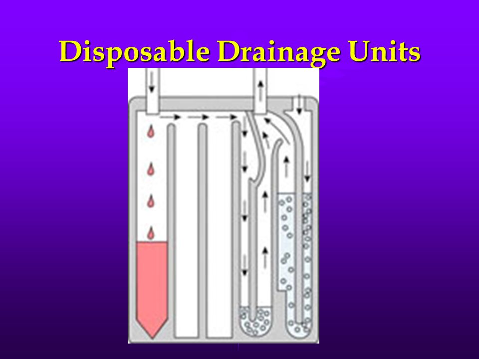
Disposable Drainage Units

20
Removing the Chest Tube CriteriaCriteria No air leak in 24 hoursNo air leak in 24 hours No fresh bleeding in 24 hoursNo fresh bleeding in 24 hours Fluid loss of < 200 ml/dayFluid loss of < 200 ml/day Clinical and Xray evidence of resolution of empyemaClinical and Xray evidence of resolution of empyema

21
Removal of Chest Tube Prepare and drape the sitePrepare and drape the site Inject local anesthesia around suture siteInject local anesthesia around suture site Insert a purse string suture at the site and leave it looseInsert a purse string suture at the site and leave it loose Cut the suture holding the chest tube in placeCut the suture holding the chest tube in place Ask the patient to take a deep breath and perform a Valsalva maneuverAsk the patient to take a deep breath and perform a Valsalva maneuver

22
Removal of Chest Tube Pull the drain tube out quickly while an assistant pulls the purse string suture tight to close the wound.Pull the drain tube out quickly while an assistant pulls the purse string suture tight to close the wound. Cover the site with an occlusive dressingCover the site with an occlusive dressing Observe the patient and order a chest xray in 4-6 hoursObserve the patient and order a chest xray in 4-6 hours

23
Video http://vimeo.com/2016659http://vimeo.com/2016659http://vimeo.com/2016659

Similar presentations


>")
