Download presentation
Presentation is loading. Please wait.

1
Chronic (gradual) Loss of Vision Definition: Symptoms which have come on gradually over the course of weeks or months rather than minutes, hours or a few days
 Loss of Vision Definition: Symptoms which have come on gradually over the course of weeks or months rather than minutes, hours or a few days")
2
History Chronic loss of vision is almost always painless Determine whether the symptoms are uniocular or binocular Duration of history History of previous ocular disease Present medications, ocular and systemic Diabetes and hypertension. A positive family history of glaucoma prompts careful exclusion of this disorder.

3
The Elderly patient with chronic loss of vision Chronic loss of vision in the elderly is often binocular but asymmetrical Aim to exclude each of the ‘big three’ Cataract, Macular degeneration Chronic open angle glauoma Cataract (COMMON) Glaucomatous optic atrophy (COMMON) Age-related macular degeneration (ARMD) (COMMON) Diabetic maculopathy (COMMON) Corneal scarring (RARE) Optic atrophy (RARE)
 Glaucomatous optic atrophy (COMMON) Age-related macular degeneration (ARMD) (COMMON) Diabetic maculopathy (COMMON) Corneal scarring (RARE) Optic atrophy (RARE)")
4
Systematic Approach Refractive Errors Cornea Anterior Chamber Lens Vitreous Macula Retina Optic Nerve

5
Systematic Approach Refractive Errors

6
Systematic Approach Refractive Errors Cornea Dystrophies Epithelium Stroma Endothelium Infective agents Others

7
Systematic Approach Refractive Errors Cornea Anterior Chamber Lens Vitreous Macula Retina Optic Nerve

8
Systematic Approach Refractive Errors Cornea Anterior Chamber Lens Vitreous Macula Retina Optic Nerve

9
Systematic Approach Refractive Errors Cornea Anterior Chamber Lens Vitreous Macula Retina Optic Nerve

12
Systematic Approach Refractive Errors Cornea Anterior Chamber Lens Vitreous Macula Retina Optic Nerve

13
Phacoemulsification Of Cataract

14
Posterior capsule opacification

15
Capsulotomy

16
Systematic Approach Refractive Errors Cornea Anterior Chamber Lens Vitreous Macula Retina Optic Nerve

17
Normal retina

18
Drusen

19
Systematic Approach Refractive Errors Cornea Anterior Chamber Lens Vitreous Macula Retina Optic Nerve

21
What do patients with AMD see?

23
Macular hole

24
Diabetic pathology

26
Capillary microaneurysms

28
Proliferative Diabetic Retinopathy

29
Optic Disc

30
Chronic Loss of Vision In the Elderly History (one eye or both) ▼ Acuities and visual Fields (cortical field defects) ▼ Check cornea (Scarring or oedema) ▼ Red Reflex (Cataract or vitreous opacities) ▼ Pupils and IOP (Glaucoma) ▼ Dilate for Fundoscopy (ARMD,diabetic retionopathy,POAG,optic atrophy)
 ▼ Acuities and visual Fields (cortical field defects) ▼ Check cornea (Scarring or oedema) ▼ Red Reflex (Cataract or vitreous opacities) ▼ Pupils and IOP (Glaucoma) ▼ Dilate for Fundoscopy (ARMD,diabetic retionopathy,POAG,optic atrophy)")
31
Chronic Loss of Vision Adult working Age Early-onset, age-related changes (COMMON) (e.g. cataract and macula degeneration and diabetic retinopathy) must be considered IN ALL CASES as age increases, but another collection of pathologies must be kept in mind.
 must be considered IN ALL CASES as age increases, but another collection of pathologies must be kept in mind..")
32
Hypertensive Retinopathy

33
Uniocular symptoms Cataract (COMMON) Corneal opacities * (RARE) Opacities in the vitreous (RARE) Severe field loss in one eye (FAIRLY RARE) Chronic uniocular visual loss from macular disease (RARE) Optic nerve-related uniocular visual loss (RARE)
 Corneal opacities * (RARE) Opacities in the vitreous (RARE) Severe field loss in one eye (FAIRLY RARE) Chronic uniocular visual loss from macular disease (RARE) Optic nerve-related uniocular visual loss (RARE)")
34
Keratoconus

35
Binocular Symptoms Cataract (COMMON) Maculopathies (COMMON) GIaucoma in the under-65s (RARE, but COMMONER in some races) Opacities in the vitreous of both eyes (RARE) Binocular field defects causing chronic visual loss (RARE) Bilateral optic atrophy (RARE) Corneal disease (RARE) Bilateral chronic Papilloedema (RARE)
 Maculopathies (COMMON) GIaucoma in the under-65s (RARE, but COMMONER in some races) Opacities in the vitreous of both eyes (RARE) Binocular field defects causing chronic visual loss (RARE) Bilateral optic atrophy (RARE) Corneal disease (RARE) Bilateral chronic Papilloedema (RARE)")
36
Chronic Loss of Vision Adult working Age Uniocular system Binocular System History and acuities History and acuities ▼ ▼ Cornea and red reflex Cornea and red reflex (keratoconus, cataract,uveitis) (corneal dystrophies,cataract,uveitis) ▼ ▼ Visual Fields Visual fields (retinal detachment,meningioma) (retinitis pigmentosa,pituitary lesions) ▼ ▼ Pupils and IOP (glaucoma) (glaucoma) ▼ ▼ Dilate for fundoscopy Dilate for fundoscopy (malignant melanoma, optic atrophy) (maculopathies,optic atrophy,papilloedema)
 (corneal dystrophies,cataract,uveitis) ▼ ▼ Visual Fields Visual fields (retinal detachment,meningioma) (retinitis pigmentosa,pituitary lesions) ▼ ▼ Pupils and IOP (glaucoma) (glaucoma) ▼ ▼ Dilate for fundoscopy Dilate for fundoscopy (malignant melanoma, optic atrophy) (maculopathies,optic atrophy,papilloedema)")
37
Acute Painless Visual Disturbance Definition Disorders of visual function which are painless and which have a well-defined onset (minutes,hours)
")
38
Pupil Reactions

39
Sympathetic system

40
Uniocular predominantly central visual loss: normal pupils Macular and paramacular disease (COMMON) Acute media opacity (RELATIVELY COMMON)
 Acute media opacity (RELATIVELY COMMON)")
41
Uniocular predominantly central visual loss: abnormal pupil reaction Optic nerve disease (COMMON) Retinal disease * (COMMON) Adie’s syndrome (RARE)
 Retinal disease * (COMMON) Adie’s syndrome (RARE)")
44
Acute uniocular peripheral visual loss Acute rhegmatogenous retinal detachment* Retinal branch artery occlusion* Anterior Ischaemic Optic Neuropathy (AION)
")
47
Uniocular acute visual disturbance Amaurosis fugax Visual intrusions benign floater vitreous heamorrhage posterior vitreous detachment Migraine

48
Binocular acute central visual loss Retinal causes (RARE) Cytomegalovirus retinitis Acute rises in blood pressure may induce macular oedema e.g. pregnancy Optic nerve causes (RARE) Bilateral optic neuritis Cortical causes (RARE)
 Bilateral optic neuritis Cortical causes (RARE).")
49
Papilloedema Definition: Swelling of the OPTIC DISK, usually in association with increased intracranial pressure, characterized by - Blind spot enlargement - Hyperemia, - Blurring of the disk margins - Microhaemorrhages - Engorgement of retinal veins. Chronic papilloedema may cause OPTIC ATROPHY and visual loss. (Miller et al., Clinical Neuro- Ophthalmology, 4th ed, p175).
..")
50
Binocular acute peripheral visual loss Homonymous defects retrochiasmal (COMMON) Bitemporal defects – (chiasmal)
 Bitemporal defects – (chiasmal)")
51
Acute Painless Visual Disturbance UniocularBinocular CentralPeripheralCentralPeripheral Retinal ARMD (age related macular degeneration CMV retinitis (cytomegalovirus) Vein and arterial occlusion CSR (central serous retnopathy Retinal Detachment Retinal Migrane CMV retinitis Retinal CMV retinitis Hypertension Retinal Detachment Optic Nerve Optic neuritis Ischaemic ON Optic Nerve Ischeamic Optic Neuropathy Optic Nerve Infiltrative papilloedema Optic Nerve Drusen Media opacity Vitreous heamorrhage Choroiditis Cortical Infarction Hysteria Cortical Homonymous Bitemporal Adie’s syndrome
 Vein and arterial occlusion CSR (central serous retnopathy Retinal Detachment Retinal Migrane CMV retinitis Retinal CMV retinitis Hypertension Retinal Detachment Optic Nerve Optic neuritis Ischaemic ON Optic Nerve Ischeamic Optic Neuropathy Optic Nerve Infiltrative papilloedema Optic Nerve Drusen Media opacity Vitreous heamorrhage Choroiditis Cortical Infarction Hysteria Cortical Homonymous Bitemporal Adie’s syndrome")
52
THE ACUTE RED EYE

54
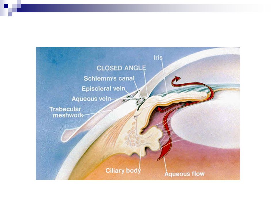
ANGLE CLOSURE GLAUCOMA Symptoms Previous history Episodes of blurring pain or haloes for an hour or two in some early evenings for a few weeks Pain severe, radiating to forehead, with vomiting often Slight photophobia Watery secretion or discharge Visual acuity -Bad, usually Onset 2-3 hours Systemic symptoms- Often prostration and vomiting because of pain Laterality- Unilateral usually Age Usually 50 +

55
Signs Hyperaemia Circumcorneal purple + diffuse, conjunctival Corneal epithelial oedema (fogged view of iris) Anterior chamber shallow (N.B. see fellow eye) Iris - Oedematous and hyperaemic Pupil - Dilated, oval Pupil light reflex - Absent or reduced Tension - Very high Tenderness - Marked
 Iris - Oedematous and hyperaemic Pupil - Dilated, oval Pupil light reflex - Absent or reduced Tension - Very high Tenderness - Marked.")
57
ACUTE IRIDOCYCLITIS Symptoms Previous history- Any previous attack protracted for weeks Pain- moderate, localized to eye. Dull Photophobia - Moderate Secretion or discharge - Watery Visual acuity - Poor or slightly reduced Onset -Gradual (1-2 days) Systemic symptoms -Usually none Laterality? - Unilateral usually Age -Usually 15-25
 Systemic symptoms -Usually none Laterality. - Unilateral usually Age -Usually")
58
Signs Hyperaemia -Circumcorneal purple + diffuse conjunctival Cornea -(Keratic) precipitates Anterior chamber -Exudate (flare, cells) often deep. Sometimes hypopyon Iris -Often hyperaemic and ‘muddy’ Pupil -Contracted (due to Synechiae) Pupil light reflex - Reduced or absent Tension -High, normal or low Tenderness -Moderate to marked Other points ankylosing spondylitis in males sometimes
 Pupil light reflex - Reduced or absent Tension -High, normal or low Tenderness -Moderate to marked Other points ankylosing spondylitis in males sometimes.")
61
CONJUNCTIVITIS Acute bacterial Symptoms Previous history -Possible Pain -Gritty, especially on blinking Photophobia -Slight Secretion or discharge -Muco-purulent. heavy. Neutrophils + Visual acuity -Normal Onset -Within 1-2 days Systemic symptoms -None Laterality -Invariably bilateral Age -Any, but usually in children

62
Signs Hyperaemia -Conjunctival,severe and diffuse.Brick red Cornea -Clear and sparkling Anterior -Chamber Normal Iris -Normal Pupil -Normal Pupil light reflex -Normal Tension -Normal Tendernss -Slight Other points -Epidemic in school or family?

64
Acute adenovirus Symptoms Previous history -Sometimes Pain -Gritty, especially on blinking Photophobia -Slight/moderate Secretion or discharge -Watery. Monocytes Visual acuity -Normal Onset -Several days Systemic symptoms -None Bilateral -Unilateral or bilateral Age -Any, but usually up to 25

65
Signs Hyperaemia -Conjunctival, mild. Often restricted to a sector next to limbus Cornea -Looks clear but fluorescein stain ‘superficial punctate’ spots, seen with slitlamp microscope Anterior chamber -Normal Iris -Normal Pupil -Normal Pupil light reflex -Normal Tension -Normal Tendemess -Slight Other points -Pre-auricular lymph node swollen and tender. Epidemic school or work?

67
Chlamydial Symptoms Previous history -Frequent recurrences: often chronic Pain -Variable discomfort, gritty Photophobia -Variable Secretion or discharge -Watery ± pus Neutrophils Inclusion bodies Visual acuity -Normal at first. If chronic, often eventual blindness Onset -Several days Systemic symptoms -None laterality -Bilateral (at first may be unilateral) Age -Any
 Age -Any.")
68
Signs Hyperaemia –Diffuse conjunctival Cornea –clear. Late: pannus and/or diffuse fibrosis, also of conjunctiva Anterior chamber -Normal Iris -Normal Pupil -Normal Pupil light reflex -Normal Tension -Normal Tendrness -Slight Other points -In newborn, mother +/- father have GU infection.

70
KERATITIS & CORNEAL ULCERS Symptoms Previous history -Previous attacks frequent in viral types foregin body or other injury. Contact lenses Pain -Moderate to severe. Sharp on blinking Photophobia -Marked Secretion or discharge -Watery + + Monocytes in herpes simplex Visual acuity -Poor to bad, usually Onset -Gradual (1-2 day) Systemic symptoms -None laterality? -Unilateral usually Age -Any
 Systemic symptoms -None laterality. -Unilateral usually Age -Any.")
71
Signs Hyperaemia -Circumcorneal purple Cornea -Grey area and/or stains with fluorescein. Dendritic pattern in herpes simplex Anterior chamber -Exudate (flare; cells) Often deep. Sometimes hypopyon Iris -Usually hyperaemic Pupil -Can have synechiae Pupil light reflex -Reduced or absent if visible Tension -Usually normal to low Tenderness -Marked Other points -History of injury often present
 Often deep. Sometimes hypopyon Iris -Usually hyperaemic Pupil -Can have synechiae Pupil light reflex -Reduced or absent if visible Tension -Usually normal to low Tenderness -Marked Other points -History of injury often present.")
74
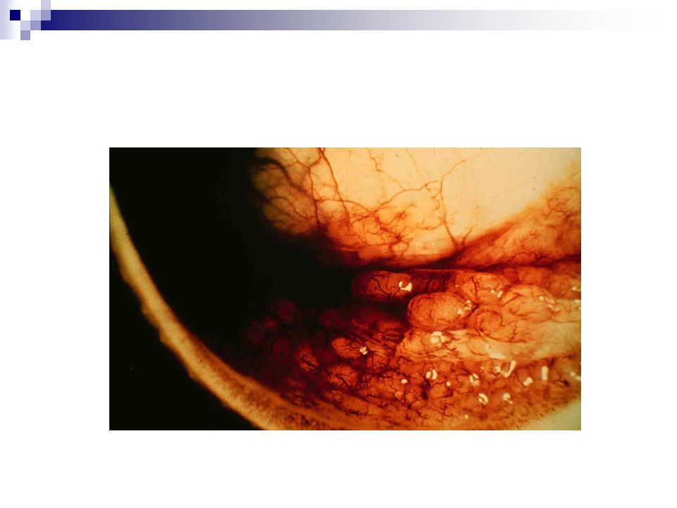
PTERYGIUM A pterygium is a triangular sheet of fibrovascular tissue which invades the cornea. Pterygia typically develop in patients who have been living in hot climates and may represent a response to chronic dryness and exposure to the sun. These may become inflamed and cause an acute red eye.

75
PINGECULA A yellow-white deposit on the bulbar conjunctiva adjacent to the nasal or temporal aspect of the limbus These may become inflamed and cause an acute red eye Histological examination shows degeneration of the collagen fibres of the conjunctival stroma, thinning of the overlying epithelium.

76
DIFFUSE SCLERITIS Scleritis is frequently bilateral and, characteristically, associated with pain. Purplish hue with involvement of the deep episcleral vessels Svstemic diseases, such as collagen vascular, ulcerative colitis, Crohn’s disease, and sarcoidosis, are present in 50% of patients. A topical steroid (such as Pred Forte) may be prescribed to reduce the inflammation A systemic nonsteroidal anti- inflammatory medication is recommended (indocid 25 mg orally)
 may be prescribed to reduce the inflammation A systemic nonsteroidal anti- inflammatory medication is recommended (indocid 25 mg orally).")
77
The Acute Red Eye Uniocular No PainPain Vision normal Vision NormalVision Reduced Corneal staining ? NoYesNoYes Subconjunctival haemorrhage Episcleritis Pterygium Pingeculum Conjunctivitis Anterior uveitis Scleritis HZO Herpes Simplex Marginal ulcer Severe uveitis Angle closure glaucoma Secondary glaucoma Herpes Simplex Bacterial keratitis HZO Binocular No Pain/good visionPain/vision good or poor Bacterial conjunctivitis Viral conjunctivitis Allergic conjunctivitis Chlamydial keratoconjunctivitis Viral keratoconjunctivitis Photophthalmia

78
Thank You

Similar presentations


 Waxman MD PhD>")


 and associated interneurones and sensory neurones. BiologyMad.com.>")


