Download presentation
Presentation is loading. Please wait.

1
Hamid Tavakkoli, MD Associate Prof. of Gastroenterology

2
Recommendations HCC screening 1. Patients at high risk for developing HCC should be entered into surveillance programs (LevelI). 2. Patients on the transplant waiting list should be screened for HCC because in the USA the development of HCC gives increased priority for OLT, and because failure to screen for HCC means that patients may develop HCC that may progress beyond listing criteria without the physician being aware
. 2. Patients on the transplant waiting list should be screened for HCC because in the USA the development of HCC gives increased priority for OLT, and because failure to screen for HCC means that patients may develop HCC that may progress beyond listing criteria without the physician being aware.")
3
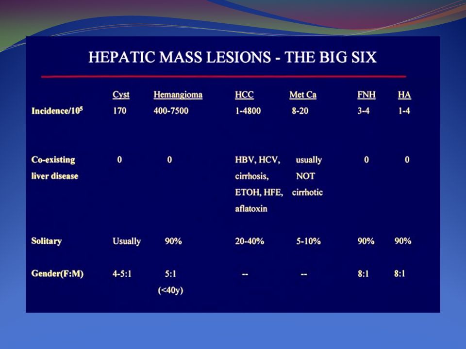
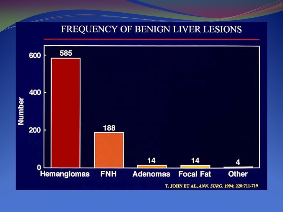
HCC surveillance in recommended Asian male hepatitis B carriers over age 40 Asian female hepatitis B carriers over age 50 Hepatitis B carrier with family history of HCC African/North American Blacks with hepatitis B Cirrhotic hepatitis B carriers Hepatitis C cirrhosis Stage 4 primary biliary cirrhosis Genetic hemachromatosis and cirrhosis Alpha 1-antitrypsin deficiency and cirrhosis Other cirrhosis

4
Surveillance benefit uncertain Hepatitis B carriers younger than 40 (males) or 50 (females) Hepatitis C and stage 3 fibrosis Non-cirrhotic NAFLD
 or 50 (females) Hepatitis C and stage 3 fibrosis Non-cirrhotic NAFLD")
5
Recommendations 3. Surveillance for HCC should be performed using ultrasonography. 4. Patients should be screened at 6 month intervals. 5. The surveillance interval does not need to be shortened for patients at higher risk of HCC.

6
Recommendations 6. Nodules found on ultrasound surveillance that are smaller than 1 cm should be followed with ultrasound at intervals from 3-6 months. If there has been no growth over a period of up to 2 years, one can revert to routine surveillance

7
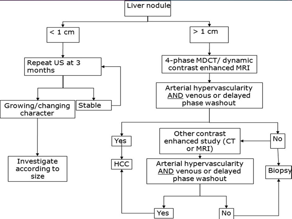
Recommendations 7. Nodules larger than 1 cm found on ultrasound screening of a cirrhotic liver should be investigated further with either 4-phase multidetector CT scan or dynamic contrast enhanced MRI. If the appearances are typical of HCC (i.e., hypervascular in the arterial phase with washout in the portal venous or delayed phase), the lesion should be treated as HCC. If the findings are not characteristic or the vascular profile is not typical, a second contrast enhanced study with the other imaging modality should be performed, or the lesion should be biopsied
, the lesion should be treated as HCC. If the findings are not characteristic or the vascular profile is not typical, a second contrast enhanced study with the other imaging modality should be performed, or the lesion should be biopsied.")
8
Role of AFP in Diagnosis Alphafetoprotein has long been used for the diagnosis of HCC AFP is insufficiently sensitive or specific for use as a surveillance assay. AFP can be elevated in intrahepatic cholangiocarcinoma (ICC) and in some metastases from colon cancer Therefore, the finding of a mass in the liver with an elevated AFP does not automatically indicate HCC.
 and in some metastases from colon cancer Therefore, the finding of a mass in the liver with an elevated AFP does not automatically indicate HCC..")
9
AFP and HCC Since AFP can be elevated in either condition, it is recommended that it no longer be used. Thus, the diagnosis of HCC must rest on radiological appearances and on histology

10
Radiological Diagnosis of HCC HCC can be diagnosed radiologically, without the need for biopsy if the typical imaging features are present.

11
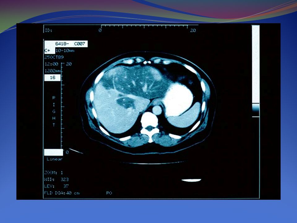
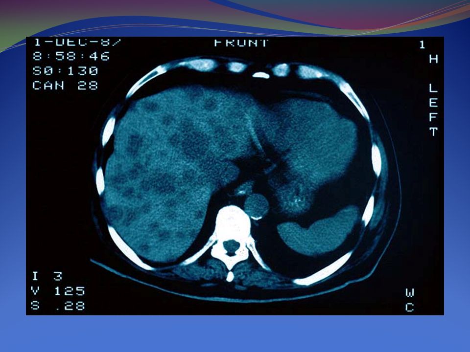
HCC CT, MR

12
HCC CT, US, MR

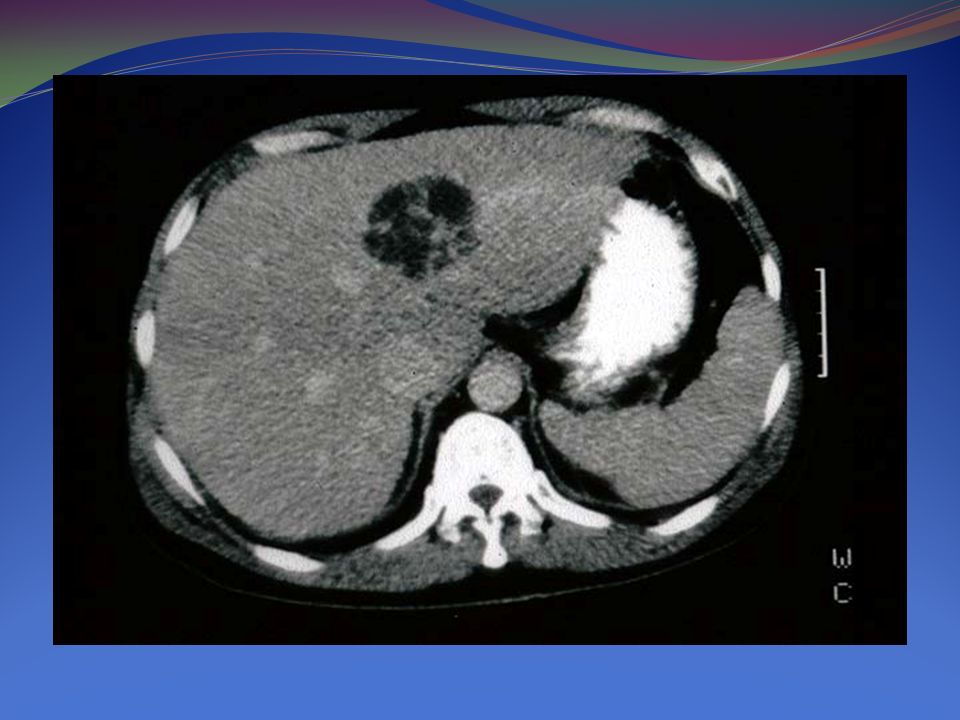
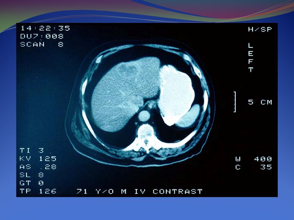
13
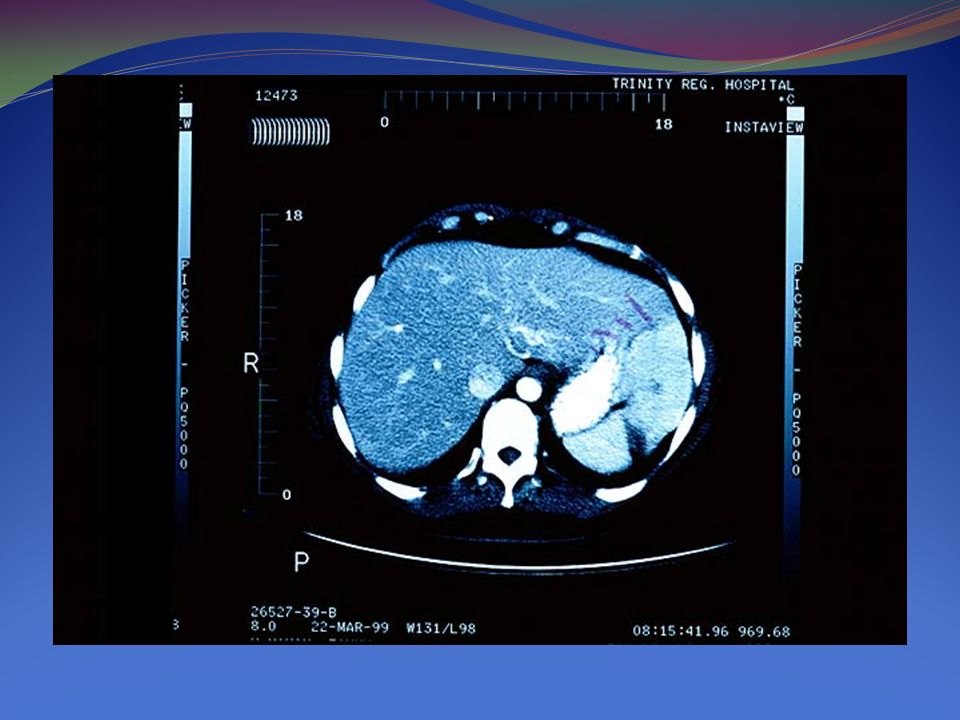
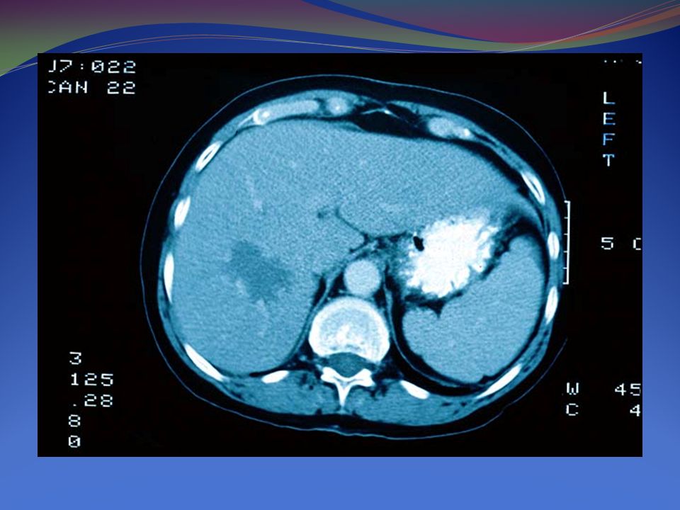
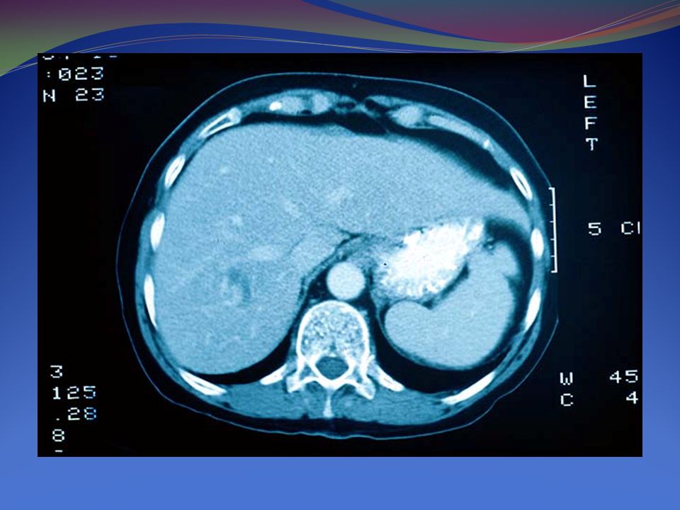
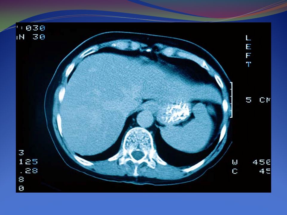
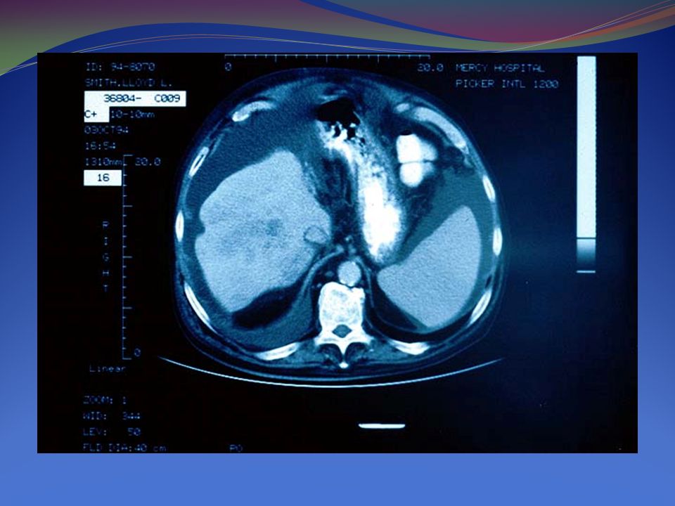
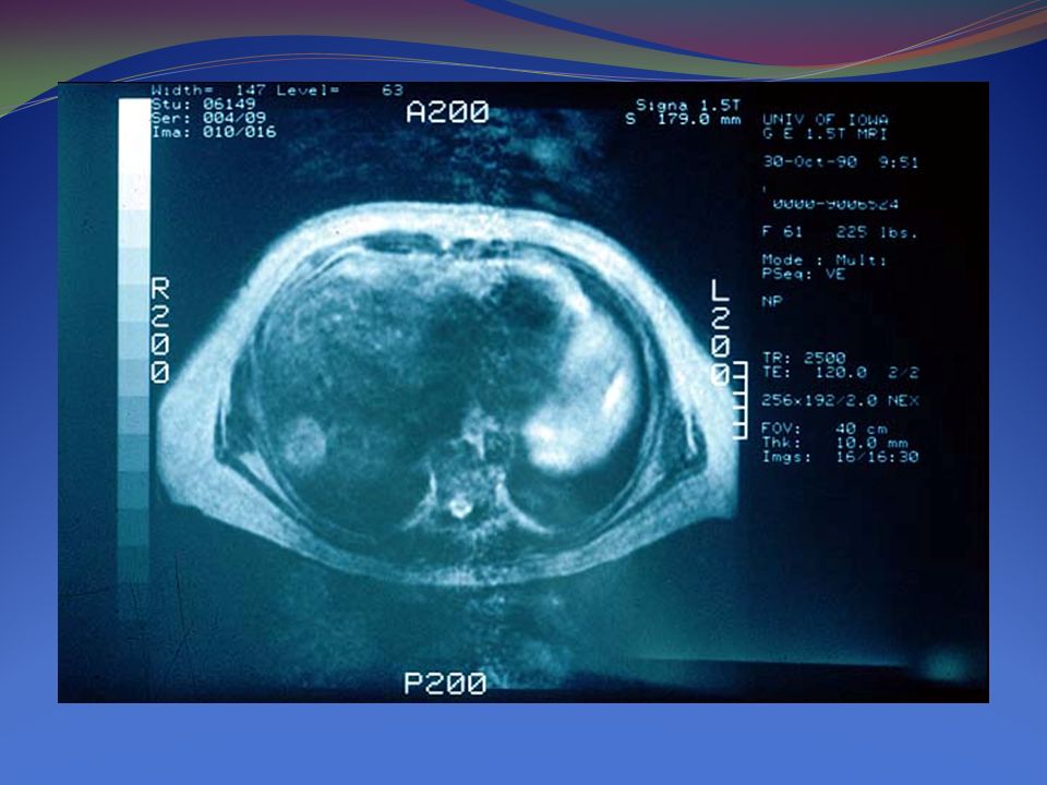
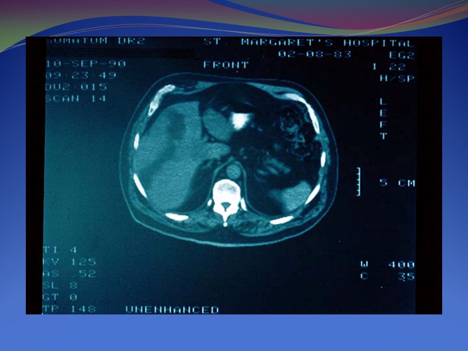
69-year-old man with HCV-related cirrhosis and HCC

16
Recommendations 8. Biopsies of small lesions should be evaluated by expert pathologists. Tissue that is not clearly HCC should be stained with all the available markers including CD34, CK7, glypican 3, HSP-70, and glutamine synthetase to improve diagnostic accuracy

17
Recommendations 9. If the biopsy is negative for patients with HCC, the lesion should be followed by imaging at 3-6 monthly intervals until the nodule either disappears, enlarges, or displays diagnostic characteristics of HCC. If the lesion enlarges but remains atypical for HCC a repeat biopsy is recommended

18
Recommendations 10. To best assess the prognosis of HCC patients it is recommended that the staging system take into account tumour stage, liver function and physical status. The impact of treatment should also be considered when estimating life expectancy. Currently, the BCLC system is the only staging system that accomplishes these aims

20
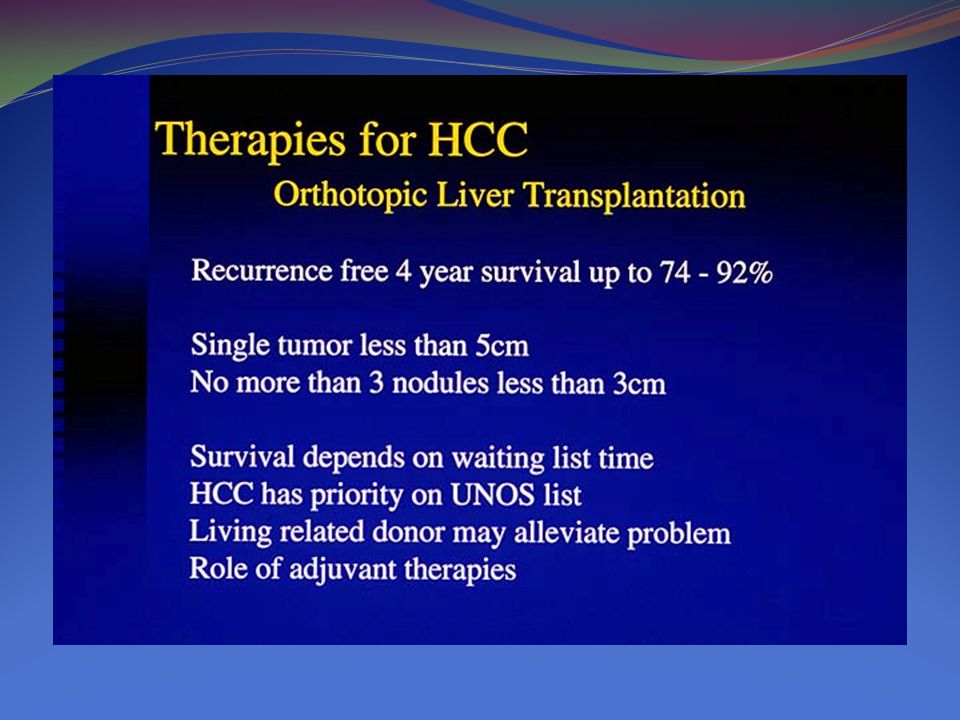
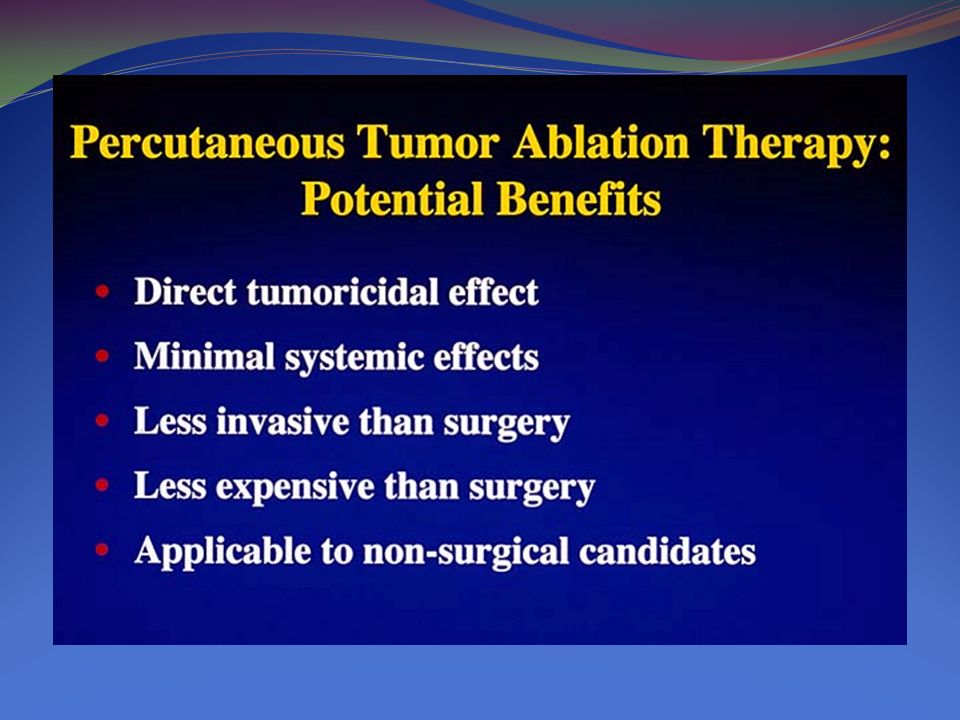
HCC Rx Early stage disease includes patients with preserved liver function (Child– Pugh A and B) with solitary HCC or up to 3 nodules 3 cm in size. These patients can be effectively treated by resection, liver transplantation or percutaneous ablation

21
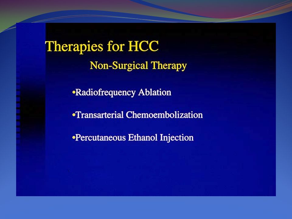
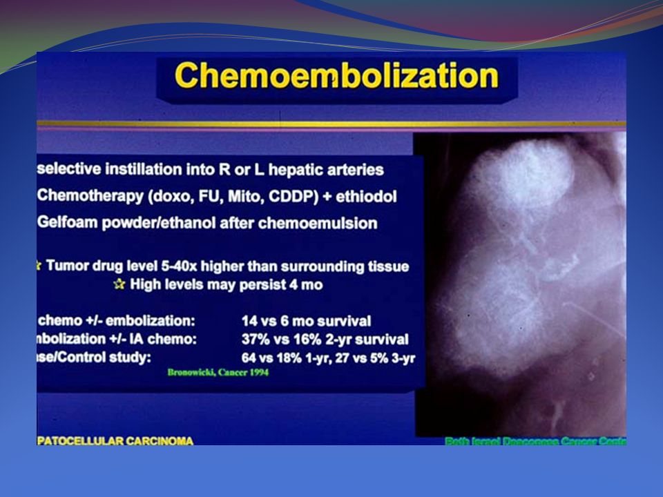
HCC Rx The intermediate stage consists of Child–Pugh A and B patients with large/multifocal HCC who do not have cancer related symptoms and do not have macrovascular invasion or extrahepatic spread. optimal candidates for transarterial chemoembolization (TACE).
..")
22
HCC Rx Patients who present with cancer symptoms and/or with vascular invasion or extrahepatic spread comprise the advanced stage. They have a shorter life expectancy (50% survival at 1 year) and are candidates for sorafenib
 and are candidates for sorafenib.")
23
HCC Rx Finally, patients with extensive tumor involvement leading to severe deterioration of their physical capacity [WHO performance status >2] and/or major impairment of liver function (Child–Pugh C) are considered end stage. Their median survival is less than 3 months.
![HCC Rx Finally, patients with extensive tumor involvement leading to severe deterioration of their physical capacity [WHO performance status >2] and/or major impairment of liver function (Child–Pugh C) are considered end stage.](http://images.slideplayer.com/35/10525114/slides/slide_23.jpg "Their median survival is less than 3 months..")
33
Fibrolamellar HCC Generally in young individuals Lacking a background of chronic liver disease and other risk factors for HCC The clinical presentations generally nonspecifi c Alpha-fetoprotein level is typically within the normal range in most cases

34
F HCC Imaging studies have a major role in clinical diagnosis, but pathology is the gold standard in confi rming diagnosis. Pathological characteristics of FLHCC include the presence of tumor cells with a deeply eosinophilic cytoplasm and macronucleoli surrounded by abundant fi brous bands. The most effective treatment for FLHCC is aggressivesurgical resection.

35
FHCC

45
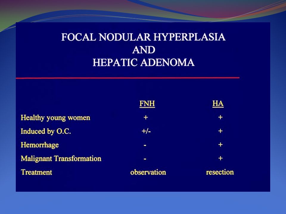
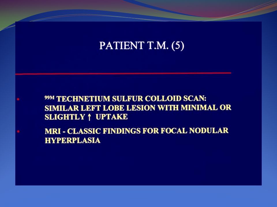
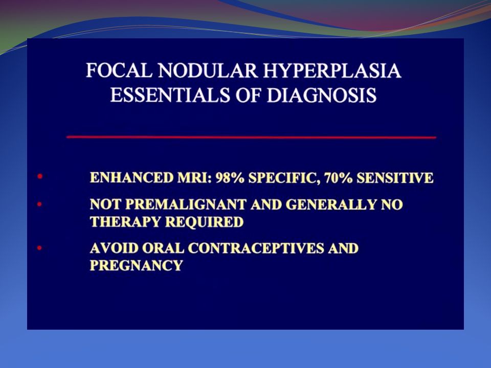
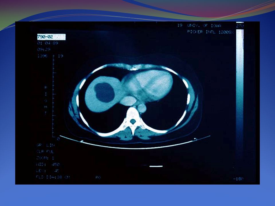
Focal Nodular Hyperplasia

Similar presentations


 for Liver Tumour Dr Dai Wing Chiu Queen Mary Hospital.>")


 New mass/nodule NoYes Alternative imaging technique Atypical featureTypical.>")


 is the most common type of primary liver cancer. Worldwide, its prevalence follows that of hepatitis B.>")


: Principal.>")
